3
DNA content evaluation for epithelial ovarian cancer identification
Analiza zawartości DNA w rozpoznaniu nabłonkowego raka jajnika
1 Centre of Gynecological Oncology and Breast, Hospital Santo Antonio – Centro Hospitalar do Porto (CHP), Porto, Portugal2 Department of Pathology, Hospital Santo Antonio – Centro Hospitalar do Porto (CHP), Porto, Portugal
3 Department of Immunology and Pathology, Portuguese Institute of Oncology (Instituto Português de Oncologia, IPO), Porto, Portugal 4 Ex-High Technician of Community Health, Instituto de Ciências Biomédicas de Abel Salazar (ICBAS), Porto University, Portugal
5 Universidade Católica Portuguesa – Centre for Interdisciplinary Research in Health (Universidade Católica Portuguesa – Centro de Investigação Interdisciplinar em Saúde, UCP – CIIS), Porto, Portugal 6 Department of Medicine (Oncology), Portuguese Institute of Oncology (Instituto Português de Oncologia, IPO), Porto, Portugal
7 Ex-Director of Department of Pathology, Hospital Santo António – Centro Hospitalar do Porto (CHP), Porto, Portugal
Correspondence: Prof. Doutor João C. Amado, Universidade Católica Portuguesa, Campus Foz, Rua Diogo Botelho, 1327 – 4169-005 Porto, Portugal, ORCID: 0000-0003-0358-7970, e-mail: [email protected]
Objective: To assess the cellular DNA status of epithelial ovarian cancer cells for clinical stage identification and its effect on survival. Methods: Sixty-two patients treated by primary surgery and six courses of platinum-based chemotherapy were enrolled. The surgical stage was analyzed in correlation with DNA ploidy, S-phase fraction and DNA index. DNA analysis was performed via image cytometry. Results: From the 62 cases, 38 were International Federation of Gynecology and Obstetrics (Fédération Internationale de Gynécologie et d’Obstétrique, FIGO) stage I and II, 24 – stage III and IV. In the DNA histograms obtained, the DNA index ranged from 0.85 to 3.02. Sixteen were classified as diploid and 46 as aneuploid (18 multiploid). S-phase fraction ranged from 9.8 to 51%. The aneuploid cells with DNA content above 5C ranged from 0.0 to 77.2%. Patients diagnosed with FIGO III and IV (vs. I and II) were 3.3 times more likely to die. Only in FIGO stage I and II the survival differed significantly for the different groups of ploidy. The risk of death for the multiploid (vs. diploid) group is 6.4 times and for aneuploid (vs. diploid) 2.3 times. Overall survival was better in the group with low DNA index. The low percentage compared with a high percentage of 5C cells ploidy groups showed association with mortality. The death hazard for the S-phase >33 median group is 4.9 times the hazard in relation to the S-phase <33. Conclusions: DNA ploidy, DNA index, S-phase, and 5C cells are important prognosticators for epithelial ovarian cancer mainly in early stages.
Keywords: DNA content, epithelial ovarian cancer, prognosis
Cel: Ocena statusu DNA komórek nabłonkowego raka jajnika w różnych stopniach zaawansowania klinicznego i jego wpływ na przeżycie. Metoda: Do badania zakwalifikowano 62 pacjentki leczone operacyjnie i za pomocą chemioterapii opartej na platynie (6 kursów). Stopień zaawansowania klinicznego nowotworu analizowano w odniesieniu do ploidalności DNA, frakcji fazy S oraz indeksu DNA. Analizę DNA przeprowadzono z zastosowaniem cytometrii obrazowej. Wyniki: Spośród 62 pacjentek 38 zakwalifikowano jako stopień zaawansowania I i II, natomiast 24 – jako stopień III i IV wg FIGO (International Federation of Gynecology and Obstetrics). W otrzymanych histogramach DNA indeks DNA wynosił 0,85–3,02. Szesnaście przypadków raka zaklasyfikowano jako diploidalne, natomiast 46 – jako aneuploidalne (18 multiploidalnych). Frakcja fazy S mieściła się w przedziale 9,8–51%. Odsetek komórek aneuploidalnych z zawartością DNA powyżej poziomu 5C wynosił 0,0–77,2%. Ryzyko śmierci było 3,3-krotnie większe w przypadku pacjentek z chorobą w stopniu zaawansowania FIGO III i IV (w porównaniu z I i II). Jedynie w przypadku stopni zaawansowania FIGO I i II odnotowano istotnie różnice w przeżywalności między poszczególnymi grupami ploidalności. Ryzyko śmierci było 6,4-krotnie większe w przypadku multiploidalności i 2,3-krotnie większe w przypadku aneuploidalności (w porównaniu z diploidalnością). Dłuższe przeżycie ogólne odnotowano w grupie o niskim indeksie DNA. Wykazano związek między niskim odsetkiem komórek 5C, w porównaniu z wysokim odsetkiem tych komórek w grupach poliploidalnych, a śmiertelnością. Ryzyko śmierci było 4,9-krotnie większe w grupie z medianą liczby komórek w fazie S >33 w porównaniu z medianą liczby komórek w fazie S <33. Wnioski: Ploidalność DNA, indeks DNA, faza S oraz obecność komórek 5C to ważne czynniki prognostyczne u pacjentek z nabłonkowym rakiem jajnika, głównie we wczesnym stadium.
Słowa kluczowe: zawartość DNA, nabłonkowy rak jajnika, rokowanie
Abstract
Streszczenie
António Tomé
1, Irene Leal
2, Carlos Palmeiras
3,
Eduarda Matos
4, João Amado
5, Miguel Abreu
6, Carlos Lopes
7Received: 15.02.2018 Accepted: 17.04.2018 Published: 29.05.2018
4
INTRODUCTION
E
pithelial ovarian cancer (EOC) is the fifth most com-mon female malignancy worldwide; more than 70% of patients are diagnosed with advanced disease, and 5-year survival rates are less than 30% with approximately 141,000 new cases and 106,000 deaths annually(1,2). The main prognostic factors considered in the evaluation of ovarian cancer are International Federation of Gynecology and Obstetrics (Fédération Internationale de Gynécologie et d’Obstétrique, FIGO) stage, the presence of a residual tumor, histological grade, histological type, and age(3–6). Most of them have significant shortcomings due to their subjectivity and lack of reproducibility, and low tic power. Therefore, the search for an additional prognos-tic factor that can be more objectively measured and with better reproducibility has been intensified. One of the more promising candidates in this regard is DNA content mea-surement(7–26). While there have been reports on a correla-tion between ploidy pattern and clinical pathological find-ings and therapeutic results, some other reports have shown no correlation between these factors(12). Thus, the results are variable and no definite conclusions may be drawn. DNA aneuploidy, which indicates a state with an abnor-mal DNA and chromosome content, has been identified or mentioned in various human cancers. This parameter is considered to be an important biological and prognostic variable in ovarian cancer(17,18,22,23).Numerous studies showed a close association between tumor ploidy and outcome (prognosis). Among patients with advanced ovarian cancer, those with diploid tumors have a significantly better survival than those with aneu-ploid tumors(8,11,24).
A multivariate analysis has shown that ploidy is a pow-erful and independent prognosis variable. These findings have been confirmed in most studies and ploidy/DNA index (DI) is a prognostic value in patients with both early- and advanced-stage ovarian tumors as well as in those with borderline ovarian tumors(25,26).
In the case of ovarian carcinoma, some authors have reported an objective assessment of grading by cytophotometry, achieving a good correlation between the histological grade of ovarian tumors and DNA content(7,10). Borderline and early-stage ovarian tumors had a significantly lower number of aneuploid cells, whereas advanced-stage invariant tumors had a significantly high number of aneuploid cells.
Image cytometry studies report that it is possible to deter-mine the ploidy level in a given tumor using the parameters obtained from DNA histogram, assessed based on Feulgen-stained nuclei. The percentage of aneuploid cells with DNA content above 5C (5CER) is another ploidy parameter and is important in assessing ovarian cancer aggressiveness, being a reliable biomarker in the risk assessment for this type of tumor(23–26).
This aim of the study was to evaluate the value of DNA content (DNA ploidy/DI/S-phase, 5CER), using image
cytometry, for predicting long-term survival of patients with epithelial ovarian cancer.
MATERIAL AND METHODS
The study comprised 62 patients with ovarian cancer diag-nosis who were treated from January 1982 to December 1997 in the Hospital Geral de Santo Antonio, Porto, Portugal.
All patients were treated by multidisciplinary medical surgi-cal teams and following international protocols (defined by the corresponding FIGO stage), and were subjected to che-motherapy based on platinum when indicated.
All patients had invasive tumors. All histological sections were reviewed by a reference pathologist and histological classifications were performed using the criteria defined by the World Health Organization (WHO). The tumors were graded according to the WHO histological grading system as grade 1, 2 or 3. Clinical information was available for all patients (date of initial diagnosis, surgical stage, tumor grade, initial tumor volume, residual tumor volume, histo-logical differentiation, age, treatment, follow-up) and the date of death confirmed. The study ended in June 2006.
Quantification of DNA content
Ovary sections of 6 µm were cut from paraffin-embed-ded blocks, deparaffinized and rehydrated. The slides were then Feulgen-stained with CAS DNA Staining Kit (Cell Analysis Systems, Elmhurst, IL), according to the manu-facturer’s instructions. Briefly, the sections were hydrolyzed in 5N HCL solution for 60 minutes and then transferred to the CAS DNA stain solution for 1 hour. After staining, the slides were placed in three consecutive CAS rinse solu-tions for 30 seconds, 5 minutes and 10 minutes, respec-tively. The slides were then washed in deionized water and placed in acid alcohol solution for 5 minutes. Finally, the slides were dehydrated and mounted. The Feulgen reaction produced a blue staining of nuclear DNA reflecting stoi-chiometric binding of the stain to the DNA. CAS staining of rat hepatocytes was used as a control slide. These cells, having a known quantity of DNA, were used as the exter-nal control, allowing for instrument calibration prior to DNA image analysis. The nuclear DNA content of cells was measured with the CAS 200 image analysis system (Cell Analysis Systems, Elmhurst, IL). The quantitative DNA analysis system is well suited for DNA content evaluation in human cells. However, if the diploid value of the specimens studied is different from human diploid cells, an “internal control” must be used, such as lymphocytes. For this reason in each tumor in addition to a minimum of 130 non-over-lapping and well preserved ovarian nuclei, 20 to 30 normal lymphocytes were measured. The resultant DNA histo-grams were analyzed by previously described methods(12). For each tumor, G0/G1 peak was visually identified, mean, standard deviation (SD) and coefficient of variation (CV)
5
values were calculated. The DI describes the relative DNA content of the study population and was defined as the ratio of the mean DNA content of the ovarian G0/G1 peak divided by the mean DNA content of the resting diploid lymphocyte G0/G1 peak. The 5CER was also evaluated and defined as the percentage of cells with DNA values above 5n. Tumors were considered aneuploid only if a separate G0/G1 peak was distinguishable on the histogram and its DI was different from the reference lymphocyte population by more than 2 SD, mean standard deviation. A DNA dip-loid tumor showed a single distinct G0/G1 peak with a DI within 2 SD from the control lymphocytes and usually with less than 1% of 5CER.
Statistical analysis
The probability for the risk of death after diagnosis was cal-culated according to the Kaplan–Meier method. The influ-ence of concomitant covariates on the cumulative probabil-ity rates was analysed using the proportional hazard model described by Cox.
RESULTS
All tumors were analyzed by image cytometry for ploidy, the percentage of cells in S-phase, and 5CER (5C).
Evaluable image cytometry DNA histograms were obtained from all 62 patients with ovarian carcinoma.
The DI ranged from 0.85 to 3.02. Sixteen (25.8%) ovar-ian carcinomas were classified as diploid and 46 (74.2%) as aneuploid including 18 multiploid tumors.
The S-phase fraction (SPF) ranged from 9.8 to 51% with a median value of 33%. This value was not observed in 20 patients.
The percentage of 5C ranged from 0.0 to 77.2% with a median value of 3.15%.
Survival by FIGO
FIGO staging was performed in 62 patients. For the pur-pose of survival analysis, tumors were classified as early stage (FIGO I + FIGO II) or advanced stage (FIGO III and FIGO IV).
Patients diagnosed with FIGO III and IV were 3.3 times more likely to die than patients with FIGO I and II (95% confidence interval, CI: 1.667–6.564).
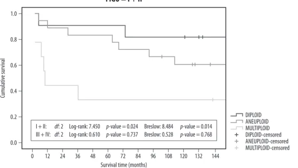
Survival for types of ploidy
by stage of FIGO
It was shown (Fig. 1) that only in FIGO I and II the sur-vival differed significantly for the different groups of ploidy: I + II: df: 2 Log-rank: 7.450 p-value = 0.024 Breslow: 8.484 p-value = 0.014
III + IV: df: 2 Log-rank: 0.610 p-value = 0.737 Breslow: 0.528 p-value = 0.768
The hazard for the multiploid group is 6.4 times the risk of death in relation to the diploid group (95% CI: 1.283–32.171) with statistical significance (p = 0.024). The hazard for the aneuploid is 2.3 times the risk of death for the diploid, without significance (95% CI: 0.474–11.015).
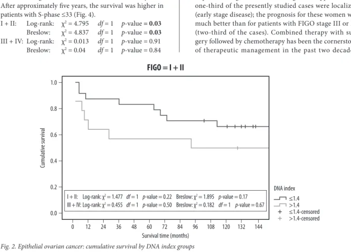
Survival for DNA median value
Considering the test in each stratum (Fig. 2) it can be seen that for FIGO I and II the survival does not differ signifi-cantly between the groups of DNA, but the curves are well separated:
FIGO = I + II
Survival time (months) 1.0 0.8 0.6 0.4 Cumula tiv e sur viv al 0.2 0.0 0 12 24 36 48 60 72 84 96 108 120 132 144
I + II: df: 2 Log-rank: 7.450 p-value = 0.024 Breslow: 8.484 p-value = 0.014 III + IV: df: 2 Log-rank: 0.610 p-value = 0.737 Breslow: 0.528 p-value = 0.768
DIPLOID DIPLOID-censored ANEUPLOID ANEUPLOID-censored MULTIPLOID MULTIPLOID-censored
6
I + II: Log-rank: χ2 = 1.477 df = 1 p-value = 0.22 Breslow: χ2 = 1.895 p-value = 0.17 III + IV: Log-rank: χ2 = 0.455 df = 1 p-value = 0.50
Breslow: χ2 = 0.182 df = 1 p-value = 0.67 Overall, the survival is higher in the group with low DNA index (Tab. 1) compared with the group with high DNA index, however with no statistical significance.
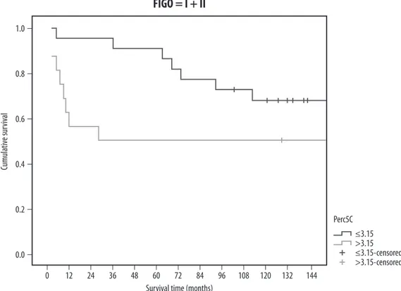
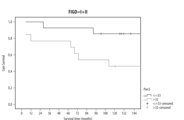
The analysis in each stratum shows that in FIGO I and II the survival does not differ significantly between the groups of the 5C percentage (Fig. 3), but the curves are well separated: I + II: Log-rank: χ2 = 2.414 df = 1 p-value = 0.12 Breslow: χ2 = 3.58 df = 1 p-value = 0.06 III + IV: Log-rank: χ2 = 0.331 df = 1 p-value = 0.56 Breslow: χ2 = 0.252 df = 1 p-value = 0.62 Results for the low percentage of 5C cells ploidy groups (≤3.15) compared with high percentage of 5C cells (>3.15) (Tab. 2 and Fig. 4) showed association with mortality. After approximately five years, the survival was higher in patients with S-phase ≤33 (Fig. 4).
I + II: Log-rank: χ2 = 4.795 df = 1 p-value = 0.03 Breslow: χ2 = 4.837 df = 1 p-value = 0.03 III + IV: Log-rank: χ2 = 0.013 df = 1 p-value = 0.91 Breslow: χ2 = 0.04 df = 1 p-value = 0.84
The hazard for the S-phase >33 (median) group is 4.9 times the risk in relation to the S-phase <33 (95% CI: 1.009–23.572) with statistical significance (p = 0.049). The clinical stage was the strongest covariate, with FIGO III and IV patients being, 3 times more likely to die than patients with FIGO I and II (95% CI: 1.571–5.610).
DISCUSSION
Ovarian cancer is the leading cause of death from gyne-cologic malignancies in the Western world. The majority of patients are diagnosed with advanced stage disease pri-marily due to peritoneal seeding from the primary tumor, which leads to peritoneal carcinomatosis without specific signs or symptoms. In addition, screening strategies have not yet been proven effective in increasing the number of patients diagnosed at earlier stages(2,27). Approximately one-third of the presently studied cases were localized (early stage disease); the prognosis for these women was much better than for patients with FIGO stage III or IV (two-third of the cases). Combined therapy with sur-gery followed by chemotherapy has been the cornerstone of therapeutic management in the past two decades.
FIGO = I + II
Survival time (months) 1.0 0.8 0.6 0.4 Cumula tiv e sur viv al 0.2 0.0 0 12 24 36 48 60 72 84 96 108 120 132 144
I + II: Log-rank: χ2 = 1.477 df = 1 p-value = 0.22 Breslow: χ2 = 1.895 p-value = 0.17
III + IV: Log-rank: χ2 = 0.455 df = 1 p-value = 0.50 Breslow: χ2 = 0.182 df = 1 p-value = 0.67
≤1.4-censored ≤1.4 DNA index
>1.4-censored >1.4
Fig. 2. Epithelial ovarian cancer: cumulative survival by DNA index groups
FIGO N % censored Median confidence interval95% median Upper quartile
I + II Perc5C ≤ 3.15Perc5C > 3.15 2216 68.250.0 28- -- 927
III + IV Perc5C ≤ 3.15Perc5C > 3.15 159 22.213.3 1914 0.000–42.3747.688–20.312 88
Per5C – percentage of 5C.
Log rank: χ2 = 2.102, df = 1, p-value = 0.15; Breslow: χ2 = 3.557, df = 1, p-value = 0.06.
7
Substantial progress has been made and although an increasing number of patients live longer with their dis-ease, the majority of patients with advanced ovarian can-cer are not cured. The prognosis in ovarian cancan-cer is dis-couraging compared to other malignancies of the female genital tract. Despite aggressive surgery and intensified chemotherapy, the outcome of patients with stage III and IV is poor. Relevant prognostic factors are necessary to estimate the course of the disease and to define biolog-ically similar subgroups for the analysis of therapeutic efficacy(10,28). The prognosis of patients with ovarian can-cer was suggested to depend on individual tumor charac-teristics rather than on therapy(10,13). Many studies have been devoted to finding “prognostic factors,” and numer-ous features have been described that can help predict the prognosis of early and advanced ovarian cancer with varying degrees of accuracy. One of the most promis-ing candidates in this regard is DNA content measure-ment. In the early 1970s, DNA ploidy in ovarian cancer was suggested to be of prognostic impact. It has been con-firmed in most studies that DNA content (DNA ploidy/ S-phase/DI/5C) is of prognostic value in patients with both early and advanced stage ovarian tumors as well as in those with borderline ovarian tumors(15,17,18,25,29–31), results that we could associate with recent EGFL7 expres-sion studies(32).
In this study, unlike in the great majority of other stud-ies, the percentage of early stage (I–II) cancers was higher than the advanced stage cancers. This is due to the fact that many of these cases were referred to hospital.
Fig. 3. Epithelial ovarian cancer cumulative survival: low vs. high percentage of 5C cells groups
Most of the patients were diagnosed in routine gyneco-logic exams or by other specialists after imaging exams for different clinical indications. In fact, nowadays, an increased number ovarian cancers are diagnosed at early stages, also as a result of improved primary care provided by general practitioners. Nevertheless, we still diagnose a great number of advanced stage cancers, although this number is decreasing. In our study, 38 cases were FIGO stages I and II and 24 cases were stage III and IV. Concerning the survival rate, groups I/II and III/IV were formed. In the first group, the survival rate was 3.3 times higher when compared with the second one. The stage is considered the most important prognostic factor in epi-thelial ovarian cancer. There are subgroups of patients with significant differences in survival rates: patients with surgically documented early disease (localized) versus patients with advanced disseminated disease. The 5-year survival rate is about 70–90% for the first groups and 10–18% for the second one, confirming the results of different studies(27,32).
The survival rate in relation to ploidy and stages showed that significant differences were present only in FIGO stages I and II, for the different ploidy groups. The group of aneuploid tumors was most frequent. The frequency of aneuploidy increases, among other things, with age, advanced disease, histologic type(3). As in other studies, the non-diploid tumors were more frequent in advanced stage tumors. The survival rate for diploid tumors was 4–6 times greater when compared with multiploid tumors. So, in the same stage, the DNA ploidy showed
FIGO = I + II
Survival time (months) 1.0 0.8 0.6 0.4 Cumula tiv e sur viv al 0.2 0.0 0 12 24 36 48 60 72 84 96 108 120 132 144 ≤3.15-censored ≤3.15 Perc5C >3.15-censored >3.15
8
to be an inadequate prognostic factor, with lower sur-vival rates for the non-diploid tumors than the diploid tumors. Tropé et al. showed that in FIGO stage I DNA ploidy was a significant independent prognostic factor for disease-free survival with p < 0.0001(22). It was reported that a DNA index of more than 1.3 was the most impor-tant prognostic factor in multivariate analysis(29), in con-trast to the report of Friedlander et al., who found that in the group of periploid tumors only the exact DNA-diploid tumors are relevant for a better prognosis(8,9). In this investigation, tumors with DNA index less than 1.4 had better survival rate compared with tumors with a DNA index higher than 1.4, although no statistical dif-ference was observed in this study. Tumors with a higher DNA index have a more aggressive biological nature(33–37). There is a correlation between DNA ploidy/DI and other factors, such as histological grade, with higher aggressive-ness in high grade tumors. Aneuploid tumors were more often poorly differentiated than euploid tumors.
Multiploid tumors differ in survival rate, with lower survival versus diploid tumors. Patients with aneuploid
tumors with a high percentage of 5C cells (multiploid) had significantly poorer 2 and 5-year corrected surviv-als than patients with diploid tumors. In this study, the tumors with 5C ploidy lower than 3.15 showed associa-tion with better survival rates when compared to tumors with an elevated percentage of 5C cells, with advanced stage cancers having a higher number of 5C cells. The cell kinetic data are also important indicators of tumor aggressiveness and treatment response. Tumor growth is determined by a balance between tumor cell proliferation and cell loss, and proliferation is, in turn, dependent on the fraction of proliferating cells and cell cycling time. Determination of ovarian tumor proliferating activity will be of prognostic value. Several methods exist for this purpose, including Ki-67 expression and cytomet-ric SPF. DNA ploidy and SPF showed a significant corre-lation with survival in malignant tumor, while SPF > 10% showed significant correlation with decreased survival in stage I and II cancer(29,30,38). There are some data to sug-gest that the SPF may be of prognostic significance in ovarian cancer, but there are also a number of studies
Fig. 4. Epithelial ovarian cancer cumulative survival: S-phase >33 (median) vs. S-phase <33 groups
FIGO=I+II
Survival time (months) 1,0 0,8 0,6 0,4 Cum S ur viv al 0,2 0,0 0 12 24 36 48 60 72 84 96 108 120 132 144 <=33-censored <=33 PercS >33-censored >33
FIGO N % censored Median confidence interval95% median Upper quartile
I + II S-phase ≤ 33S-phase > 33 1413 85.746.2 112- -- 63
-III + IV S-phase ≤ 33S-phase > 33 114 25.09.1 1511 2.053–27.9470.000–25.700 85
Log rank: χ2 = 2.095, df =1, p-value = 0.15; Breslow: χ2 = 4.38, df =1, p-value = 0.036.
9
that contradict these results(25). Coley et al. published that tumors with a high SPF showed shorter median sur-vival compared to those with a low SPF(13). The aneuploid tumor also showed the highest SPFs, which was indica-tive of a high proliferation rate(13). In Norwegian Radium Hospital series, the diploid tumors had a significantly lower SPF than the aneuploid tumors. One of the reasons why the results from previous works have been contro-versial is probably the tumor heterogeneity(30,39). In our study patients with S-phase ≤33 had greater survival rate when compared with patients with S-phase >33, with a risk of survival less than 4.9 showing statistical signifi-cance in the present study.
CONCLUSION
In this study we can conclude that all the variables that differed in mortality occurred in patients with early stage cancers (I and II), although these are increasingly detected with higher sensibility and specificity of screening using CA-125 plus HE4 and biomarkers(2,27,28). In these stages, the multiploid tumors with elevated S-phase showed a predic-tive value for higher risk of mortality.
The DNA content is an important prognostic factor for epi-thelial ovarian cancer. Other important factors like resid-ual disease, age, histology type and grade were not evalu-ated as well as other imunohistochemical factors including ARID1A, CA-125 and its new cut-off of 18 U/mL(32,40). The study of these other factors would contribute to a bet-ter understanding of this cancer.
Conflict of interest
No potential conflict of interest relevant to this article was reported.
Acknowledgements
To all contributors for this paper, we must register our acknowledgment.
References
1. Kim YH, Kim SC: Recent advances in the biomarkers for epithe-lial ovarian cancer. J Gynecol Oncol 2011; 22: 219–221. 2. Chiang YC, Chen CA, Chiang CJ et al.: Trends in incidence and
survival outcome of epithelial ovarian cancer: 30-year national population-based registry in Taiwan. J Gynecol Oncol 2013; 24: 342–351.
3. Vergote I: Prognostic factors in stage I ovarian carcinoma. Verh K Acad Geneeskd Belg 2001; 63: 257–271.
4. DiSilvestro P, Peipert JF, Hogan JW et al.: Prognostic value of clinical variables in ovarian cancer. J Clin Epidemiol 1997; 50: 501–505.
5. Clark TG, Stewart ME, Altman DG et al.: A prognostic model for ovarian cancer. Br J Cancer 2001; 85: 944–952.
6. Friedlander ML: Prognostic factors in ovarian cancer. Semin Oncol 1998; 25: 305–314.
7. Friedlander ML, Hedley DW, Taylor IW et al.: Influence of cel-lular DNA content on survival in advanced ovarian cancer. Cancer Res 1984; 44: 397–400.
8. Friedlander ML, Hedley DW, Swanson C et al.: Prediction of long-term survival by flow cytometric analysis of cellular DNA content in patients with advanced ovarian cancer. J Clin Oncol 1988; 6: 282–290.
9. Rice LW, Mark SD, Berkowitz RS et al.: Clinicopathologic vari-ables, operative characteristics, and DNA ploidy in predicting outcome in ovarian epithelial carcinoma. Obstet Gynecol 1995; 86: 379–385.
10. Ozalp S, Yalcin OT, Gulbas Z et al.: Effect of cellular DNA con-tent on the prognosis of epithelial ovarian cancers. Gynecol Obstet Invest 2001; 52: 93–97.
11. El-Naggar AK, Vielh P: Solid tumor DNA content analysis. Methods Mol Biol 2004; 263: 355–370.
12. Silvestrini R: Relevance of DNA-ploidy as a prognostic instru-ment for solid tumors. Ann Oncol 2000; 11: 259–261. 13. Coley HM, Sargent JM, Titley J et al.: Lack of prognostic
signif-icance of ploidy and S-phase measurements in advanced ovarian cancer. Anticancer Res 1999; 19: 2111–2116.
14. Winter WE 3rd, Maxwell GL, Tian C et al.; Gynecologic Oncology Group Study: Prognostic factors for stage III epithelial ovarian cancer: a Gynecologic Oncology Group Study. J Clin Oncol 2007; 25: 3621–3627.
15. Skirnisdóttir I, Sorbe B, Karlsson M et al.: Prognostic importance of DNA ploidy and p53 in early stages of epithelial ovarian car-cinoma. Int J Oncol 2001; 19: 1295–1302.
16. Curling M, Stenning S, Hudson CN et al.: Multivariate analyses of DNA index, p62c-myc, and clinicopathological status of patients with ovarian cancer. J Clin Pathol 1998; 51: 455–461. 17. Schueler JA, Trimbos JB, vd Burg M et al.: DNA index reflects the
biological behavior of ovarian carcinoma stage I–IIa. Gynecol Oncol 1996; 62: 59–66.
18. Kim YT, Zhao M, Kim SH et al.: Prognostic significance of DNA quantification by flow cytometry in ovarian tumors. Int J Gynaecol Obstet 2005; 88: 286–291.
19. Milczek T, Klasa-Mazurkiewicz D, Emerich J et al.: [Prognostic significance of Sphase fraction in ovarian cancer patients]. Ginekol Pol 2006; 77: 840–847.
20. Yoon BS, Kim YT, Kim S et al.: Prognostic value of nuclear DNA quantification and cyclin A expression in epithelial ovarian car-cinoma. Eur J Obstet Gynecol Reprod Biol 2008; 136: 110–115. 21. Novik VI, Gevorkian VA, Maksimov SIa: [Prognostic
signifi-cance of tumor cell ploidy in advanced ovarian carcinoma]. Vopr Onkol 2006; 52: 54–58.
22. Tropé CG, Abeler V, Baekelandt M et al.: [DNA ploidy in epithe-lial ovarian cancer – an independent prognostic factor]. Tidsskr Nor Laegeforen 2000; 120: 43–49.
23. Pietrzak K, Olszewski W: DNA ploidy as a prognostic factor in patients with ovarian carcinoma. Pol J Pathol 1998; 49: 141–144.
24. Lodhi S, Najam S, Pervez S: DNA ploidy analysis of borderline epithelial ovarian tumours. J Pak Med Assoc 2000; 50: 349–351. 25. Flezar MS, But I, Kavalar R et al.: Flow and image cytometric
DNA ploidy, including 5c exceeding cells, of serous borderline malignant ovarian tumors. Correlation with clinicopathologic characteristics. Anal Quant Cytol Histol 2003; 25: 139–145. 26. Hwang J, Na S, Lee H et al.: Correlation between preoperative
serum levels of five biomarkers and relationships between these biomarkers and cancer stage in epithelial ovarian cancer. J Gynecol Oncol 2009; 20: 169–175.
27. Oh J, Park SH, Lee TS et al.: High expression of epidermal growth factor-like domain 7 is correlated with poor differentia-tion and poor prognosis in patients with epithelial ovarian cancer. J Gynecol Oncol 2014; 25: 334–341.
28. Chiang AJ, Chen J, Chung YC et al.: A longitudinal analysis with CA-125 to predict overall survival in patients with ovarian cancer. J Gynecol Oncol 2014; 25: 51–57.
29. Klemi PJ, Joensuu H, Mäenpää J et al.: Influence of cellular DNA content on survival in ovarian carcinoma. Obstet Gynecol 1989; 74: 200–204.
10
30. Kaern J, Tropé CG, Kristensen GB et al.: Evaluation of deoxyri-bonucleic acid ploidy and S-phase fraction as prognostic param-eters in advanced epithelial ovarian carcinoma: a prospective study. Am J Obstet Gynecol 1994; 170: 479–487.
31. Khoo SK, Hurst T, Kearsley J et al.: Prognostic significance of tumor ploidy in patients with advanced ovarian carcinoma. Gynecol Oncol 1990; 39: 284–288.
32. Wagner TMU, Adler A, Sevelda P et al.: Prognostic significance of cell DNA content in early-stage ovarian cancer (FIGO stages I and II/A) by means of automatic image cytometry. Int J Cancer 1994; 56: 167–172.
33. Gajewski WH, Fuller AF Jr, Pastel-Ley C et al.: Prognostic signif-icance of DNA content in epithelial ovarian cancer. Gynecol Oncol 1994; 53: 5–12.
34. Pfisterer J, Kommoss F, Sauerbrei W et al.: Cellular DNA content and survival in advanced ovarian carcinoma. Cancer 1994; 74: 2509–2515.
35. Brescia RJ, Barakat RA, Beller U et al.: The prognostic signifi-cance of nuclear DNA content in malignant epithelial tumors of the ovary. Cancer 1990; 65: 141–147.
36. Resnik E, Trujillo YP, Taxy JB: Long-term survival and DNA ploidy in advanced epithelial ovarian cancer. J Surg Oncol 1997; 64: 299–303.
37. Reles AE, Gee C, Schellschmidt I et al.: Prognostic significance of DNA content and S-phase fraction in epithelial ovarian carci-nomas analyzed by image cytometry. Gynecol Oncol 1998; 71: 3–13.
38. Yokoyama Y, Matsushita Y, Shigeto T et al.: Decreased ARID1A expression is correlated with chemoresistance in epithelial ovar-ian cancer. J Gynecol Oncol 2014; 25: 58–63.
39. Kallioniemi OP, Punnonen R, Mattila J et al.: Prognostic signifi-cance of DNA index, multiploidy, and S-Phase fraction in ovar-ian cancer. Cancer 1988; 61: 334–339.
40. Kang S, Kim TJ, Seo SS et al.: Prediction of a high-risk group based on postoperative nadir CA-125 levels in patients with advanced epithelial ovarian cancer. J Gynecol Oncol 2011; 22: 269–274.
Zasady prenumeraty kwartalnika
„Current Gynecologic Oncology”
1. Prenumeratę można rozpocząć od dowolnego numeru pisma. Prenumerujący otrzyma zamówione numery kwartalnika pocztą na podany adres. 2. Pojedynczy egzemplarz kwartalnika kosztuje 40 zł.
Przy zamówieniu rocznej prenumeraty (4 kolejne numery) koszt całorocznej prenumeraty wynosi 120 zł. Koszt całorocznej prenumeraty zagranicznej wynosi 40 euro. 3. Istnieje możliwość zamówienia numerów
archiwalnych (do wyczerpania nakładu). Cena numeru archiwalnego – 40 zł. 4. Zamówienie można złożyć:
• Dokonując przelewu z własnego konta bankowego (ROR) – wpłaty należy kierować na konto: Medical Communications Sp. z o.o., ul. Powsińska 34, 02-903 Warszawa Deutsche Bank PBC SA
42 1910 1048 2215 9954 5473 0001 Prosimy o podanie dokładnych danych imiennych i adresowych.
• Drogą mailową: [email protected]. • Telefonicznie: 22 651 97 83.
• Wypełniając formularz prenumeraty
zamieszczony na stronie www.ginekologia.com.pl. 5. Zamawiający, którzy chcą otrzymać fakturę VAT,
proszeni są o kontakt z redakcją.
Rules of subscription to the quarterly
“Current Gynecologic Oncology”
1. Subscription may begin at any time. Subscribers will receive ordered volumes of the journal to the address provided.
2. A single volume of the quarterly costs 40 PLN (10 EUR). The cost of annual subscription (4 consecutive
volumes) is 120 PLN. The cost of annual subscription for foreign subscribers is 40 EUR.
3. Archival volumes may be ordered at a price of 40 PLN per volume until the stock lasts. 4. Orders may be placed:
• By making a money transfer from own bank account – payments should be made payable to: Medical Communications Sp. z o.o.,
ul. Powsińska 34, 02-903 Warszawa Deutsche Bank PBC SA
42 1910 1048 2215 9954 5473 0001 For foreign subscribers:
Account Name: Medical Communications Sp. z o.o. Bank Name: Deutsche Bank PBC S.A.
Bank Address: 02-903 Warszawa, ul. Powsińska 42/44 Account number: 15 1910 1048 2215 9954 5473 0002 SWIFT Code/IBAN: DEUTPLPK
Please provide a precise address and nominative data. • By e-mail: [email protected].
• Filling-in a subscription form, which may be found on the page www.ginekologia.com.pl. 5. Customers wishing a VAT invoice, are