R E S E A R C H
Open Access
Purification and characterization of a hyaluronidase
from venom of the spider
Vitalius dubius
(Araneae,
Theraphosidae)
Rafael Sutti
1, Mariana Leite Tamascia
2, Stephen Hyslop
2and Thomaz Augusto Alves Rocha-e-Silva
1*Abstract
Background:Venom hyaluronidase (Hyase) contributes to the diffusion of venom from the inoculation site. In this work, we purified and characterized Hyase from the venom ofVitalius dubius(Araneae, Theraphosidae), a large theraphosid found in southeastern Brazil. Venom obtained by electrical stimulation of adult male and female V. dubiuswas initially fractionated by gel filtration on a Superdex® 75 column. Active fractions were pooled and applied to a heparin-sepharose affinity column. The proteins were eluted with a linear NaCl gradient.
Results:Active fractions were pooled and assessed for purity by SDS-PAGE and RP-HPLC. The physicochemical tests included optimum pH, heat stability, presence of isoforms, neutralization by flavonoids and assessment of commercial antivenoms. Hyase was purified and presented a specific activity of 148 turbidity-reducing units (TRU)/mg (venom: 36 TRU/mg; purification factor of ~4). Hyase displayed a molecular mass of 43 kDa by SDS-PAGE. Zymography in
hyaluronic-acid-containing gels indicated an absence of enzyme isoforms. The optimum pH was 4-5, with highest activity at 37°C. Hyase was stable up to 60°C; but its activity was lost at higher temperatures and maintained after several freeze-thaw cycles. The NaCl concentration (up to 1 M) did not influence activity. Hyase had greater action towards hyaluronic acid compared to chondroitin sulfate, and was completely neutralized by polyvalent antiarachnid sera, but not by caterpillar, scorpion or snakes antivenoms.
Conclusion:The neutralization by arachnid but not scorpion antivenom indicates that this enzyme shares antigenic epitopes with similar enzymes in other spider venoms. The biochemical properties of this Hyase are comparable to others described.
Keywords:Hyaluronidase, Venom, Purification,Vitalius dubius
Background
Spider venoms comprise complex mixtures of compo-nents of low molecular weight, peptides and proteins [1]. Among the enzymatic activities, collagenase and hy-aluronidase (Hyase) are often found and were formerly attributed to matrix degradation [1].
Hyase activity is found in several spider species, such as Cupiennius salei, Lycosa godeffroy, Lampona cylin-drata/murina, Loxosceles recluse, Loxosceles rufescens, Loxosceles deserta, Loxosceles gaucho, Loxosceles laeta, Loxosceles recluse, and Loxosceles intermedia [1-6]. The
first report of hyaluronidasic activity was in venoms of
the Brazilian spiders Lycosa raptorial and Phoneutria
nigriventer [7,8]. The first Hyase to be purified from a Theraphosidae spider was the one from the tarantula Dugesiella hentzi (Girard), which was characterized and identified as the major venom component [9].
The clinical relevance of Hyases is not confined to a toxin-spreading factor, since it acts as an allergen in a manner similar to hymenoptera venoms. Hyase, phospho-lipase A2 and melitin were identified as the tree major causes of allergic reactions involved in bee stings, and phospholipase A1 and antigen 5 are the major allergens in wasp venoms [10,11].
Although tarantula bites to humans are relatively rare [12-14], these spider venoms represent a rich source of
* Correspondence:[email protected]
1Department of Physiological Sciences, Santa Casa de São Paulo Medical
School, Rua Cesário Motta Jr., 61, Vila Buarque, CEP 01.221-020 São Paulo, SP, Brasil
Full list of author information is available at the end of the article
bioactive molecules for scientific interest, basic research and possible therapeutic applications [15-18].
The tarantula Vitalius dubius (Mello-Leitão, 1923) is
characterized as nonaggressive and is found in the very populous area of southeastern Brazil [19]. Recent studies
have shown that V. dubius venom has a complex
bio-chemical and pharmacological composition, with differ-ent biological activities [20,21]. In the presdiffer-ent work we describe the purification and characterization of the hy-aluronidase present in its venom.
Methods Reagents
Refined chemicals were purchased from Sigma Chemical Co. (USA) whereas heparin was purchased from Labora-tório Cristália (Brazil). Molecular mass markers for SDS-PAGE were acquired from BioRad (USA). The resins for column chromatography were purchased from GE Health-care Life Sciences (Sweden). The HPLC column was a
Jupiter C18 (4.6 mm×250 mm×12 μm) acquired from
Phenomenex (USA).
Spiders and venom extraction
Specimens ofV. dubiusand Phoneutria nigriventerwere
provided by the Centers for Zoonosis Control for the cities in the region of Campinas, São Paulo state. The spiders were identified and kept in captivity, where they
were fed cockroaches and water ad libitum. The
taran-tula venom was extracted as described by Rocha-e-Silva et al.[20] then lyophilized and stored at−80°C until use.
Antisera
We used six antivenom sera produced by the Butantan Institute in São Paulo: (1) antiarachnid (Phoneutria nigriventer,Loxosceles gauchoandTityus serrulatus); (2) antiscorpionic (T. serrulatus); (3) antilonomic (Lonomia obliqua); (4) antibothropic (Bothrops alternatus,B. jar-araca, B. jararacussu, B. moojeni and B. neuwiedi); (5)
anticrotalic (Crotalus durissus terrificus and Crotalus
durissus collilineatus); and (6) antielapidic (Micrurus frontalisandM. corallinus).
Enzymatic activity
Hyaluronidase activity was determined by a turbidimet-ric method [22]. The working solution for this trial con-sisted of a 200μL buffer (0.2 M sodium acetate, pH 6.0,
containing 0.15 M NaCl), 200 μL of substrate
(hyalur-onic acid from human umbilical cord 1 mg/mL in acet-ate buffer) and 100μL of enzyme (20 μg) to give a total
reaction volume of 500μL. This mixture was incubated
for 15 minutes at 37°C after which the reaction was stopped by adding 2 mL of hexadecyltrimethylammo-nium bromide in 2.5% NaOH. The resulting turbidity was read at 400 nm in a SpectraMax® 340 microplate
reader (Molecular Devices, USA) after 30 minutes of in-cubation at room temperature. One unit of activity cor-responded to the amount of enzyme that produced a 50% reduction in turbidity caused by 200μg of substrate under the conditions described above.
Purification of Hyaluronidase
The venom (10 mg) ofV. dubiuswas dissolved in 0.1 M
sodium acetate buffer, pH 6.0, containing 0.15 M NaCl, and fractionated by a Superdex® 75 gel filtration column (10 mm × 30 cm) balanced and eluted with the same buffer. Fractions of 1.5 mL each were collected at a flow rate of 1 mL per minute using an ÄKTA-Purifier chro-matographic system (Pharmacia, Sweden). The elution profile was determined by monitoring the absorbance at 280 nm.
The hyaluronidase-containing fractions eluted from the previous step were applied to a heparin-sepharose column equilibrated with 0.1 M sodium acetate buffer, pH 6.0. The fractions were washed and eluted under a gradient of NaCl (0 to 1.0 M). The elution profile was determined by monitoring the absorbance at 280 nm whereas fractions (1.5 mL) containing the enzyme were stored at 8°C.
Hyase purity was evaluated by RP-HPLC chromatog-raphy using a resource RPC column (4.6 mm × 250 mm × 12μm). The column was balanced with 0.1% trifluoracetic acid (TFA) and the enzyme was eluted with a linear gradi-ent (0-100%) of 66% acetonitrile in 0.1% TFA. The elution profile was monitored at 280 nm.
SDS-polyacrylamide gel electrophoresis (SDS-PAGE) and zymography
Electrophoresis was performed using 12% acrylamide or 5-20% gradients gels [23]. The existence of isoforms of
Hyase was investigated as described by Cevallos et al. [24]. The hyaluronic acid was mixed into the SDS-polyacrylamide gels to obtain a final concentration of 0.6 mg/mL of non-polymerized solution.
During zymography, the electrophoretic separation phase was performed under the same conditions previously de-scribed (gels, buffers etc.), except for the samples, that were not warmed, to conserve their enzymatic activities. After Stains all staining (Sigma-Aldrich Co.), the gel was washed with a buffer containing 0.015 M Tris–HCl, 5% formamide and 20% isopropyl alcohol, pH 7.9, and photo documented.
Physicochemical characterization of the enzyme
The optimum pH was determined by changing the buffers of the enzymatic turbidimetric assay as follows: 0.1 M sodium citrate, pH 3.0 to 6.0, 0.1 M sodium
acet-ate, pH 4.0 to 6.0 and 0.1 M Tris–HCl, pH 5.5 to 8.0
(0.15 M NaCl was added to all buffers).
The optimal working temperature of the enzyme was evaluated by adjusting assay temperature between 10 and 70°C. The thermal stability of the enzyme was tested by pre-incubating enzyme solution for 15 minutes under a temperature ranging from 25 to 80°C.
To evaluate freezing longevity of the enzyme,
sepa-rated aliquots were stored at−20°C and enzyme activity
measured at the times 1, 2, 3, 6, 24, 72 hours and 7 and 15 days. Each aliquot was thawed once before the assay. To evaluate freezing cycles stability, a stock so-lution was stored, then thawed and refrozen for each enzyme assay, following same intervals in the freezing longevity test.
We investigated substrate specificity of the enzyme to-wards hyaluronic acid and chondroitin sulfate A. For this assay the Hyase activity was measured by quantifying the released sugars according to the colorimetric method of Reissiget al.[25].
The maximum reaction velocity (Vmax) and the substrate
concentration that results in the half of the maximum vel-ocity (Km) were assessed with fixed amounts of enzyme and
variable substrate concentrations. The N-acetylamine re-lease was quantified by the method of Reissig et al. [25]. The parameters were calculated using Lineweaver-Burk graphs, as previously described by Segel [26].
Immunological comparisons
An ELISA test was used to assess cross-reactivity be-tween the enzyme and the commercial sera against sev-eral venomous species of spiders, scorpions and snakes.
The ability of Instituto Butantan’s commercial
anti-venom to neutralize the enzymatic activity of V. dubius
was assessed using the turbidimetric assay. The enzyme was pre-incubated with different volumes of each anti-venom for 30 minutes at 37°C before measuring the enzyme activity. Control assays were performed using venom only.
Statistics
The results were expressed as the mean ± SEM for the number of assays. Statistical comparisons were made using ANOVA followed by the Tukey test, with p < 0.05 indicating significance.
Results
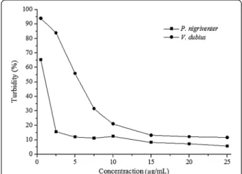
The Hyase activity of V. dubius venom was lower than
that of P. nigriventer (with respective turbidities of 11.6 and 5.6%). The venom ofP. nigriventerreached assay
sat-uration at a concentration of 5 μg/mL, while V. dubius
venom showed maximum activity at 15μg/mL (Figure 1).
Hyase purification was initiated through a size ex-clusion chromatography, which provided three active
fractions (fractions 7, 8 and 9), pooled for the second stage (Figure 2A). In the following affinity chromatography, Hyase activity was detected in one fraction (Figure 2B) (fraction 5). Reversed-phase HPLC provided the purified Hyase (Figure 2C). The enzyme purity was confirmed by a 12% SDS-PAGE and zymography, which showed only one band around 43 kDa (Figure 2D).
About 850 μg of purified Hyase was obtained from
18 mg of crude venom protein, with an activity corre-sponding to 19.5% of the venom. The total activity sig-nificantly dwindled in the second stage (from 615.3 TRU to 124.4), but the specific activity augmented gradually during the purification process (148 U/mg). The purifi-cation factor had also increased (4.1).
Figure 3Properties of hyaluronidase fromV. dubius.The purified enzyme showed maximum activity at pH between 4 and 5 and temperature from 35 and 40°C. Lower activity was observed starting from 25°C, decreasing gradually from 45°C onwards with total loss above 60°C. The points represent the mean ± S.E. (n = 6).
Figure 4Stability of purified Hyase after freeze-thaw cycles (−20°C): the activity remained at maximum after the first three
freeze-thaw cycles (0, 1 and 3 hours), with a slight decrease after six hours, unchanged until 15 days.Aliquots maintained their activity until the seventh day, losing activity after 15 days of freezing. The points represent mean ± SEM (n = 6), * p < 0.05.
The investigation of the physicochemical characteristics of the purified enzyme showed maximum activity at pH between 4 and 5 and temperature from 35 and 40°C. Lower activity was observed at 25°C, decreasing gradually from 45°C onwards with total loss above 60°C (Figure 3).
Enzyme samples submitted to consecutive freeze-thaw cycles remained at maximum activity after the first three cycles (0, 1 and 3 hours), with a slight decrease after six hours, remaining stable until 15 days. Otherwise, individual frozen aliquots maintained their activity until the seventh day, losing activity after 15 days of freezing (Figure 4).
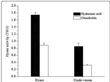
The substrate specificity was evaluated in both purified enzyme and crude venom; it was observed that the Hyase has a higher specificity to hyaluronic acid, but a lower activity in the presence of chondroitin (Figure 5).
The values obtained for Vmax and Km from V.
dubius-purified Hyase were 11.4 μg/min and 677.0 μg/mL,
respectively, as shown in Figure 6.
Antiophidic, antiscorpionic and antilonomic serum were inefficient at neutralizing the enzymatic activity (Figure 7A). The antiarachnid serum against enzyme and crude venom neutralized Hyase activity in a dose-dependent manner (Figure 7B).
Discussion
The hyaluronidases (Hyases) that have been isolated from various sources and extensively characterized com-prise a group of enzymes described as 4-hyaluronate glu-canohydrolase (EC 3.2.1.35, hyaluronoglucosamidase), hyaluronate-3-glucanohydrolase (EC 3.2.1.36, hyalurono-glucuronidase) and hyaluronatelyase (EC 4.2.2.1) [27]. The presence of this enzyme has been described in several human tissues, animal venoms, pathogenic organisms and cancers. Animal venom Hyases are structurally similar to those from the acrosome membrane of sperm, which play a fundamental role in mammalian fertilization, and to Hyases Hyal-1 and Hyal-2 of mammalian lysosomes [28-30]. Thus, venom Hyases are attributed the scattering function by facilitating the spread of toxins through glucosamine glycan hydrolysis in connective tissue, thereby yielding systemic poisoning [31].
As expected, the venom ofP. nigriventerpresents higher Hyase activity than that ofV. dubius, due to the former’s abundant high-molecular-weight molecules, thus requir-ing a higher permeability to promote the dissemination of
such components [1]. Moreover, the venom ofV. dubius
has few components of high molecular weight, but rather a predominance of low-weight molecules, such as pep-tides, thus less need of Hyase [20].
That molecular profile ofV. dubiusHyase venom
con-tributed to the enzyme purification process. In three chromatographic steps, we obtained the pure enzyme,
while in the venoms from other animals–namely Tityus
serrulatus,Buthus martensi, Apis mellifera,Vespula vul-garis, Synanceja horrida and Lonomia obliqua–more
Figure 7Immunological comparisons. (A)Dose-dependent neutralization of Hyase by arachnidic serum. Antiscorpionic, antilonomic, anti-bothropic, anticrotalic and antielapidic sera were inefficient at neutralizing the enzymatic activity.(B)Reactivity ofV. dubiusvenom (10μg) and
purified Hyase (5μg) compared to anti-arachnidic serum on ELISA. The points represent mean ± SEM (n = 6).
Figure 6Lineweaver-Burk graphs (1/S vs. 1/V) to Hyase Vmax
chromatographic steps were necessary for Hyase purifi-cation [11,27,28,32-34].
The enzyme yield (4.8%) was considered low after purifi-cation, mainly because the enzyme is present at small con-centrations in the total venom. Similar yields (4.9%) can be obtained during the Hyase purification of the scorpion venom ofTityus serrulatus [27]. Otherwise, the
purifica-tion from the venom of the scorpionPalamneus
gravima-nus, provided a higher yield, about 40% [31].
The optimum working temperature of purified V.
dubiusHyase is around 37°C, which matches the physio-logical range of some of its largest prey, such as small rodents [35]. The enzyme loses its activity at 60°C, per-haps due to losses in the molecular structure, thus com-promising its activity. Same hyaluronidase temperature range can be found in the venom activity presented by Agkistrodon contortrixandTityus serrulatus[27,36].
The optimum pH found in V. dubius Hyase was
be-tween 4 and 5, the same described for Buthus martensi
and Palamneus gravimanus enzymes [31,32]. In the
Hyase ofHippasa partita, the best pH was observed to
be 6 [37]. The finding of a pH between 4 and 5 can be explained by the preferential degradation of an acid sub-strate by the enzyme.
The V. dubius Hyase has a Km of 677.0 μg/mL, indi-cating a comparatively low affinity of the substrate for the enzyme catalytic site, in contrast to the Hyases ofT. serrulatus (69.7 μg/mL) and P. gravimanus (47.61 μg/
mL) scorpions, but similar to the stonefish Synanceja
horrida(709μg/mL) [27,31,33].
Testing the specificity of substrates, we observed that V. dubius Hyase exerts greater activity on hyaluronic acid compared to chondroitin, although still positive for the latter. The chondroitin test was positive for bovine hyaluronidase but negative for Hyases purified from the
venoms of Hippasa partita and Agkistrodon contortrix
[36,37].
The neutralization of Hyase activity by antivenoms has not been widely studied. The experiments conducted in
our laboratory showed that the venom Hyase of V.
dubiuswas recognized by antiarachnid serum, in a dose-dependent manner. The lack of influence of the
antiscor-pionic sera, obtained from Tytius venoms that are used
for antiarachnid production, attest that the above neutralization is specifically due to antibodies raised
against Loxosceles and Phoneutria sp., pooled for
anti-venom preparation against the latter. Neutralization also failed with antilonomic and antisnake sera, reinforcing the reaction specificity ofV. dubiusHyase against spider antivenoms. This indicates that immunological identities may differ between the Hyase of arachnids and those of snakes and caterpillars. This immunological relationship has also been reported for several other enzymes and toxins of venoms [38].
Conclusions
Based on these findings, we conclude that the venom of V. dubius contains a Hyase with physicochemical and biochemical characteristics similar to other Hyases of venoms, although less potent. Nevertheless, the studied Hyase shares immunological features specifically with other spiders’ enzymes, rather than those of caterpillars and snakes.
Ethics committee approval
The present study was approved by the Ethical Committee on Animal Research (UNICAMP) under the registration number 2167-1.
Competing interests
The authors declare that there are no competing interests.
Authors’contributions
This work was developed by RS, with the assistance of MLT in the experiments and was mentored by TAARS and SH. All authors read and approved the final manuscript.
Acknowledgements
The authors would like to thank the Center for Control of Zoonoses in Itu, São Paulo state, Brazil for providing the spiders for this study, José Ilton dos Santos for his support in the laboratory and CNPq and FAPESP for financial support and scholarships.
Author details
1
Department of Physiological Sciences, Santa Casa de São Paulo Medical School, Rua Cesário Motta Jr., 61, Vila Buarque, CEP 01.221-020 São Paulo, SP, Brasil.2Department of Pharmacology, School of Medical Sciences, Federal
University of Campinas (UNICAMP), Campinas São Paulo state, Brazil.
Received: 13 August 2013 Accepted: 31 January 2014 Published: 4 February 2014
References
1. Rash LD, Hodgson WC:Pharmacology and biochemistry of spider venoms.Toxicon2002,40(3):225–254.
2. Kuhn-Nentwig L, Schaller J, Nentwig W:Purification of toxic peptides and the amino acid sequence of CSTX-1 from the multicomponent venom of Cupiennius salei(Areneae: Ctenidae).Toxicon1994,32(3):287–302. 3. Wright RP, Elgert KD, Campbell BJ, Barrett JT:Hyaluronidase and esterase
activities of the venom of the poisonous brown recluse spider.
Arch Biochem Biophys1973,159(1):415–426.
4. Young AR, Pincus SJ:Comparison of enzymatic activity from three species of necrotizing arachnids in Australia:Loxosceles rufescens, Badumma insignisandLampona cylindrata.Toxicon2001,39(2-3):391–400. 5. Barbaro KC, Knysak I, Martins R, Hogan C, Winkel K:Enzymatic characterization, antigenic cross-reactivity and neutralization of dermonecrotic activity of five Loxoscelesspider venoms of medical importance in the Americas.Toxicon 2005,45(4):489–499.
6. da Silveira RB, Chaim OM, Mangili OC, Gremski W, Dietrich CP, Nader HB, Veiga SS:Hyaluronidases inLoxosceles intermedia(Brown spider) venom are endo-β-N-acetyl-d-hexosaminidases hydrolases.Toxicon2007,
49(6):758–768.
7. Kaiser E:The enzymatic activity of spider venom on the influence of sulfonated polysaccharides on the proteolytic and hyaluronic acid splitting activity of spider venom.Mem Inst Butantan1953,25(1):35–39. 8. Kaiser E:Enzymatic activity of spider venoms.InVenoms.Edited by
Buckley EE, Porges N. Washington, DC: American Association for the Advancement of Science; 1956:91–93.
10. Lu G, Kochoumian L, King TP:Sequence identity and antigenic cross-reactivity of white face hornet venom allergen, also a hyaluronidase, with other proteins.J Biol Chem1995,270(9):4457–4465.
11. Kolarich D, Léonard R, Hemmer W, Altmann F:The N-glycans of yellow jacket venom hyaluronidases and the protein sequence of its major iso-form inVespula vulgaris.FEBS J2005,272(20):5182–5190.
12. Lucas S:Spiders in Brazil.Toxicon1988,26(9):759–772.
13. Lucas SM, Da Silva PI Jr, Bertani R, Costa Cardoso JL:Mygalomorph spider bites: a report on 91 cases in the state of São Paulo.Brazil Toxicon1994,
32(10):1211–1215.
14. Isbister GK, Seymour JE, Gray MR, Raven RJ:Bites by spiders of the family Theraphosidae in humans and canines.Toxicon2003,41(4):519–524. 15. Escoubas P, Rash L:Tarantulas: eight-legged pharmacists and combinatorial
chemists.Toxicon2004,43(5):555–574.
16. Windley MJ, Escoubas P, Valenzuela SM, Nicholson GM:A novel family of insect-selective peptide neurotoxins targeting insect BKCa channels isolated from the venom of the theraphosid spider: eucratoscelus constrictus.Mol Pharmacol2011,80(1):1–13.
17. Edgerton GB, Blumenthal KM, Hanck DA:Inhibition of the activation pathway of the T-type calcium channel CaV3.1 by ProTxII.Toxicon2010,
56(4):624–636.
18. Escoubas P:Molecular diversification in spider venoms: a web of combinatorial peptide libraries.Mol Divers2006,10(4):545–554. 19. Bertani R:Revision, cladistic analysis, and zoogeography ofVitalius,
Nhandu, andProshapalopus; with notes on other theraphosine genera (Araneae, Theraphosidae).Arq Zool S Paulo2001,36(3):266–350. 20. Rocha-e-Silva TAA, Sutti R, Hyslop S:Milking and partial characterization of
venom from the Brazilian spiderVitalius dubius(Theraphosidae).Toxicon 2009,53(1):153–161.
21. Rocha-E-Silva TAA, Rostelato-Ferreira S, Leite GB, da Silva PI, Jr HS, Rodrigues-Simioni L:VdTX-1, a reversible nicotinic receptor antagonist isolated from venom of the spiderVitalius dubius(Theraphosidae).
Toxicon2013,70:135–141.
22. Di Ferrante N:Turbidimetric measurement of acid mucopolysaccharides and hyaluronidase activity.J Biol Chem1956,220(1):303–306.
23. Hames BD:One-dimensional polyacrylamide gel electrophoresis.InGel Electrophoresis of Proteins: a Practical Approach.2nd edition. Edited by Hames BD, Rickwood D. New York: Oxford University Press; 1990:1–147. 24. Cevallos MA, Navarro-Duque C, Varela-Julia M, Alagon AC:Molecular mass
determination and assay of venom hyaluronidases by sodium dodecyl sulfate-polyacrylamide gel electrophoresis.Toxicon1992,30(8):925–930. 25. Reissig JL, Strominger JL, Leloir LF:A modified colorimetric method for
the estimation of N-acetylamino sugars.J Biol Chem1955,217(2):959–966. 26. Segel I:Biochemical Calculations.New York: John Wiley & Sons; 1972:454. 27. Pessini AC, Takao TT, Cavalheiro EC, Vichnewski W, Sampaio SV, Giglio JR, Arantes EC:A hyaluronidase fromTityus serrulatusscorpion venom: isolation, characterization and inhibition by flavonoids.Toxicon2001,
39(10):1495–1504.
28. Gmachl M, Kreil G:Bee venom hyaluronidase is homologous to a membrane protein of mammalian sperm.Proc Natl Acad Sci USA1993,
90(8):3569–3573.
29. Frost GI, Csóka TB, Wong T, Stern R:Purification, cloning, and expression of human plasma hyaluronidase.Biochem Biophys Res Commun1997,
236(1):10–15.
30. Lepperdinger G, Stroble B, Kreil G:Hyal2, a human gene expressed in many cells, encodes a lysosomal hyaluronidase with a novel type of specificity.J Biol Chem1998,273(35):22466–22470.
31. Morey SS, Kiran KM, Gadag JR:Purification and properties of hyaluronidase fromPalamneus gravimanus(Indian black scorpion) venom.Toxicon2006,47(2):188–195.
32. Feng L, Gao R, Gopalakrishnakone P:Isolation and characterization of a hyaluronidase from the venom of Chinese red scorpionButhus martensi.
Comp Biochem Physiol C Toxicol Pharmacol2008,148(3):250–257. 33. Poh CH, Yuen R, Chung MC, Khoo HE:Purification and partial
characterization of hyaluronidase from stonefish (Synanceja horrida) venom.Comp Biochem Physiol B1992,101(1-2):159–163.
34. Da CB Gouveia AI, Da Silveira RB, Nader HB, Dietrich CP, Gremski W, Veiga SS:Identification and partial characterization of hyaluronidases in Lonomia obliquavenom.Toxicon2005,45(4):403–410.
35. Dias SC, Brescovit AD:Notes on the behavior ofPachistopelma rufonigrum Pocock (Araneae, Theraphosidae, Aviculariinae).Rev Bras Zool2003,
20(1):13–17.
36. Kudo K, Tu AT:Characterization of hyaluronidase isolatedfrom Agkistrodon contortrix contortrix(Southern copperhead) venom.Arch Biochem Biophys 2001,386(2):154–162.
37. Nagaraju S, Devaraja S, Kemparaju K:Purification and properties of hyaluronidase fromHippasa partita(funnel web spider) venom gland extract.Toxicon2007,50(3):383–393.
38. Elliott WB:Chemistry and immunology of reptilian venoms.InBiology of the reptilia: physiology B.8th edition. Edited by Gans C, Gans K. London: Academic Press; 1978:163–436.
doi:10.1186/1678-9199-20-2
Cite this article as:Suttiet al.:Purification and characterization of a hyaluronidase from venom of the spiderVitalius dubius(Araneae, Theraphosidae).Journal of Venomous Animals and Toxins including Tropical Diseases201420:2.
Submit your next manuscript to BioMed Central and take full advantage of:
• Convenient online submission
• Thorough peer review
• No space constraints or color figure charges
• Immediate publication on acceptance
• Inclusion in PubMed, CAS, Scopus and Google Scholar
• Research which is freely available for redistribution