Kinase from
Clonorchis sinensis
: A Candidate
Chemotherapeutic Target
Jing-ying Xiao1,2., Ji-Yun Lee3., Shinji Tokuhiro1
, Mitsuru Nagataki1, Blanca R. Jarilla1, Haruka Nomura1, Tae Im Kim3, Sung-Jong Hong3*, Takeshi Agatsuma1*
1Department of Environmental Health Sciences, Kochi Medical School, Nankoku, Kochi, Japan,2Department of Parasitology, Basic Medical College, Jiamusi University, Jiamusi, China,3Department of Medical Environmental Biology, Chung-Ang University College of Medicine, Seoul, Republic of Korea
Abstract
Background: AdultClonorchis sinensis lives in the bile duct and causes endemic clonorchiasis in East Asian countries. Phosphagen kinases (PK) constitute a highly conserved family of enzymes, which play a role in ATP buffering in cells, and are potential targets for chemotherapeutic agents, since variants of PK are found only in invertebrate animals, including helminthic parasites. This work is conducted to characterize a PK fromC. sinensisand to address further investigation for future drug development.
Methology/Principal findings:A cDNA clone encoding a putative polypeptide of 717 amino acids was retrieved from aC. sinensistranscriptome. This polypeptide was homologous to taurocyamine kinase (TK) of the invertebrate animals and consisted of two contiguous domains.C. sinensisTK (CsTK) gene was reported and found consist of 13 exons intercalated with 12 introns. This suggested an evolutionary pathway originating from an arginine kinase gene group, and distinguished annelid TK from the general CK phylogenetic group. CsTK was found not to have a homologous counterpart in sequences analysis of its mammalian hosts from public databases. Individual domains of CsTK, as well as the whole two-domain enzyme, showed enzymatic activity and specificity toward taurocyamine substrate. Of the CsTK residues, R58, I60 and Y84 of domain 1, and H60, I63 and Y87 of domain 2 were found to participate in binding taurocyamine. CsTK expression was distributed in locomotive and reproductive organs of adultC. sinensis. Developmentally, CsTK was stably expressed in both the adult and metacercariae stages. Recombinant CsTK protein was found to have low sensitivity and specificity towardC. sinensisand platyhelminth-infected human sera on ELISA.
Conclusion:CsTK is a promising anti-C. sinensisdrug target since the enzyme is found only in the C. sinensisand has a substrate specificity for taurocyamine, which is different from its mammalian counterpart, creatine.
Citation:Xiao J-y, Lee J-Y, Tokuhiro S, Nagataki M, Jarilla BR, et al. (2013) Molecular Cloning and Characterization of Taurocyamine Kinase fromClonorchis sinensis: A Candidate Chemotherapeutic Target. PLoS Negl Trop Dis 7(11): e2548. doi:10.1371/journal.pntd.0002548
Editor:Banchob Sripa, Khon Kaen University, Thailand
ReceivedJuly 14, 2013;AcceptedOctober 7, 2013;PublishedNovember 21, 2013
Copyright:ß2013 Xiao et al. This is an open-access article distributed under the terms of the Creative Commons Attribution License, which permits unrestricted use, distribution, and reproduction in any medium, provided the original author and source are credited.
Funding:This work was supported by a grant from Japan Society for the Promotion of Science (No. csc-10916), and by the Scientific Research Fund of Heilongjiang Provincial Science and Technology Department (No. LC2009C15). The funders had no role in study design, data collection and analysis, decision to publish, or preparation of the manuscript.
Competing Interests:The authors have declared that no competing financial interests exist. * E-mail: hongsj@cau.ac.kr (SJH); agatsuma@kochi.u.ac.jp (TA)
.These authors contributed equally to this work.
Introduction
Clonorchis sinensisis an important food-borne trematode parasite, which causes clonorchiasis in human and mammalian animals. This parasite is endemic in China, Korea, Taiwan, and northern Vietnam. Globally, 35 million people are estimated to be infected byC. sinensis, with approximately 15 million of these cases being in China [1]. The highest prevalence ofC. sinensishuman infection is reported in southern and northeastern parts of China, especially in Guangdong, Guangxi, and Heilongjiang provinces [2].
In humans, clonorchiasis can provoke severe pathologic changes in the hepatobiliary tract, and was recently recognized as belonging to the group of biological carcinogenic agents that cause cholangiocarcinoma [3], [4]. The public health and
economic impact of clonorchiasis is considerable and has inspired the development of vaccines and drugs in addition to other public health measures of control and eradication the parasite itself [5].
kinasing enzyme is creatine kinase (CK). In invertebrates, seven unique phosphagens and corresponding kinases were identified in addition to phosphocreatine [7], [8], [9]: glycocyamine kinase (GK), taurocyamine kinase (TK), lombricine kinase (LK), opheline kinase (OK), hypotaurocyamine kinase (HTK), thalessemine kinase (ThK), and arginine kinase (AK). Several studies have also described the presence of phosphagen kinases from parasites [10]. Studies on arginine kinase from the protozoa Trypanosoma cruzi
have identified AK as a potential target for novel drug development for Chagas’ disease [11], [12]. AK has also been presented inToxocara canis [13], andAscaris suum[14]. Recently, two-domain TKs were reported from parasitic trematodes
Paragonimus westermani[15] andSchistosoma japonicum[16]. Absence of these invertebrate PKs from the mammalian hosts, including human, imply that these kinases area possible target for new candidate drug against parasites and for development of new diagnostic reagent to detect infections.
We found a cDNA clone encoding a polypeptide (CsTK) from theC. sinensistranscriptome database, which was homologous with the two-domain TKs as well as the PKs of other organisms. This study was performed to elucidate biomolecular functions of CsTK such as catalytic activity comparing with TKs and PKs, tissue localization, and developmental expression.
Materials and Methods
Ethics statement
BALB/c mice (female, 7-week-old) and rabbits (New Zealand White, male, 2.2–2.5 kg) were handled in an accredited animal facility at Chung-Ang University (Korea FDA; Unit Number 36). Approval for animal experiments was obtained from the Institu-tional Animal Care and Use Committee at Chung-Ang University (Permit Number: CAU-2011-0052 and CAU-2013-0005). This study was carried out in strict accordance with the recommenda-tions provided in the Guide for the Care and Use of Laboratory Animals by the US National Institutes of Health.
Synthesis ofC. sinensistotal cDNA
The adult worms of C. sinensis were recovered from infected rabbits. Total RNA was isolated from adult worms by acid guanidinium thiocyanate-phenol-chloroform extraction method [17]. Messenger RNA (mRNA) was purified from total RNA using a poly (A)+
isolation kit (Nippon Gene, Tokyo, Japan). Single-stranded cDNA was synthesized with Ready-To-Go You-Prime First-Strand Beads (Amersham Pharmacia Biotech, NJ, USA) with a lock-docking oligo-dT primer [18].
Amplification of 39-cDNA end ofC. sinensisPK D2 domain Polymerase chain reaction (PCR) was carried out with reaction mixture containing cDNA, 10 pmol of each primer, 2ml of
2.5 mM of dNTPs, 1 U of Ex Taq polymerase, 2.5ml of 106Ex Taq buffer (TaKaRa, Tokyo, Japan). Thermal cycles were prepared as follows: initial denaturation at 94uC for 2 min, followed by 35 cycles of 94uC for 30 s, annealing at 50uC for 35 s, and extension at 72uC for 2 min, and a final extension at 72uC for 4 min. PCR was done in a thermal cycler, MyCycler (BioRad, Foster, USA). The 39-half of the cDNA was first amplified with lock-docking oligo (dT) primer and an ‘‘universal’’ redundant oligonucleotide primer 59-GT(ACGT) TGG(AG) T(ACGT) AA(TC)GA(AG) GA(AG) GA (TC) CA-39, designed for amplifi-cation of PK [19]. PCR products were purified using GENE CLEAN Kit (Funakoshi, Tokyo, Japan).
Purified PCR product (400 bp) was ligated into pGEMT-vector (Promega, USA) and transformed intoEscherichia coliJM109 cells. Positive clones were obtained and plasmid DNA was extracted. Nucleotide sequences were determined with an ABI PRISM 3100-Avant DNA sequencer using a BigDye Terminators v3.1 Cycle Sequencing Kit (Applied Bio-systems, Foster, CA, USA) with two T-vector-specific primers, T7 and SP6.
PCR amplification of 59-half and internal region ofC. sinensisPK D1D2 cDNA
The 59-half of the cDNA was amplified as follows: A poly (G)+ tail was added to the 59-end of the C. sinensiscDNA pool with terminal deoxynucleotidy transferase (Promega, Madison, WI, USA). 59-half of cDNA ofC. sinensisPK was amplified using oligo-dC primer (59-GAA TTC18-39) and PK-csR0 primer (59-CCA AAT TAC TCG GGC AAC AA -39) designed on the sequence of 39-half.
To confirm a tandem connection of CsTK D1 and D2 domains, central region of the cDNA was amplified using inner specific primers that were designed on the sequences of cDNA obtained by 59-RACE and 39-RACE PCR until full sequence of the cDNA was obtained [20]. With the PCR products, T-vector cloning and 39 -sequence determination of C. sinensis PK were performed as described above.
Expression and purification of truncated and contiguous two-domainC. sinensisPKs
PCRs were done in total volumes of 50ml. The reaction mixture contained cDNA of D1D2, 10 pmol of csPKXbaI forward primer (59-TCT AGA ATG CAG GTC GAA CCA CTG AAA TC-39), 10 pmol of csPKPstI reverse primer (59-CTG CAG CTA TGG CAA GGA TTT TTC AAT AGC -39), 1 U of KOD Plus DNA polymerase (Toyobo Co., Ltd., Tokyo, Japan), 5ml of 106KOD Plus buffer, 5ml of 2 mM KOD dNTPs, and 4ml of 25 mM
MgSO4. The amplified products were purified using QIA quick
PCR purification columns (QIAGEN GmbH, Hilden, Germany). A-tail was added to 39-end of the purified PCR fragments (blunt-ended). A-tailing was done in a total volume of 30ml
Author Summary
The food-borne clonorchiasis imposes public health problems on inhabitants in endemic areas. Praziquantel has been employed as an efficacious anthelminthic in large-scale campaigns as well as for individual treatment of
Clonorchis sinensishuman infections. Although praziquan-tel continues to have good efficacy, new drug develop-ment for this parasite has been recognized as a crucial issue to be investigated intensively. Clonorchis sinensis
containing purified KOD PCR product, 15 U of Gene Taq DNA polymerase (Wako Nippon Gene), 3ml of 106Gene Taq buffer and 1.2ml of 5 mM dNTP. This mixture was incubated at 70uC for 30 min. The resulting product was purified, subcloned into pGEMT-vector, and sequenced as described above.
Coding region ofC. sinensisPK cDNA of D1D2 was cloned into XbaI/PstI site of pMAL-c2 (New England Biolabs, Ipswich, MA, USA). Maltose binding protein (MBP)-C. sinensisPK fusion protein was expressed inE. coliTB1 cells by induction with 1 mM IPTG at 25uC for 24 h. The cells were resuspended and sonicated in 56 TE buffer. Soluble recombinant fusion protein was purified by affinity chromatography using amylose resin (New England Biolabs, Ipswich, MA, USA). Homogeneity of the purified recombinant enzyme was verified by SDS–PAGE and placed on ice until assayed for enzymatic activity within 12 h. MBP-tagged CsTK D1 and D2 expressed and purified as described above.
Amplification ofC. sinensistaurocyamine kinase gene Using Easy-DNA Kit (Invitrogen, Carlsbad, USA), genomic DNA was isolated from a C. sinensis adult worm. PCR was performed with Ex Taq polymerase (TAKARA) and primers designed on the ORF. PCR conditions were as follows: initial denaturation at 94uC for 2 min, followed by 35 cycles of 94uC for 30 s, annealing at 50uC for 30 s and extension at 72uC for 3 min and a final extension at 72uC for 4 min. PCR product was purified and sequenced as described above.
Multiple alignment and phylogenetic analysis
Using programs CLUSTAL W (http://www.ddbj.nig.ac.jp) and GENETYXMAX (ver. 6.0), multiple sequence alignment was performed. Phylogenetic analysis was done using the distance method in MEGA (ver. 5.0). For distance analyses, the Kimura 2-parameter model was used to construct the distance matrix, and the tree was inferred from this using the Neighbor-Joining (NJ) approach. Bootstrap re-sampling was performed to assess the degree of support for groupings on the tree. Accession numbers of other amino acid sequences used in the present study are shown in Table S1.
Site-directed mutagenesis ofC. sinensistaurocyamine kinase
The following amino acid substitutions were amplified in the template of pMAL/C. sinensisTK wild type (WT): R58A, I60A, Y84A and Y84R of TKD1; H61A, I63A, Y87A and Y87R of TKD2; R58A, I60A, Y84A and Y84R of TKD1D2 in D1 region; H61A, I63A, Y87A and Y87R of TKD1D2 in D2 region. Substitution was made using KOD+
-DNA polymerase under the subsequent PCR conditions: initial denaturation at 94uC for 2 min, followed by 35 cycles of 94uC for 15 s, annealing at 60uC for 30 s and extension at 68uC for 9 min and a final extension at 72uC for 5 min. The primer sequences designed were as follows: CsPKMutD1R58Af: 59-GCT TGC ATC CTT CCT CGC G-39, CsPKMutD1R58Ar: 59- CGG ATT ACG AGC ATT GTG ACT GAC-39; CsPKMutD1I60Af: 59-GCT CTT CCT CGC GCT TGT GAT TTG-39, CsPKMutD1I60Ar: 59-GCA CCG CGG ATT ACG AGC ATT GTG-39; CsPKMutD1Y84Af: 59-GCT CAT AAG GTG AAA GGA GAC-39, CsPKMutD1Y84RAr: 59 -GTC TAT AAT AAC GGC -GTC AAA G-39; CsPKMut-D1Y84Rf: 59-CGA CAT AAG GTG AAA GGA GAC-39; CsPKMutD2H61Af: 59-GCT TCA ATC TGT CCA CGG TAC TGG-39, CsPKMutD2H61Ar: 59-TGG GTT GTA AGC ACC GTT ACG-39; CsPKMutD2I63Af: 59-GCT TGT CCA CGT ACT GGA GAA GC-39, CsPKMutD2I63Ar: 59-TGA ATG
TGG GTT GTA AGC ACC GT-39; CsPKMutD2Y87Af: 59 -GCT CAT GGA GTG AGT GAC CCA -GCT T-39, CsPKMut-D2Y87RAr: 59-GTC CAA AAT CAC TGC ATC CAG GTA GTC-39; CsPKMutD2Y87Rf: 59-CGA CAT GGA GTG AGT GAC CCA GCT T-39. PCR products were purified by QIAquick PCR purification column (Qiagen GmbH, Hilden, Germany). After blunting and phosphorylation, the DNA was self-ligated. Expression and enzyme assay of the mutated proteins were performed as described above.
Enzyme assays for substrate specificity ofC. sinensisTK Enzyme activity was measured by absorbance at a wavelength of 340 nm (with MBP, UV/Visible Spectrophotometer 4300 Pro, Amersham, Biosciences) with an NADH-linked assay at 25uC [21], [22]. The reaction mixture (total 1.0 ml) contained 0.65 ml of 100 mM Tris–HCl (pH 8), 0.05 ml of 750 mM KCl, 0.05 ml of 250 mM Mg-acetate, 0.05 ml of 25 mM phosphoenolpyruvate prepared in 100 mM imidazole/HCl (pH 7), 0.05 ml of 5 mM NADH prepared in Tris–HCl (pH 8), 0.05 ml of pyruvate kinase/ lactate dehydrogenase mixture prepared in 100 mM imidazole/ HCl (pH 7), 0.05 ml of an appropriate concentration of ATP prepare in 100 mM imidazole/HCl (pH 7), and 0.05 ml of recombinant enzyme. The reaction was started by adding 0.05 ml of an appropriate concentration of guanidine substrate made up in 100 mM Tris–HCl (pH 8). Initial velocity values were obtained by varying the concentration of guanidine substrate under fixed concentrations of ATP. Protein concentration was estimated from an absorbance at 280 nm (0.77 AU at 280 nm in a 1 cm cuvette corresponds to 1 mg protein/ml).
Quantitative real-time PCR of Cs TK
To measure mRNA transcript level in developmental stages of
C. sinensis, quantitative real-time PCR (qRT-PCR) was performed using SYBR Green I dye with LightCycler Carousel-Based System (Roche Applied Science, Indianapolis, IN, USA). cDNAs of C. sinensisadults and metacercariae were employed as templates of qRT-PCR. Four pairs of forward and reverse primers were designed on each 59- and 39-end of CsTK D1 and CsTK D2 using Oligo6 program (Figure S1 Panel A). For qRT-PCR, forward primer 59- TTT CCA CAA TGC CAA CAA GAC -39 and reverse primer 59- GCT TGA ATA CCC TGG ATG AGT -39on 39-end of CsTK D2 were employed, producing a 442 bp amplicon (Figure S1, Panels B and C). qRT-PCR mix was prepared as of 16 SYBR green master mix, 1mM gene-specific primers, and 80 ng total cDNAs. Reference genes employed wereb-actin, phospho-glycerate kinase, and calcyphosine [23]. Thermal cycling of qRT-PCR started with pre-incubation at 95uC for 15 min, then continued 40 cycles of 95uC for 10 sec, 48uC for 10 sec, and 72uC for 30 sec. To verify a specific amplication of target mRNA, one melting cycle was run, 65uC for 1 min, and increase at 0.1uC/sec to 95uC to dissociate double-stranded amplicons. LightCycler software 4.05 (Roche Applied Science, Penzberg, Germany) was used to analyze melting curves and to calculate CTvalues. ADCT
of target gene was calculated by subtracting an average CT of
three reference genes from an average CTof the target gene. The
DDCT
of target gene was analyzed using equationDDCT
= (DC T tar-get
2average of DC
Tadult). The 22
DDCT
shows relative gene expression level [24].
Purification of recombinant CsTK D1 protein
transformed with the expression construct and induced to produce the fusion protein using IPTG at 0.1 mM for 3 hrs. The fusion protein was absorbed to glutathione sepharose 4B column (GE Healthcare, Uppsala, Sweden) and washed with PBS. Recombi-nant CsTK D1 was cleaved off from Cs28GST tag on bead with 10 U/ml thrombin protease (GE Healthcare, Buckinghamshire, UK) overnight, and then eluted in PBS. Residual tagged protein was eluted in 5 mM reduced glutathione/PBS.
Production of anti-recombinant CsTK D1 mouse immune sera
Recombinant CsTK D1 was mixed with same amount of either complete or incomplete Freund adjuvant. BALB/c mice were injected peritoneally once with 40mg antigen/200ml of complete adjuvant mix and again with the same amount of incomplete adjuvant mix 2 weeks later. A final buster, 1.2ml antigen in 30ml PBS each mouse, was injected into a tail vein after 2 weeks. After 4–5 days, blood was taken and checked for antibody production toward recombinant CsTK D1 by western blotting.
Immunoblot of native CsTK in adultC. sinensis
For soluble extract,Clonorchis sinensis adults were washed with PBS several times and homogenized in PBS/1% Triton X-100/ proteases inhibitor (16 Complete Mini, EDTA-free, Roche Diagnostics). After keeping 4uC overnight, the homogenate was spun at 20,0006gfor 60 min at 4uC and supernatant was saved as soluble extract or crude antigen ofC. sinensis. The soluble extract, 20ml, was deployed in 12% SDS-PAGE and transferred onto Hybond ECL membrane (GE Healthcare, Uppsala, Sweden). The blotted membrane was incubated overnight in CsTK D1-immune mouse serum at 1:5,000 dilution in skim milk/PBS, then in the secondary antibody, alkaline phosphatase-conjugated goat anti-mouse IgG at 1:5,000. Color was developed using BCIP/NBT substrate (Sigma Co., St. Louis, MO, USA) and stopped in water.
Immunohistochemical staining
AdultC. sinensisflukes within a rabbit liver were fixed in 10% neutral formalin and processed for paraffin blocks. The sectioned flukes in paraffin ribbons were deparaffinized and rehydrated. The
C. sinensisribbons were incubated in CsTKD1-immune mouse sera at 1:200 dilution for 30 min at room temperature. Then, the sections were incubated in peroxidase- and antimouse IgG antibody-conjugated dextran polymer (to the dextran backbone, about 70 enzyme molecules and 10 primary antibodies were conjugated; Dako, Glostrup, Denmark) for 30 min at room temperature. Color was developed in AEC+substrate for 5 min. Normal mouse sera were used as negative control.
ELISA
Recombinant CsTK D1 protein, 1mg/ml in carbonated buffer, was coated on 96-well plate at 4uC overnight. The wells were washed three times with PBS containing 0.1% Tween-20 (PBS/T) and incubated with human sera at 1:100 dilution at 37uC for 1 hr. A secondary antibody, peroxidase -conjugated anti-human IgG (MP Biomedicals, Santa Ana, CA, USA) of 1:4000 dilution was added to the wells and incubated at 37uC for 1 hr. Color was developed with a substrate, ophenylene diamine (Sigma Co., St. Louis, MO, USA), and optical density was measured at a wavelength of 490 nm.
Human sera used were from 47 patients with clonorchiasis, 20 with opisthorchiasis viverinii, 14 with cysticercosis cellulosae, 14 with sparganosis erinacei and from 14 patients with paragonimi-asis westermani. As a control group, serum samples from 28
parasite-free human subjects were employed. A cut-off line was set at an average+doubled standard deviation which was calculated with OD values of the control group.
Results
Identification of theC. sinensisTK cDNA and Bioinformatics analysis
During large-scale cDNA sequencing of an adult C. sinensis
cDNA library, a 2,154 bp long cDNA was successfully amplified by PCR, which encoded for a polypeptide of 717 amino acids (Fig. S2) and had 59-UTR of 52 bp and 39-UTR of 288 bp. Molecular mass of the polypeptide was estimated to be 80,274 Da with a pI of 7.88, using ProtParam (http://www.expasy.ch/tools/ protparam.html). cDNA sequence analysis showed that this polypeptide consisted of two repetitive domains (D1 and D2) homologous to known sequences of TKs (Fig. S2). D1 contained 360 amino acids with calculated molecular mass 40,573 Da and a pI of 8.09, and D2 consisted of 357 amino acids with calculated mass 39,719 Da and a pI of 7.39. This cDNA sequence was archived in GenBank under accession number JX435779.
cDNA sequence analysis using BLASTn revealed thatC. sinensis
TK D1 and D2 had 72.3% identity withP. westermaniTK D1 and D2. Further, the peptide sequence analysis, using BLASTp, showed that CsTK D1 and D2 were homologous with the respective domain in many TKs of different species (Fig. S2).C. sinensisTK polypeptide shared 79.2% sequence identity with P. westermaniTK. CsTK D1 shared 77.7% identity withP. westermani
TK D1 and 71.7% identity withS. mansoniTK D1. Meanwhile, CsTK D2 shared 82.3% identity withP. westermaniTK D2 and 67.9% identity with S. mansoni TK D2 (Table 1). With this sequence information, the polypeptide conceptually deduced from theC. sinensiscDNA clone was considered as a new member of PK ofC. sinensis. Residues in GS region are highly conserved across animal PKs. I60 in GS region ofC. sinensisTK was replaced by Val in all CKs. CsTK had not a mitochondrial targeting signal peptide in N-terminus (Fig. S2).
A phylogenetic tree, constructed using NJ algorithm (Fig. 1), indicated that PKs can be grouped into two clusters. Trematode TKs including CsTK were grouped in Cluster 1 with mulluscan AK group and nematode, protozoan, and arthropod AK group. Cluster 2 was comprised of annelid PK group and CK group of CKs, GKs, LKs, and TKs from protozoan and various insect species.
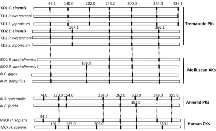
Exon/intron organization ofC. sinensisTK gene
C. sinensisTK gene had 13 exons and 12 introns, encoding D1 and D2 domains. Introns ofC. sinensisTK were located at amino acid positions 97.1, 140.0, 202.0, 243.2, 300.0, 354.0, 426.1 (bridge intron) in D1, and at 97.1.1, 167.1, 243.2, 300.0, 366.1 in D2 (Fig. 2). The introns of C. sinensis TK began with GT and ended with AG (GT–AG pattern), except for introns at position 202.0 (GC–AG pattern) in D1. Size of the introns was variable from 115 bp to more than 4,000 bp (Table S2). Positions ofC. sinensisTK introns were conserved betweenP. westermaniTK and
S. mansoniTK. Introns of C. sinensis TK 97.1, 243.2, and 300.0 shared equivalent positions withP. westermaniTK andS. mansoni
Enzyme activity of recombinantC. sinensisTK
Recombinant whole and truncated variants of CsTK were successfully expressed as MBP-fusion proteins. Each set of recombinant fusion proteins was purified by affinity column chromatography to homogeneity, and appeared as a single band in SDS-PAGE CsTK D1 and D2 (truncated domain+MBP) about 80 kDa and whole CsTK 120 kDa (Fig. 3). Enzymatic activity of the recombinant CsTK was measured by NADH-linked assay for substrates taurocyamine, glycocyamine, creatine, D-arginine, and L-arginine. Whole and truncated D1 and D2 of CsTK showed enzyme activity 0.84–1.36mmol/min?mg protein toward tauro-cyamine (Table 2).
Kinetic parameter ofC. sinensisTK Kinetic parameter of whole CsTK Km
Tc
0.49 mM was higher than that of individual domain D1 or D2, 0.35 and 0.48 mM each, indicating that whole CsTK had lower affinity for substrate
taurocyamine. CsTK D1 had stronger affinity for ATP as its
KmATP, 0.46 mM, was lower when compared to that of CsTK D2,
0.75 mM, and of whole CsTK, 0.78 mM.Kcatvalue of CsTK D1,
22.59 s21, was higher than that of D2, 4.50 s21, and of whole CsTK, 17.78 s21. Similar results were also recorded forVmaxand kcat/KmTc, reflecting that CsTK D1 had more efficient catalytic
activity than D2 domain or whole CsTK did. This enzymatic feature was different from that ofP. westermaniTK, of which whole
P. westermaniTK is catalytically more efficient than either of the truncated individual domains, D1 or D2 (Table 3).
Kinetic constant and catalytic efficiency ofC. sinensisTK As appeared in the alignment of multiple PK polypeptide sequences (Fig. S2), guanidino specificity (GS) region of CsTK had 2–3 less amino acids than that of the other known AKs, which is common feature of trematode TKs. To characterize the substrate recognition system in GS region of C. sinensis TK, residue substitution was introduced in CsTK (Fig. S2). Parameters of affinity, activity, and catalytic efficiency are presented in Table 4. For CsTK D1, mutations in GS region decreased its affinity for taurocyamine as evidenced by the increase ofKm
Tc
values. Most significant decrease was observed in the Y84R mutant showing no enzymatic activity. Substitutions of equivalent residues in truncat-ed D2 domain (Y84A, Y84R), whole CsTK (Y84A, Y84R in D1 and Y87A, Y87R in D2) resulted in the loss of detectable enzyme activity. Substitution of tyrosine in GS region might have affected stabilization of the closed structure of PK, suggesting that this amino acid plays an important role in taurocyamine binding. Another substitutions in D1 (I58A, R60A), and whole CsTK (I58A, R60A in D1 and H61A, I63A in D2) also decreased enzyme activity. However, mutation of equivalent position (H61A and I63A mutants) in truncated D2 increased catalytic efficiency, as evidenced by higher values ofkcat,kcat/KmTcandVmax.
Developmental expression of CsTK D1D2
To compare relative gene expression level between develop-mental stages by usingDDCTequation, three reference genes were employed such as of b-actin, phosphoglycerate kinase, and calcyphosine. CsTK D1D2 mRNA level was 1.2-fold higher in the metacercariae than in the adults ofC. sinensis(Fig. 4)
Recombinant Cs28GST-CsTK D1 protein
The recombinant Cs28GST-CsTK D1 fusion protein was produced as a major component and soluble form in theE. coli
host. The recombinant CsTK D1 protein was cleaved off efficiently from the tag, Cs28GST, by thrombin treatment on bead. The cleaved CsTK D1 protein was eluted at high concentration and purity with a molecular mass of 42 kDa in SDS-PAGE gel (Fig. 5). This CsTK D1 was used for downstream experiments such as immune serum production and antigenicity tests on ELISA.
Native CsTK protein
Anti-CsTK D1 mouse immune sera reacted strongly to recombinant CsTKD1 protein. These mouse sera reacted to and detected native CsTKD1D2 protein from adultC. sinensissoluble extract. The native CsTKD1D2 protein was revealed as a distinctive major band of 80 kDa protein. Anti-CsTK D1 antibody was able to detect CsTK D1D2 as well as CsTK D1, since CsTK D1 and CsTK D2 are fused in tandem (Fig. 6).
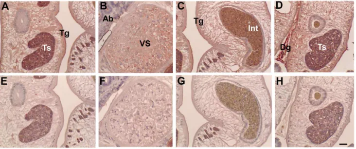
Tissue localization
Anti-CsTK D1 mouse immune sera were used for localization of CsTK D1D2 in the adult C. sinensis by immunohistochemical Table 1.Sequence identity ofC. sinensisTK D1 and D2
domains to other phosphagen kinases*.
C. sinensis TK D1 (%)
C. sinensis TK D2 (%)
Trematode TKs
C. sinensisTK D1 - 51.5
C. sinensisTK D2 51.5
-P. westermaniTK D1 77.7 50.1
P. westermaniTK D2 50.3 82.3
S. mansoniTK D1 71.7 60.0
S. mansoniTK D2 47.1 67.9
Sipunculid HTK
S. cumanenseHTK 46.6 48.3
Molluscan AKs
C. kaikoiAKD1 43.3 42.6
C. kaikoiAKD2 46.7 40.6
T. cornutusAK 48.9 45.1
H. madakaAK 51.7 46.5
Nematode AKs
T. canisAK 39.2 39.8
A. suumAK 41.7 42.0
Protozoan AKs
T. cruziAK 41.7 46.5
T. bruceiAK 41.3 47.5
Arthropod AKs
B. malayiAK 43.7 35.5
L. polyphemusAK 43.1 45.7
Annelid PKs
A. brasiliensisTK 34.2 32.8
R. pachyptilaTK 36.1 31.7
E. fetidaLK 34.4 35.6
N. diversicolorGK 33.1 33.6
Mammalian CK
H. sapiensMCK 33.6 34.5
*Identity was calculated using ClustalW2 (http://www.ebi.ac.uk/Tools/msa/ clustalw2/).
staining. The CsTKD1D2 was localized in tegument, oral and ventral suckers, testes, seminal vesicle and sperms, intra-uterine eggs and intestinal contents in adultC. sinensis. Dregs betweenC. sinensis were stained with strong positive color (Fig. 7). Acini of biliary epithelium had contents reacting positively to the immune sera.
ELISA
Recombinant CsTK D1 protein was evaluated for serodiag-nostic antigen toward IgG antibody inC. sinensis-infected human sera by ELISA. A positive cut-off value, derived from the normal control sera, was set at A490= 0.21. The CsTK D1 protein gave
29.7% positive rate from sera of clonorchiasis patients. On the
Figure 1. Neighbor-Joining tree for the amino acid sequences of phosphagen kinases using a program in MEGA version 5.Bootstrap values were shown at the branching point (1,000 replications). Amino acid sequences were retrieved from DDBJ and GenBank.
other hand, this positive rate was 65.0% from sera of opisthor-chiasis patients, 14.3% of paragonimiasis patients, 0% from cysticercosis, 35.7% from sparganosis and 7.1% from the normal control sera (Fig. 8, Table S3).
Discussion
Taurocyamine kinase (TK) is a member of the phosphagen kinase family, which was first isolated from the body wall muscle of polychaete lugworm, Arenicola marina [25]. Two types of TK, cytoplasmic TK and mitochondrial (Mi) TK were previously purified from Arenicola brasiliensis [26] and from deep-sea Riftia pachyptila [27]. As such, TKs were believed to be restricted to certain marine annelids [26]. However, P. westermani and S. japonicum TK showed activity only towards taurocyamine [15], [28]. Additionally, a two-domain phosphagen kinase with enzyme
activity to taurocyamine was also reported fromS. mansoni[29]. Thus, it was found that TK was not exclusive to marine annelids. In the present study,C. sinensisTK also showed exclusive activity towards taurocyamine.
In this study, we identified a cDNA clone encoding a polypeptide of 717 amino acids. The deduced amino acid sequence showed a high similarity to TKs previously reported from P. westermani and other helminthic parasites. It should be noted that C. sinensis TK had higher sequence identity with molluscan AKs than with other annelid TKs. The neighbor-joining tree revealed two major groups: CK and AK isoenzyme groups. Both domains ofC. sinensisTK fell in AK cluster, together with P. westermani TK, S. mansoni TK, molluscan AKs and sipunculid HTK. Recombinant C. sinensis TK showed high
Figure 2. Comparison intron/exon organization ofC. sinensisTK with other phosphagen kinases.Intron positions ofC. sinensisTK were based on aligned amino acid sequences. Nomenclature for the intron positions was taken from Uda et al. (2006) and Jarilla (2010). Intron phase is indicated by ‘‘.0’’, ‘‘.1’’, or ‘‘.2’’ following the amino acid sequence position. Conserved introns are shown by vertical dashes.
doi:10.1371/journal.pntd.0002548.g002
Figure 3. Expression and purification of recombinantC. sinensis TK whole, D1 and D2 domains fused to MBP in E. coli. M: molecular weight marker, 1, uninduced pellet; 2, uninduced superna-tant; 3, IPTG-induced pellet; 4, IPTG-induced supernasuperna-tant; 5, purifiedC. sinensisTK. Arrow indicates recombinant protein.
doi:10.1371/journal.pntd.0002548.g003
Table 2.Enzyme activity ofC. sinensisphosphagen kinase for various guanidine derivatives.
Substrate PK activity (mmol/min.mg21
protein)
D1 D2 D1D2
Taurocyamine 1.08 0.84 1.36
L-arginine 0.05 0.014 0.08
D-arginine 0.05 0.020 0.09
Creatine 0.06 0.016 0.09
Glycocyamine 0.06 0.013 0.06
enzymatic activity toward the taurocyamine substrate. With these results, it was identified for the first time that theC. sinensiscDNA encoded a phophagen kinase.C. sinensisTK was considered to be a cytoplasmic TK, since it did not have the N-terminal signal peptide of 40 residues which were the probable mitochondrial targeting sequence present in characterized mitochondrial CKs [30].
Annelid PKs had considerable catalytic efficiencies for the guanidino substrates, glycocyamine and lombricine, in addition to
its original target substrate, taurocyamine [26], [27]. This was in contrast toC. sinensisTK,P. westermaniTK, andS. mansoniTK, all of which showed exclusive enzyme activity for taurocyamine. Low degree of substrate specificity in annelid enzymes supported flexibility in substrate recognition, which might have been a driving force in the evolution of phaphagen kinases. EiseniaTK had obtained remarkable diversity supported by the mutation K95Y LK, dramatically changing guanidino substrate specificity Table 3.Comparison ofC. sinensisrecombinant TK kinetic parameters with other TKs*.
Source Ref KmTc(mM) KdTc(mM)
KdTc/
KmTc
KmATP
(mM)
KdATP
(mM)
KdATP/
KmATP kcat(S21)
kcat/
KmTc
Vmax(umol/min.
mg protein)
C. sinensisTK D1 Present study 0.3560.01 2.6560.65 7.57 0.4660.12 2.3360.64 5.05 22.5960.15 54.54 33.8962.01
C. sinensisTK D2 Present study 0.4860.05 1.9960.15 4.15 0.7560.13 2.5060.52 3.33 4.5060.11 9.38 6.7160.61
C. sinensisTK whole Present study 0.4960.02 1.9260.43 3.92 0.7860.07 3.4660.70 4.44 17.7860.98 36.26 26.6862.11
S.japonicumTK D1 Tokuhiro (2012) 1.3060.01 3.0060.58 2.31 1.1160.18 2.5760.27 2.32 52.9160.68 40.70 82.9063.17
S.japonicumTK D2 Tokuhiro (2012) 0.5360.06 1.1560.21 2.17 1.6060.20 3.4760.58 2.17 15.3960.10 29.04 28.8361.06
S.japonicumTK whole Tokuhiro (2012) 0.4760.03 1.1760.18 2.49 0.9760.10 0.4360.30 2.51 39.0060.36 82.98 67.8261.50
P. westermaniTK D1 Blanca et al (2009) 0.7560.07 4.2261.12 5.63 0.6660.11 3.5860.27 5.42 24.1661.54 32.21 40.3162.51
P. westermaniTK D2 Blanca et al (2009) 0.5160.04 1.4960.29 2.92 1.4360.36 4.0360.76 2.82 11.5660.45 22.67 21.4361.75
P. westermaniTK whole Blanca et al (2009) 0.5760.10 1.9560.43 3.42 0.9860.16 3.3760.70 3.44 33.4461.01 58.67 60.0163.01
A. brasiliensisTK Uda et al (2005) 4.0160.41 NA NA NA NA NA 9.4360.45 2.35 28.7161.06
A. brasiliensisMiTK Uda et al (2005) 0.8860.08 NA NA NA NA NA 14.361.01 16.23 17.8261.24
R. pachyptilaMiTK Uda et al (2005) 2.1260.45 NA NA NA NA NA 12.561.52 5.90 10.460.59
*Value: mean of three assays6SD. NA, data not available.
doi:10.1371/journal.pntd.0002548.t003
Table 4.Comparison of kinetic parameters ofC.sinensismutant TKs*.
Source KmTc(mM) KdTc(mM)
KdTc/
KmTc
KmATP
(mM)
KdATP
(mM) kcat(s21) kcat/KmTc
Vmax(umol/
min?mg protein)
TK D1 0.3560.01 2.6560.65 7.57 0.4660.12 2.3360.64 22.5960.15 54.54 33.8962.01
R58A of TK D1 0.8560.06 2.0260.18 2.38 0.6660.11 1.0660.17 33.0361.14 38.86 30.5461.71
I60A of TK D1 0.5160.07 2.4560.53 4.80 0.5260.08 2.5060.70 22.4360.38 44.02 32.6462.00
Y84A of TK D1 0.8260.07 2.0760.12 2.76 0.6260.11 1.1860.27 32.9061.54 40.12 29.6662.51
Y84R of TK D1 0 0 0 0 0 0 0 0
TK D2 0.4860.05 1.9960.15 4.15 0.7560.13 2.5060.52 4.5060.11 9.38 6.7160.61
H61A of TK D2 0.3660.07 2.6560.43 7.36 0.5860.23 2.8060.42 5.9261.23 15.58 20.8960.81
I63A of TK D2 0.3060.05 2.9960.17 9.90 0.6160.10 2.7860.27 6.4660.45 21.53 26.1962.06
Y87A of TK D2 3.6460.21 3.8660.33 1.05 NA NA 6.45 1.77 5.1960.24
Y87R of TK D2 3.0160.25 2.4160.17 0.80 NA NA 0.9960.52 0.33 1.1960.59
TK whole 0.4960.02 1.9260.43 3.92 0.7860.07 3.4660.70 17.7860.98 36.26 26.6862.11
R58A of D1 in TK whole 0.4560.19 1.7260.43 3.82 0.7660.12 3.4360.70 15.0060.25 33.33 24.1561.11
I60A of D1 in TK whole 0.3960.21 1.4360.13 3.24 0.6960.07 3.2660.70 12.0460.18 30.87 23.7562.12
Y84A of D1 in TK whole 0.3560.02 1.3260.43 3.77 1.2560.11 1.8160.80 13.0060.04 16.25 19.4461.24
Y84R of D1 in TK whole 0.5460.04 1.7260.26 3.12 1.0160.23 3.5060.32 10.9260.37 20.22 16.3861.32
H61A of D2 in TK whole 0.4060.02 1.2260.43 3.05 0.6660.07 3.2360.70 12.0160.45 30.03 23.2561.21
I63A of D2 in TK whole 0.5460.03 0.8660.04 1.59 0.9160.07 1.4560.42 11.2060.28 13.02 16.8161.31
Y87A of D2 in TK whole 0.4460.02 0.7360.05 1.66 1.1260.02 1.8660.30 11.1960.61 25.43 16.7961.07
Y87R of D2 in TK whole 0.4660.05 1.1560.02 2.50 0.5460.07 2.3060.70 10.6760.38 23.20 16.0162.01
*Value: mean of three assays6SD. NA, data not available.
from lombricine to taurocyamine [31]. Amino acid sequences of GS region of mitochondrial TK were quite different from cytoplasmic TK [32], which reflects independent evolutionary processes. Sequential difference of these two enzymes did translate to differences in enzymatic activity and substrate specificity toward the guanidino substrates, taurocyamine, lombricine, glycocya-mine, and arginine [26], [27]. Number of amino acids in GS region ofC. sinensisTK,P. westermaniTK, andS. mansoniTK was smaller than those of annelid TKs (Fig. S2). Cytoplasmic TKs ofA. brasiliensis and R. pachyptilawere missing five residues [26], [27], and this might affect the differences in guanidino substrate specificity.
The GS region was described as a possible candidate for the guanidine recognition site, and a number of amino acid deletion in this region correlated with the size of phosphagen substrates utilized [20]. Amino acid substitutitions in the GS region resulted in a significant decrease of enzyme activity for arginine [11], [33].
However, functional properties and substrate binding mechanism of TK is not well defined yet.
To characterize substrate recognition property ofC. sinensisTK, amino acid substitutions were put in the GS region of truncated D1 and D2 domains, and in the D1 or D2 domain of whole enzyme. The residue 140 (Fig. S2) was conserved across phosphagen kinases: even Tyr was replaced by Arg in CK, Ile in GK, His in TK, and Lys in LK [20], [26]. This residue was not directly associated with substrate binding, as revealed by the CK and AK crystal structures. However, its position was close to the guanidine substrate-binding site [33], [34], [35], and functionally, this residue determines guanidino substrate specificity [31], [36]. The residue 140 was replaced by His and Lys in cytoplasmic and mitochondrial TKs, respectively in nature. The equivalent residues in other phosphagen kinases, which correspond with the residue 95 inDanioCK, had the roles of distinguishing guanidino substrates and organizing the hydrogen-bond network around this position, which offered an appropriate active center for high catalytic turnover. The mode of development of this network appeared to be unique in each phosphagen kinase, reflecting the evolution of each enzyme [27]. The equivalent residue was replaced by Tyr at position 84 and 87 inC. sinensisTK D1 and D2 domains, and similarly in TK ofP. westermaniand S. mansoni. A substitution of Y84R inC. sinensis TK D1 caused loss of affinity for taurocyamine. However, Y84R in D1 domain of whole CsTK still retained low enzyme activity, suggesting that two-domain struc-ture of TK have synergistic role for enzymatic activity. Moreover, substitutions of Y84A in TK D1, Y87R and Y87A in D2, Y84A and Y84R in D1 of whole TK, Y87A and Y87R in D2 of whole TK decreased affinity for taurocyamine. It is suggested that Tyr84 in D1 domain was not a key residue for substrate recognition since replacement of this amino acid residue did not alter substrate specificity from taurocyamine to glycocyamine, but the residue is still important for taurocyamine binding.
Amino acid residues in GS region were conserved among phosphagen kinase subgroups [37]. R58 and I60 were in GS region of C. sinensis TK, which were key residues for catalytic activity or substrate binding among other PKs. I60 ofC. sinensis
TK was replaced by Val in all CKs, which showed low enzymatic activity for glycocyamine. The fact that the equivalent amino position ofArenicolaMiTK V71A mutant revealed strong activity for glycocyamine suggested that the Val71 in CK might minimize its kinase activity for this substrate [38]. Substitution of H61A or
Figure 4. Developmental mRNA level of CsTK in C. sinensis. mRNA level was measured by quantitative real time PCR using a gene-specific primer pair.
doi:10.1371/journal.pntd.0002548.g004
Figure 5. Purification of recombinant CsTKD1 by on-bead cleavage.Cs28GST-CsTKD1 was loaded to a glutathione sepharose 4B column and cleaved with thrombin. The cleaved-off CsTKD1 was eluted with PBS.
doi:10.1371/journal.pntd.0002548.g005
Figure 6. Reactivity of anti-CsTKD1 mouse immune serum toC. sinensis CsTK D1D2 and recombinant CsTK D1 proteins by immunoblotting.
I63A in CsTK D2 enhanced the enzyme’s affinity for taurocya-mine by about 2-fold increase. These results suggested that substitution of these two residues in the GS region affected stability of the closed structure, and that these amino acids were important for taurocyamine binding. High catalytic efficiency and strong affinity ofC. sinensisTK toward the substrate suggested that it had a significant role in the energy metabolism for the parasite organism.
A previous study [39] reported that major AK and CK clusters could be categorized. Through phylogenetic analysis, a broad spectrum of animal PKs grouped into either an annelid-specific phosphagen kinase cluster (lombricine kinase, glycocyamine kinase, and cytoplasmic and mitochondrial TKs) or a sister-group of CKs from vertebrate and invertebrate animals. It appeared that the annelid-specific phosphagen kinases, including cytoplasmic
and mitochondrial TKs, evolved from a CK-like ancestor(s) early in the divergence of the protostome metazoans. Furthermore, these results suggested that the cytoplasmic and mitochondrial isoforms of TK evolved independently [27]. It was proposed from tree topology and sequence identities thatC. sinensisTK was in the AK subcluster with P. westermani TK, S. mansoni TK, molluscan AKs, and sipunculid HTK. Genomic organization ofC. sinensis
TK DNA, 13 exons interrupted by 12 introns, was remarkably conserved with those ofP. westermaniTK andS. mansoniTK. C. sinensisTK shared more intron positions with molluscan AKs than trematode TKs, and did not share any intron position with taurocyamine kinase from the annelidA. brasiliensisnor with other representative PKs belonging to the CK cluster. This result suggested that C. sinensis TK evolved from AK gene clad and supported the phylogeny or evolution of CsTK as shown in Fig. 1, which was different from annelid TK evolved from CK group. Phylogenetic and gene structure analyses showed that trematode TKs had evolved from a different lineage of taurocyamine kinase. Close phylogenetic relationship had been reported between flatworms and mollusks as molecular data grouped them together in the Lopotrochozoa [40]. It was hypothesized that through horizontal gene transfer and exon shuffling trematodes acquired arginine kinase from gastropod intermediate hosts, which eventu-ally became the class of annelid taurocyamine kinases. It was hypothesized arginine kinase of the protozoa trypanosoma was a product of horizontal gene transfer from arthropods [41]. It has also been proved thatP. westermaniTK and other trematode TKs represent a distinct lineage of TKs which evolved from a molluscan AK gene rather than from a CK gene from phylogenetic and gene structure analyses [28]. 13 extron/12 intron of C. sinensisTK had complex gene structures compared with otherC. sinensisgenes. Phospholipid hydroperoxide glutathi-one peroxidase (PHGPx) and myophilin-like protein ofC. sinensis
had three and five intron, respectively [42], [43]. The results provided further insight into the evolution of taurocyamine kinase in the family of phosphagen kinases.
In our experiments, CsTK was transcribed in the metacercaria and adults of C. sinensis. Quantitative realtime PCR analysis
Figure 7. Localization of CsTK inC. sinensisadults by immunohistochemical staining.Upper panels A–D were stained with anti-CsTKD1 mouse sera, and lower panels E-H with normal mouse serum. Mouse anti-CsTKD1 and normal sera were used at 1:100 dilution. Panels A and E are testis (Ts); B and F, ventral sucker (VS); C and G, intestine (Int) with content in full; D and H, Testis and dreg (Dg) between two flukes. Tg, tegument; Ab, acini of biliary epithelium. Scale bar = 50mm.
doi:10.1371/journal.pntd.0002548.g007
Figure 8. Antigenicity of recombinant CsTKD1 toward hel-minth-infected human sera in ELISA. Sera evaluated were of clonorchiasis (Cs), opisthorchiasis viverrini (Ov), paragonimasis wester-mani (Pw), cysticercosis cellulosae (Cy), sparganosis erinacei (Sp) and uninfected human controls (-). Each point is a mean of triplicate measurements.
revealed that the transcription level of CsTK mRNA was lower in the adult stage than in the metacercaria. This was possibly due to that the protein played an important role in growth and development of the juvenile flukes. . Moreover, immunolocaliza-tion results showed that CsTK was distributed in the tegument, testes, ventral sucker, and intestinal contents in adult C. sinensis. The extensive distribution and developmental stage-independent expression may imply that CsTK is a multifunctional molecule in the developmental biology ofC. sinensis, especially in organogen-esis. The tegument is an interface between parasite and its host, which was a dynamic host-interactive layer which played an important role in modulation of the host response and parasite survival [44], [45]. Moreover, tegument was one of the most active sites of energy metabolism involved with signal transduction, modulation, excretion, and osmoregulation. Previous studies have reported that proteins identified at the tegument of helminthes were suggested as important candidate antigens for drugs, immunological diagnosis, and vaccine [45], [46].
In this study, anti-CsTK D1 mouse immune sera reacted strongly to recombinant CsTKD1 protein, and these mouse sera reacted to and detected native CsTKD1D2 protein from adultC. sinensissoluble extract. The CsTK in the seminal vesicle, intestine and uterus of C. sinensis is passed out along sperms, eggs and intestinal contents into bile. The bile containing CsTK is percolated in the biliary acni and stagnated as dregs between the flukes in the biliary track, then could be presented as immunogen to the host. Through this way, the CsTK could evoke the host immune system and had decent immunogenicity.
Anti-C.sinensis antibody detection by enzyme-linked immuno-sorbent assay (ELISA) has been used for epidemiological surveys of clonorchiasis for convenience and celerity, but an ideal diagnostic and/or treatment-response assay using C. sinensisspecific antigen or anti-body subtype could improve diagnostic sensitivity and specificity in the clinical setting. In the present study, the sensitivities of specific IgG detection and cross-reactions were measured. Recombinant CsTK D1 had low specificity and sensitivity toward sera ofC. sinensisinfected individuals.
In conclusion, a novel gene coding of C. sinensis TK was identified from cDNA library for the first time. CsTK gene and gene products were characterized using phylogenetic analysis, gene structure, enzyme activity, mutation, immunogenicity,
antigenicity, and immunolocalization. Our current study might enhance the deduction that TK could play an important role in the growth of C. sinensis organism and provides clues for a promising novel candidate drug target in the control of clonorchiasis.
Supporting Information
Figure S1 PCR amplification of CsTK cDNA from a total cDNA ofC. sinensisadults. A, Design of PCR primers on the CsTK cDNA. B, Table of primer pairs and amplicon size. C, Amplicons electrophorated in agarose gel. Lane number as in panel B. Amplicon each in lanes 6–9 reveals the CsTK D1 and CsTK D2 cDNAs are connected in tandem.
(TIF)
Figure S2 Multiple alignment ofC. sinensistaurocyamine kinase (TK) D1 and D2 domains with animal phosphagen kinases (PKs). The guanidine specificity (GS) region is shown in the red box. Signal peptide targeting to mitochondria is underlined. Black backgrounded residue is conserved in all PKs and gray backgrounded residue is conserved in 80% of PKs. This figure was prepared with GeneDoc (http://www.psc.edu/biomed/ genedoc).
(TIF)
Table S1 Accession numbers of amino acid sequences used in present study.
(DOC)
Table S2 Intron size and the slice of boundaries sequence of
C.sinenseTK. (DOC)
Table S3 Seroreactivity of recombinant CsTKD1 against various helminth-infected patients’ and normal human sera. (DOC)
Author Contributions
Conceived and designed the experiments: TA SJH. Performed the experiments: JyX JYL ST MN BRJ. Analyzed the data: JyX JYL BRJ HN TIK SJH TA. Contributed reagents/materials/analysis tools: HN SJH TA. Wrote the paper: JyX JYL TIK SJH TA.
References
1. Lun ZR, Gasser RB, Lai DH, Li AX, Zhu XQ, et al. (2005) Clonorchiasis: A key foodborne zoonosis in china. Lancet Infect Dis 5: 31–41.
2. Jeon HK, Lee D, Park H, Min DY, Rim HJ, et al. (2012) Human infections with liver and minute intestinal flukes in Guangxi, China: analysis by DNA sequencing, ultrasonography, and immunoaffinity chromatography. Korean J Parasitol 50: 391–394.
3. Bouvard V, Baan R, Straif K, Grosse Y, Secretan B, et al. (2009) A review of human carcinogens–Part B: biological agents. Lancet Oncol 10: 321–322. 4. Kim TI, Yoo WG, Kwak BK, Seok JW, Hong SJ. (2011) Tracing of the
Bile-chemotactic migration of juvenile Clonorchis sinensis in rabbits by PET-CT. PLoS Negl Trop Dis 5:e1414.
5. Kim TI, Yoo WG, Li S, Hong ST, Keiser J, et al. (2009) Efficacy of artesunate and artemether against Clonorchis sinensis in rabbits. Parasitol Res 106:153– 156.
6. Ellington WR. (2001) Evolution and physiological roles of phosphagen systems. Ann Rev of Physiol 63: 289–325.
7. Van TN, Roche J eds. (1968) Homologous Enzymes and Biochemical Evolution, Gordon and Breach, New York pp. 199–229.
8. Morrison J. (1973) Arginine kinase and other invertebrate guanidino kinases. The Enzymes: Academic Press pp. 457–486.
9. Mcleish MJ, Kenyon GL. (2005) Relating structure to mechanism in creatine kinase. Crit Rev Biochem Mol Biol 40: 1–20.
10. Jarilla BR, Agatsuma T. (2010) Phosphagen kinases of parasites: unexplored chemotherapeutic targets. Korean J Parasitol 48: 281–284.
11. Pruett PS, Azzi A, Clark SA, Yousef MS, Gattis JL, et al. (2003) The putative catalytic bases have, at most, an accessory role in the mechanism of arginine kinase. J Biol Chem 278: 26952–26957.
12. Alonso GD, Pereira CA, Remedi MS, Paveto MC, Cochella L, et al.(2001) Arginine kinase of the flagellate protozoa Trypanosoma cruzi: Regulation of its expression and catalytic activity. FEBS Lett 498: 22–25.
13. Wickramasinghe S, Uda K, Nagataki M, Yatawara L, Rajapakse RP, et al. (2007) Toxocara canis: Molecular cloning, characterization, expression and comparison of the kinetics of cDNA-derived arginine kinase. Exp Parasitol 117: 124–132.
14. Nagataki M, Uda K, Jarilla BR, Tokuhiro S, Wickramasinghe S, et al. (2012) Molecular and catalytic properties of an arginine kinase from the nematode Ascaris suum. J Helminthol 86: 276–286.
15. Jarilla BR, Tokuhiro S, Nagataki M, Hong SJ, Uda K, et al. (2009) Molecular characterization and kinetic properties of a novel two-domain taurocyamine kinase from the lung fluke Paragonimus westermani. FEBS Lett 583: 2218–2224. 16. Tokuhiro S, Uda K, Yano H, Nagataki M, Jarilla BR, et al. (2013) Phosphagen kinase in Schistisoma japonicum: Charaterization of its enzymatic properties and determination of its gene structure. Mol Biochem Parasitol 188: 91–98. 17. Chomczynski P, Sacchi N. (1987) Single step method of RNA isolation by acid
guanidinium thiocyanate-phenol-chlorof orm extraction. Anal Biochem 162: 156–159.
18. Borson ND, Salo WL, Drewes LR. (1992) A lock-docking oligo(dT) primer for 59 and 39RACE PCR. PCR Methods Appl 2: 144–148.
19. Suzuki T, Furukohri T. (1994) Evolution of phosphagen kinase:: Primary structure of glycocyamine kinase and arginine kinase from invertebrates. J Mol Biol 237: 353–357.
identification of a possible candidate for the guanidine substrate recognition site Biochim Biophys Acta 1343: 152–159.
21. Morrison J, James E. (1965) The mechanism of the reaction catalysed by adenosine triphosphate–creatine phosphotransferase. Biochem J 97: 37–52. 22. Fujimoto N, Tanaka K, Suzuki T. (2005) Amino acid residues 62 and 193 play
the key role in regulating the synergism of substrate binding in oyster arginine kinase. FEBS Lett 579: 1688–1692.
23. Yoo WG, Kim TI, Li S, Kwon OS, Cho PY, et al. (2009) Reference genes for quantitative analysis on Clonorchis sinensis gene expression by real-time PCR. Parasitol Res 104:321–328.
24. Livak KJ, Schmittgen TD. (2001) Analysis of relative gene expression data using real-time quantitative PCR and the 2(-Delta Delta C (T)) Method. Methods 25: 402–408.
25. Kassab R, Pradel LA, Van TN. (1965) ATP: Taurocyamine and ATP: Lombricine phosphotransferases purification and study of SH groups. Biochim Biophys Acta 99: 397–405.
26. Uda K, Saishoji N, Ichinari S, Ellington WR, Suzuki T. (2005) Origin and properties of cytoplasmic and mitochondrial isoforms of taurocyamine kinase. FEBS J 272: 3521–3530.
27. Uda K, Tanaka K, Bailly X, Zal F, Suzuki T. (2005) Phosphagen kinase of the giant tubeworm Riftia pachyptila. Int J Biol Macromol 37: 54–60.
28. Jarilla BR, Tokuhiro S, Nagataki M, Uda K, Suzuki T, et al. (2013) Gene structure of the two-domain taurocyamine kinase from Paragonimus wester-mani: Evidence for a distinct lineage of trematode phosphagen kinases. FEBS Lett 587: 2278–2283.
29. Awama AM, Paracuellos P, Laurent S, Dissous C, Marcillat O, et al. (2008) Crystallization and x-ray analysis of the Schistosoma mansoni guanidino kinase. Acta Crystallogr Sect F Struct Biol Cryst Commun 64: 854–857.
30. Wyss M, Smeitink J, Wevers RA, Wallimann T. (1992) Mitochondrial creatine kinase: A key enzyme of aerobic energy metabolism. Biochim Biophys Acta 1102: 119–166.
31. Tanaka K, Suzuki T. (2004) Role of amino-acid residue 95 in substrate specificity of phosphagen kinases. FEBS Lett 573: 78–82.
32. Tanaka K, Uda K, Shimada M, Takahashi K, Gamou S, et al. (2007) Evolution of the cytoplasmic and mitochondrial phosphagen kinases unique to annelid groups. J Mol Evol 65: 616–625.
33. Yousef MS, Clark SA, Pruett PK, Somasundaram T, Ellington WR, et al. (2003) Induced fit in guanidino kinases-comparison of substrate-free and transition state analog structures of arginine kinase. Protein Sci 12: 103–111.
34. Zhou G, Somasundaram T, Blanc E, Parthasarathy G, Ellington WR, et al. (1998) Transition state structure of arginine kinase: Implications for catalysis of bimolecular reactions. Proc Natl Acad Sci USA 95: 8449–54.
35. Lahiri SD, Wang PF, Babbitt PC, McLeish MJ, Kenyon GL, et al. (2002) The 2.1 structure of torpedo californica creatine kinase complexed with the ADP-Mg(2+)-NO(3)(-)-creatine transition-state analogue complex. Biochemistry 41: 13861–13867.
36. Uda K, Suzuki T. (2004) Role of amino acid residues on the GS region of stichopus arginine kinase and danio creatine kinase. Protein J 23: 53–64. 37. Suzuki T, Uda K, Adachi M, Sanada H, Tanaka K, et al. (2009) Evolution of
the diverse array of phosphagen systems present in annelids. Comp Biochem Physiol B Biochem Mol Biol 152: 60–66.
38. Tanaka K, Matsumoto T, Suzuki T. (2011) Identification of amino acid residues responsible for taurocyamine binding in mitochondrial taurocyamine kinase from Arenicola brasiliensis. Biochim Biophys Acta 1814: 1219–1225. 39. Klein SC, Haas RC, Perryman MB, Billadello JJ, Strauss AW. (1991) Regulatory
element analysis and structural characterization of the human sarcomeric mitochondrial creatine kinase gene. J Biol Chem 266: 18058–18065. 40. Wu W, Niles EG, Hirai H, LoVerde PT. (2007) Evolution of a novel subfamily
of nuclear receptors with members that each contain two DNA binding domains. BMC Evol Biol 7: 27.
41. Pereira CA, Alonso GD, Paveto MC, Iribarren A, Cabanas ML, et al. (2000) Trypanosoma cruzi arginine kinase characterization and cloning. J Biol Chem 275: 1495–1501.
42. Cai GB, Bae YA, Kim SH, Sohn WM, Lee YS, et al. (2008) Vitellocyte-specific expression of phospholipid hydroperoxide glutathione peroxidases inClonorchis sinensis. Int J Parasitol 38: 1613–1623.
43. Huang Y, Li W, Huang L, Hu Y, Chen W, et al. (2012). Identification and characterization of myophil in-like protein: a life stage and tissue-specific antigen of Clonorchis sinensis. Parasitol Res 111:1143–1150.
44. Jones MK, Gobert GN, Zhang L, Sunderland P, McManus DP. (2004) The cytoskeleton and motor proteins of human schistosomes and their roles in surface maintenance and host-parasite interactions. Bioessays 26: 752–765.
45. Van Hellemond JJ, Retra K, Brouwers JF, van Balkom BW, Yazdanbakhsh M, et al. (2006). Functions of the tegument of schistosomes: clues from the proteome and lipidome. Int J Parasitol 36: 691–699.