Comparative genomics in Brucella suis: from intra-specific and
inter-specific distinctive features to diagnostic molecular
markers
Doutoramento em Biologia
Especialidade de Microbiologia
Ana Cristina Ribeiro Alves Ferreira Inácio
Tese orientada por:
Professor Doutor Rogério Paulo de Andrade Tenreiro
Doutora Maria Inácia Aleixo Vacas de Carvalho Corrêa de Sá
Documento especialmente elaborado para a obtenção do grau de doutor
Comparative genomics in Brucella suis: from intra-specific and
inter-specific distinctive features to diagnostic molecular
markers
Doutoramento em Biologia
Especialidade de Microbiologia
Ana Cristina Ribeiro Alves Ferreira Inácio
Tese orientada por:
Professor Doutor Rogério Paulo de Andrade Tenreiro
Doutora Maria Inácia Aleixo Vacas de Carvalho Corrêa de Sá
Júri:Presidente:
● Doutor José Manuel Gonçalves Barroso Vogais:
● Doutor João Paulo dos Santos Gomes ● Doutor Albano Gonçalo Beja-Pereira
● Doutora Maria Inácia Vacas de Carvalho Corrêa de Sá ● Doutora Lélia Mariana Marcão Chambel
● Doutor Ricardo Pedro Moreira Dias
Documento especialmente elaborado para a obtenção do grau de doutor
It is not the strongest of the species that survive, nor the most intelligent, but the one most adaptable to change.
Wrongly attributed to Charles Darwin. Leon Megginson, 1964.
First of all I would like to thank my supervisors, Professor Rogério Tenreiro and Doctor Maria Inácia Corrêa de Sá, for giving me the opportunity to work on this thesis, as well as for their friendly and good-humored mentoring, constant availability and guidance. I am very grateful for their support throughout my studies and doubts. Professor Rogério, thank you for receiving me so well at BioISI Research group M&B, and for always trying to meet the best conditions so that I could achieve my goals. Doctor Maria Inácia, I would like to express my admiration and gratitude for you and for all the opportunities of valorization and scientific training you have given me during these last 20 years. Thank you for your interest and most of all, thank you for all the advices (often personal) and words of encouragement.
A special acknowledge to Doctor Ricardo Dias from the BioISI Research group M&B for open the doors for me on the whole-genome sequencing field, and for all the support and guidance in the comparative genomic studies. Thank you for sharing knowledge and suggestions, constant availability, constructive criticism and for your friendship, having always respected the way I work. Without your support this journey would have been much more difficult!
Thanks to all the members of the M&B group (the “bugworkers family”) who in one way or another contributed for the success of this PhD work. Thanks for making me feel as a bugworker member, for all the good moments and fruitful scientific (or not) discussions. A special thanks to Doctor Lélia Chambel for being always available to assist me with BioNumerics and for all the precious advices and friendship. I also want to thank the colleagues that help me out with the Sanger sequencing in the beginning of this work.
I would like to thank my superiors, Doctor Miguel Fevereiro and Doctor Ana Botelho, and all my colleagues from the Instituto Nacional de Investigação Agrária e Veterinária (INIAV, IP) that, directly or indirectly, contributed to the accomplishment of this work. In particular, special thanks to my colleagues from the brucellosis sector, Isabel Travassos Dias and Regina Cardoso, and to my dear friends Helga Waap, Cristina Ferreira and Alice Geraldes, for their friendship, support in the most difficult moments and for always believe that I could do this.
I’m very grateful to my friends Lúcia, Teresa and Marta for making part of my life, and for always believing in my capability to go further.
I wish to thank my parents, in-laws, brothers, sisters-in-law and nephews, for being always around and for encouraging me to go on. A special thanks to my parents and in-laws for all the support. Without you it would be much more difficult.
Finally, I want to deeply thank my dearest husband, Daniel, for always being by my side, for the patience and above of all, for all the love. You will be in my heart forever! To my favorite persons
Support from Biosystems and Integrative Sciences Institute (BioISI, FCT/UID/Multi/04046/2013), FCT/MCTES/PIDDAC (Portugal) and FCT project PTDC/CVT/104050/2008 is also acknowledged.
Abstract... xiii
Resumo ... xv
Table Index ... xix
Figure Index ... xxi
Supplementary Material Index ... xxiii
List of abbreviations…………..………..………..…………. xxv
Research papers integrated into this thesis ... xxvii
Submitted research papers ... xxvii
Chapter 1. General Introduction ... 1
1.1. The Genus Brucella: a taxonomic and phylogenetic overview ... 3
1.2. The Brucella species: approaches on diversity and epidemiology ... 6
1.3. Brucella suis biovar 2 and brucellosis ... 9
Occurrence of B. suis biovar 2 infection in Portugal ... 10
1.4. From the outside-in: the cell envelope and virulence of brucellae ... 11
1.5. Comparative genomics as a tool to understand evolution in brucellae ... 17
1.6. Objectives, research strategy and thesis organization ... 21
Thesis organization ... 23
References ... 24
Chapter 2.Isolation and molecular characterization of Brucella suis strains from swine and wild boars in Portugal ... 33
Subchapter 2.1.Development and evaluation of a selective medium for Brucella suis ... 35
1.Introduction ... 37
2.Material and Methods ... 38
2.1.Brucella strains ... 38
2.2.Culture media ... 38
2.3.Culture conditions ... 38
2.4.Evaluation of the relative diagnostic performance of LNIV-M in the primary isolation of B. suis with respect to FAR and MTM ... 38
3.Results and Discussion ... 39
4.Conclusion ... 41
5.References ... 42
Subchapter 2.2. Genetic diversity of Brucella suis biovar 2 strains circulating in Europe... 43
1.Introduction ... 45
2.Material and Methods ... 46
2.1.B. suis isolates and genomic DNA bank ... 46
2.2.Suis ladder multiplex PCR and PCR-RFLP analysis ... 46
2.3.MLVA-16 assay and data analysis ... 46
Chapter 3. Whole-genome mapping reveals a large genetic inversion on Iberian Brucella suis
biovar 2 strains ... 75
1.Introduction ... 77
2.Material and Methods ... 78
2.1.Brucella suis strains ... 78
2.2.Optical map construction and sequence-to-map comparison ... 78
2.3.PCR assessment of chromosomal inversion and indel event ... 79
3.Results and Discussion ... 79
3.1.Genomic properties and comparison of the six B. suis biovar 2 optical maps ... 79
3.2.Comparison of BamH I in silico and optical maps of ATCC 23445 ... 85
4.Conclusion ... 86
5.Supplementary material ... 87
6.References ... 87
Chapter 4.Comparative Genomic Analysis of Brucella suis biovar 2 gives support to the distinctiveness of Iberian and Central-European clones ... 107
Subchapter 4.1.Full–genome sequencing of five Brucella suis biovar 2 strains representative of the two circulating clonal lineages in Iberian Peninsula ... 109
1.Introduction ... 111
2.Material and Methods ... 112
2.1.B. suis strains and DNA isolation ... 112
2.2.Sequencing and de novo assembly ... 113
2.3.Sequence-to-map comparison ... 113
2.4.Functional annotation ... 113
2.5.Nucleotide sequence accession numbers ... 114
3.Results and Discussion ... 114
4.Conclusion ... 116
5.Supplementary material ... 116
6.References ... 117
Subchapter 4.2.Evolution and genomic specialization of Brucella suis biovar 2 Iberian Lineages ... 127
1.Introduction ... 129
2.Material and Methods ... 130
2.1.Bacterial strains and genetic characterization ... 130
2.2.Comparative genomic analysis ... 130
2.3.Evolutionary whole-genome-based studies... 131
2.4.PCR assessment of INDEL events ... 132
3.Results and Discussion ... 133
3.2.3.INDELs differentiative of biovar 2 Iberian ecovar ... 143
4.Conclusion ... 145
5.Supplementary material ... 146
6.References ... 146
Chapter 5. Concluding remarks and future perspectives ...155
Concluding remarks ... 157
Brucella suis is divided into five biovars of which only biovars 1, 2 and 3 infect Suidae. Biovars 1 and 3 cause severe disease in humans and are mainly prevalent in South America and South-East Asia. In contrast, biovar 2 has been rarely isolated from humans, and its zoonotic role is questioned. Currently, B. suis biovar 2 is the unique biovar isolated in Portugal and Spain, representing an emerging disease in domestic swine throughout Europe, being associated with the increase of extensive pig farms and the high density of infected wild boars (Sus scrofa).
In the present work the genetic structure of a collection of B. suis strains was characterized. Molecular fingerprinting with restriction enzymes and Variable Number Tandem Repeats (VNTR) showed differences between strains from Iberian Peninsula and others from Central Europe. The majority of strains isolated in Portugal and Spain share specific molecular characteristics establishing an Iberian clonal lineage. However, strains isolated from wild boars in the North-East region of Spain were similar to those isolated in other Central-European countries.
In order to deeply understand genomic differences between Iberian and Central-European clones and further unveil B. suis pan-genome and evolutive history, the genomes of five B. suis biovar 2 strains isolated from wild boars and representative of both clonal lineages circulating in Iberian Peninsula were sequenced and compared with 18 publicly available sequenced genomes from eight of the 12 recognized Brucella species. This full genome comparative analysis showed that Brucella is a highly conserved genus with two chromosomes and apparently slow evolutionary rates at species level. Nevertheless, B. suis biovar 2 strains from Iberian clonal lineage can be differentiated from those from Central-European clonal lineage not only by the presence of one large inversion in Chromosome I but also by a number of specific SNPs, deletions and insertions. Furthermore, 10 different target-PCRs protocols were established and validated for the differentiation of strains from a specific clonal lineage, being useful to be used as epidemiological markers for future investigations. The mutational enrichment of Iberian lineage was associated to genes encoding membrane proteins described with potential of interaction with external stimulus, as well as to genes with impact on the metabolism of the pathogen. The genomic specialization and local adaptation of B. suis biovar 2 strains establish an Iberian ecovar, raising an important question regarding the mechanisms responsible for putative tropisms as response to adaptation to a specific host and/or pathobiological conditions. Future work should be done to better understand the consequences of these disarrangements and their impact in pathogenicity or virulence in wide range of hosts, including man.
Keywords: B. suis biovar 2; Whole-genome sequencing; Optical mapping; Comparative genomics; Genomic specialization.
A brucelose em suínos é uma infecção causada por Brucella suis. Esta espécie apresenta cinco biovares, sendo a infecção em suínos causada apenas pelos biovares 1, 2 e 3. Os biovares 1 e 3 são patogénicos para o homem e prevalecem sobretudo na América do Sul e Sudeste da Ásia. O biovar 2 não é patogénico para o homem e é enzoótico nas populações de javalis (Sus scrofa) e lebres (Lepus europaeus) do Norte, Centro e Sudeste da Europa, sendo estes animais silváticos os agentes transmissores da infeção aos suínos. De facto, a brucelose devida ao biovar 2 representa uma doença emergente em suínos domésticos em toda a Europa e está associada ao aumento de explorações extensivas e à alta densidade de javalis infetados, representando um perigo importante especialmente para a população de porcos ibéricos criados em sistemas extensivos.
Na Península Ibérica, o biovar 2 é o único biovar isolado em porcos e javalis. Apesar da escassez de estudos para compreender as relações epidemiológicas e a filogenia deste agente patogénico, a tipificação molecular baseada em polimorfismos no tamanho de fragmentos de hidrólise com enzimas de restrição (RFLPs) de genes codificantes de proteínas da membrana externa (Omp2a, Omp2b, Omp31) e em variações no número de repetições em tandem em regiões micro- ou minissatélites do genoma (VNTRs) mostraram diferenças entre as estirpes da Península Ibérica e as da Europa Central. Assim, o trabalho desenvolvido ao longo desta tese visou aumentar o conhecimento das estirpes de B. suis biovar 2 que circulam na Europa e, em particular, em Portugal, bem como evidenciar variações genómicas associadas a estirpes de origem geográfica específica.
Em Portugal, o diagnóstico bacteriológico da brucelose é apenas realizado no Laboratório de Bacteriologia e Micologia da Unidade Estratégica de Produção e Saúde Animal do Instituto Nacional de Investigação Agrária e Veterinária (INIAV, IP). No entanto, este diagnóstico está especialmente focado no isolamento de B. abortus e B. melitensis na sequência das campanhas de controlo e erradicação da brucelose em bovinos e pequenos ruminantes. Relativamente aos suínos, não é efetuada qualquer vigilância sistemática, desconhecendo-se a prevalência da infeção em Portugal.
Os estudos efetuados neste trabalho visaram alargar o conhecimento sobre a diversidade genética das estirpes de B. suis biovar 2 que circulam em Portugal. Inicialmente foi desenvolvido e avaliado um meio seletivo, LNIV-M, que foi formulado utilizando antibióticos menos inibitórios, permitindo aumentar a sensibilidade do diagnóstico bacteriológico e aumentar o número de isolados de B. suis. Uma vez que o conhecimento das estirpes predominantes é um pré-requisito para qualquer estudo epidemiológico, foram aplicados diferentes métodos moleculares a uma coleção de isolados de B. suis, que incluiu não só os isolados de Portugal como também de outros países europeus. Foram identificados cinco perfis de restrição (haplótipos) entre os isolados de biovar 2, com dois haplótipos específicos restritos a Portugal e Espanha (2d e 2e). Os três haplótipos restantes (2a, 2b e 2c),
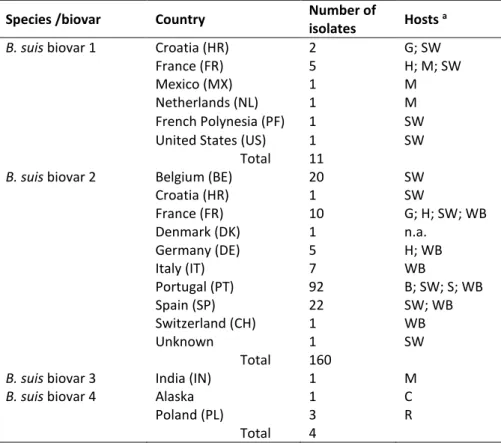
múltiplos locais do genoma, vulgarmente designada por MLVA. O ensaio de MLVA aplicado à genotipificação de estirpes de Brucella spp. utiliza um conjunto de 16 VNTRs que estão distribuídos por três painéis: painel 1, composto por oito minissatélites, painel 2A e painel 2B, compostos respetivamente por três e cinco microssatélites. A análise alocou os isolados de biovar 2 em dois clusters de acordo com as suas origens geográficas e haplótipos, definindo assim uma linhagem clonal ibérica (Portugal e Espanha) e uma linhagem clonal Central-Europeia. A análise de 350 estirpes adicionais de todas as biovariedades de B. suis revelou ainda uma elevada divergência genética entre as estirpes com base nos seus hospedeiros, realçando a estreita relação entre as estirpes de suínos, javalis e lebres. Além de corroborar a existência das duas linhagens clonais de biovar 2, os resultados obtidos sugerem que a evolução da linhagem clonal Ibérica ocorre devido a um evento de especiação alopátrica.
Para comparar a estrutura genómica e avaliar a diversidade genética entre as estirpes de B. suis biovar 2, foram construídos os mapas ópticos de cinco estirpes de campo isoladas de javalis e representativas das linhagens clonal Ibérica (PT09143, PT09172, Bs143CITA) e Central-Europeia (Bs364CITA, Bs396CITA), bem como da estirpe de referência B. suis biovar 2 ATCC 23445 (linhagem Central-Europeia, origem: Dinamarca), utilizando a tecnologia Optical Mapping (mapeamento óptico). Cada estirpe apresentou um perfil único de restrição, de 228 a 232 fragmentos, distribuídos pelos dois cromossomas, tendo-se observado baixa divergência no cromossoma II (1,6%) relativamente ao cromossoma I (2,4%). O mapeamento óptico revelou no cromossoma I a presença de um evento de inserção-deleção (INDEL, 3,5 kb) específico da estirpe de referência ATCC 23445, e uma grande inversão (944 kb) exclusiva da linhagem clonal Ibérica.
Além de validar a existência de uma inversão na linhagem clonal Ibérica, reforçando a plasticidade genómica de Brucella spp., os mapas ópticos permitiram também determinar o posicionamento e a orientação das sequências consenso (contigs), agilizando o processo de montagem e finalização dos genomas. Assim, as sequências genómicas completas e anotadas, das cinco estirpes de B. suis biovar 2 referidas anteriormente, foram prontamente obtidas utilizando-se uma estratégia que combinou: (1) a sequenciação das estirpes, utilizando a plataforma de sequenciação de nova geração (NGS), Illumina HiSeq 2000; (2) a re-sequenciação, por Sanger, das regiões de baixa qualidade ou contendo bases ambíguas; (3) a montagem de novo e finalização dos genomas guiada por Optical mapping. À semelhança do genoma da estirpe de referência de B. suis biovar 2, os cinco genomas sequenciados neste trabalho são compostos por dois cromossomas circulares (Chr I e Chr II) com aproximadamente 1,93 e 1,40 Mb. Em média o teor GC (%) dos Chr I e Chr II foi de 57,1% e 57,3%, respetivamente. Os cinco genomas contêm três operões idênticos de genes de RNA ribossomal (rRNA), um localizado no Chr I e dois no Chr II. Todos os genomas possuem 54 genes de RNA de transferência
(Bs396CITA) e o número de pseudogenes entre 87 (Bs396CITA) e 91 (Bs364CITA).
A fim de melhor compreender os mecanismos envolvidos na evolução e especialização das linhagens Ibéricas, foi realizada uma análise genómica comparativa entre os cinco genomas de B. suis biovar 2 e os 18 genomas disponíveis nas bases de dado públicas, incluindo oito espécies de Brucella. A análise efetuada confirmou que o género Brucella é altamente conservado, com dois cromossomas e taxas de evolução aparentemente lentas a nível de espécie. No entanto, as estirpes de B. suis biovar 2 da linhagem clonal Ibérica foram diferenciadas da Central-Europeia não só pela presença de uma grande inversão no Chr I, mas também por um conjunto de polimorfismos de base única (SNPs) e deleções-inserções (INDELs) específicas. Estes resultados permitiram desenvolver e validar diferentes protocolos de PCR para 10 regiões polimórficas com potencial epidemiológico para a diferenciação das estirpes de uma linhagem clonal específica.Foi ainda observado que o enriquecimento mutacional da linhagem Ibérica está associado a genes que codificam proteínas de membrana, bem como a genes com impacto no metabolismo deste agente patogénico. No entanto, outros estudos são necessários para compreender melhor as consequências destas variações genómicas e o seu impacto na patogenicidade ou virulência numa ampla gama de hospedeiros, incluindo o Homem.
Em conclusão, com este trabalho foi demonstrado que existem duas linhagens clonais de B. suis biovar 2 a circular na Europa, sendo que a especialização genómica e adaptação local das estirpes pertencentes à linhagem clonal Ibérica estabelecem um ecovar Ibérico, levantando uma importante questão sobre os mecanismos responsáveis por tropismos putativos como resposta à adaptação a um hospedeiro específico e/ou condições patobiológicas.
Palavras-chave: B. suis biovar 2; Optical Mapping; Sequenciação de Nova geração; Genómica Comparativa; Filogenómica.
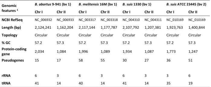
Table 1.1 Brucella species, preferred host(s) and pathogenicity for humans………... 5 Table 1.2 General characteristics of the reference genomes for B. abortus strain 9-941, B.
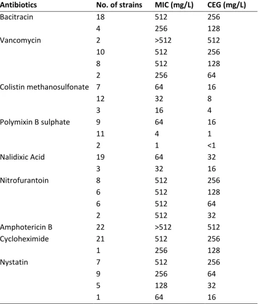
melitensis strain 16M, B. suis strain 1330 and B. suis ATCC 23445 (strain Thomsen)……….…………. 19 Table 2.1.1 Minimal Inhibitory Concentration (MIC) and Concentration Enabling Growth (CEG) for
22 B. suis field strains………..………. 40 Table 2.1.2 Susceptibility of B. suis reference and field strains to the new LNIV-M selective culture
medium, in comparison to modified Thayer-Martin (MTM) and Farrell (FAR) media……… 40 Table 2.2.1 Brucella suis isolates used in the study………. 48 Table 2.2.2 Repeat copy numbers at each locus in the MLVA-16 assay and Hunter-Gaston
Diversity Index (HGDI) for each locus and MLVA-16, MLVA-11 and MLVA-8 subsets……. 51 Table 3.1 Estimated size for each chromosome and respective number of restriction fragments
in the optical maps of B.suis strains………. 80 Table 4.1.1 Listing of strains and respective accession numbers……….. 114 Table 4.1.2 Summary statistics for assembly of five B. suis biovar 2 strains isolates from wild
boars………. 115 Table 4.1.3 Characteristics of Brucella suis biovar 2 genomes………. 116 Table 4.2.1 List of genomes used for comparative genomic and phylogenetic analysis………..…. 132 Table 4.2.2 List of the primers used for assessment of the INDEL events differentiating the two B.
suis biovar 2 clonal lineages……….. 133 Table 4.2.3 Resume of mutation analysis of Brucella spp. genomes using B. suis ATCC 23445 as
Figure 1.1 Mediterranean fever Commission (MFC) in 1904……….... 3
Figure 1.2 Phylogenetic analysis of the Brucella species……….. 7
Figure 1.3 Schematic representation of Brucellae cellular membrane……….……….. 13
Figure 1.4 Zoonotic and non-zoonotic Brucella species………. 14
Figure 1.5 Working model of brucellae intracellular trafficking in macrophage cells……… 17
Figure 1.6 Graphical circular map of the genome for B. suis 1330 and B. suis ATCC 23445…….. 19
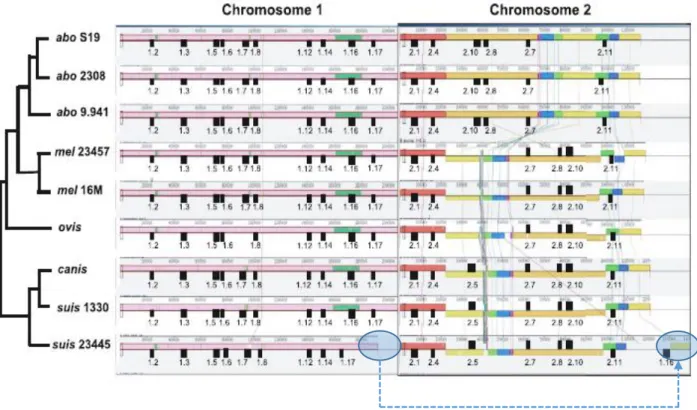
Figure 1.7. Mauve alignment of both chromosomes from nine complete Brucella genomes…… 20
Figure 2.2.1 MLVA-16 clustering analysis………. 53
Figure 2.2.2 Minimum spanning tree (MST) analysis of B. suis strains using the MLVA-11 typing data……… 55
Figure 3.1 Map similarity cluster of chromosomes I and II of B. suis strains using unweighted pair group method with arithmetic average (UPGMA)…….……… 82
Figure 3.2 Pairwise alignment of chromosome I optical maps for B. suis biovar 2 Iberian strains and in silico reference map……….………. 83
Figure 3.3 Assessment of the large genomic inversion by left- and right-junction site-specific PCR………. 84
Figure 3.4 Comparison of the BamH I optical and in silico maps of B. suis ATCC 23445………. 86
Figure 4.2.1 Comparative chromosome mapping between 23 Brucella and O. anthropi DNA sequences………. 135
Figure 4.2.2 Evolutionary relationships of the genus Brucella including 23 genomes of eight species………. 136
Figure 4.2.3 Minimum spanning tree depicting the genomic relationships of Brucella species based on WG-INDELs analysis………. 138
Figure 4.2.4 Translocation event in B. suis biovar 2 strains………. 139
Figure 4.2.5 Left and right crossover points of the large inversion present in B. suis biovar 2 Iberian ecovar………..……….. 141
Figure 4.2.6 Distribution of putative missense mutations between the two distinct B. suis biovar 2 ecovars……….……… 143
Supp. Table S2.2.1 Full information on B. suis isolates, including geographic origin and molecular typing data obtained with Suis-ladder multiplex PCR and RFLP-PCR ……… 61 Supp. Table S2.2.2 MLVA-16 allelic profile for the set of 181 B. suis strains……….. 69 Supp. Figure S3.1 Whole-genome BamH I optical maps of chromosome I and chromosome II of
the six B. suis biovar 2 strains: ATCC 23445, Bs364CITA, Bs396CITA, PT09172, PT09143 and Bs143CITA………. 89 Supp. Table S3.1 Chromosome I (A) and chromosome II (B) restriction fragments for each B. suis
biovar 2 BamH I in silico and optical maps………. 91 Supp. Table S3.2 Brucella strains used to search for the inversion in chromosome I and the large
indel event in the reference strain B. suis ATCC 23445………. 99 Supp. Table S4.1.1 Low coverage regions and indels confirmed by Sanger resequencing
(Reference positions to B. suis ATCC 23445)………. 119 Supp. Table S4.1.2 Ribosomal RNA (rRNA) operons identified using RNAmmer……….. 121 Supp. Table S4.1.3 Transference RNA (tRNA) genes identified using tRNAscan-SE 1.21………. 123 Supp. Table S4.1.4 Predicted coding sequences (CDS) and respective functional annotation for the
five B. suis biovar 2 sequenced genomes.
http://www.mediafire.com/file/ducz93rz73wmu29/Supplementary_Table_S 4.1.4_A-E.xlsx
Supp. Table S4.2.1 List of mutations disclosed in the comparative genomic analysis of 23 Brucella genomes and Ochrobactrum anthropi.
http://www.mediafire.com/file/2mm9zdwo7lyovm2/Supplementary_Table_ S4.2.1_A-C.xlsx
Supp. Figure S4.2.1 Distribution of SNPs along the genome (SNPs per 0,2 Mbp).
http://www.mediafire.com/file/3m9xsgpsfbf17tx/Supp.Figure_S4.2.1_SNPs_ distribution.pdf
ATCC American Type Culture Collection
BAB Blood agar base
BioISI Biosystems & Integrative Sciences Institute Blast Basic Local Alignment Search Tool
BMB Brucella medium base
CDS Coding DNA sequence
CEG Concentration enabling growth CFU Colony-forming unit
Chr Chromosome
DNA Deoxyribonucleic acid
Dntp Deoxyribonucleotide triphosphate
FAR Farrell’s medium
GC GC medium
GO Gene Ontology
GT Genotype
HGDI Hunter-Gaston diversity index INDEL Insertion-deletion event
INIAV Instituto Nacional de Investigação Agrária e Veterinária
IS Insertion sequence
KEGG Kyoto Encyclopedia of Genes and Genomes
LPS Lipopolysaccharide
MIC Minimal inhibitory concentration
ML Maximum likelihood
MLSA Multilocus sequencing analysis MLST Multilocus sequencing typing
MLVA Multilocus variable number tandem repeat analysis
MST Minimum spanning tree
MTM Modified Thayer-Martin’s medium
NCBI National Center for Biotechnology Information
NJ Neighbour-Joining
NCTC National Collection of Type Cultures NGS Next generation sequencing
OM Optical map
OMP Outer membrane protein
ORF Open reading frame
PATRIC Pathosystems Resource Integration Center PCR Polymerase chain reaction
PHAST Phage search tool
PM Plommet medium
tRNA Transfer RNA R-LPS Rough-LPS S-LPS Smooth- LPS
SNP Single nucleotide polymorphism TSA Trypticase soy agar
UPGMA Unweighted pair group method with arithmetic average VNTR Variable number tandem repeat
WG Whole-genome
WGM Whole-genome mapping
WG-INDEL Whole-genome insertion-deletion events distribution WG-MSA Whole-genome multiple sequence alignment
reveals a large genetic inversion on Iberian Brucella suis biovar 2 strains. Veterinary
Microbiology, 192: 220–225. doi:10.1016/j.vetmic.2016.07.024.
Ferreira, A.C., Corrêa de Sá, M.I., Tenreiro, R. and Dias, R. (2014). Complete genome sequences of three Iberian Brucella suis biovar 2 strains isolated from wild boars. Genome Announcements. 2(4):e00618-14. doi:10.1128/genomeA.00618-14.
Ferreira, A.C., Corrêa de Sá, M.I., Tenreiro, R. and Dias, R. (2014). Complete genome sequences of two Central-European Brucella suis bv. 2 haplotype 2c strains isolated from wild boars.
Genome Announcements. 2(4):e00686-14. doi:10.1128/genomeA.00686-14.
Ferreira, A.C., Almendra, C., Cardoso, R., Pereira, M.S., Beja-Pereira, A., Luikart, G., Corrêa de Sá, M.I. (2012). Development and evaluation of a selective medium for Brucella suis. Res. Vet. Sci. 93(2): 565-567.
Submitted research papers:
Ferreira, A.C., Tenreiro, R., Dias, R. and Corrêa de Sá, M.I. (2016). Genetic diversity of Brucella
suis biovar 2 strains circulating in Europe. Submitted to Veterinary Microbiology.
Ferreira, A.C., Tenreiro, R., Corrêa de Sá, M.I. and Dias, R. (2017). Evolution and genomic specialization of Brucella suis biovar 2 Iberian Lineages. Submitted to BMC Genomics.
Chapter 1
1.1. The Genus Brucella: a taxonomic and phylogenetic overview
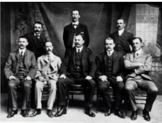
In the year of 1887 in Malta, with the isolation and identification of a bacterium by David Bruce and Themistocles Zammit, now known as Brucella melitensis (Figure 1.1), began the history of Brucellosis, one of the most extended bacterial zoonosis at a global level and a complex infection of animals and humans with a worldwide impact. This bacteria was first isolated in Malta from the spleens of soldiers with fatal cases of brucellosis, also known as undulant fever or Malta fever. The genus
Brucella, created in 1920 by Meyer and Shaw (De Ley et al., 1987), belongs to the family Brucellaceae within the order Rhizobiales of the class Alphaproteobacteria, which is one of the largest and most diverse groups within the phylum Proteobacteria (Scholz et al., 2012; Godfroid et al., 2011; Ficht, 2010; Audic et al., 2009; Bergey and Holt, 1994). The order Rhizobiales includes a variety of bacteria strategically important for their diversity in function and in niche occupancy, including animal intra or pericellular pathogens (Afipia, Anaplasma, Bartonella, Brucella, Erlichia, Ricketsia), opportunistic animal pathogens (such as Ochrobactrum), plant pathogens (e.g. Agrobacterium) and several plant endosymbionts (Carvalho et al., 2010; Velasco et al., 1998). Phylogenetic reconstructions based on whole-genome sequences have confirmed the evolutionary proximity between Brucella genus and members of the subgroup of the alpha-2 Proteobacteria, that includes soil organisms (e.g.
Ochrobactrum spp.), plant symbionts (e.g. Rhizobium spp.) and phytopathogens (e.g. Agrobacterium
spp.), and identified the Ochrobactrum, a soil living facultative human pathogen, as the most closely related genus (Bohlin et al., 2010; Whatmore et al., 2009; Wattam et al., 2009; Scholz et al., 2008b).
Figure 1.1. Mediterranean fever Commission (MFC) in 1904. Standing: Dr. T. Zammit; Capt. Crawford Kennedy; Major J.C. Weir; Seated: Major J.G. McNought; Dr. J.W.H. Eyre; Col. David Bruce.; Major T. McCulloch; Staff Surgeon E.M.A. Clayton.
The genus Brucella was created with two species, B. abortus and B. melitensis, which preferential hosts are cattle and small ruminants (sheep and goats), respectively. B. suis was isolated for the first time from aborted pigs fetus in Europe in 1909, and after in the United States. For many years, it was believed that the agent was a highly pathogenic variant of B. abortus but, in 1929, B. suis was finally considered a separate species. In the following years, new species were added to the genus:
B. ovis isolated from sheep (1956), B. neotomae (1957) from the desert wood rat, and B. canis (1968)
from dogs. Like the first two species, they were exclusively classified and characterized on the basis of their phenotype and host preference (Alton, 1990). The three major species in terms of disease and economic impact for man, B. melitensis, B. abortus and B. suis are further divided into biovars based on a range of phenotypic and serological characteristics: B. melitensis with 3 biovars, B. abortus with 8 biovars, and B. suis with 5 biovars. Since the genetic homogeneity of these “classical” species seems to support the hypothesis of a monospecific genus, B. melitensis has been proposed as the sole representative species with different biotypes (Verger et al., 1985). This classification was accepted by the Sub-committee on the Taxonomy of Brucella in 1986. However, the host range was a long-recognized biological criterion and the presence of species specific markers in outer membrane protein genes and in other genes showed that B. melitensis, B. abortus, B. suis, B. ovis, B. canis and B. neotomae were not mere pathovars (or nomenspecies) but biologically meaningful species. Consequently, in 2003, the Sub-committee approved to return to the multi-nomen species classification with the readoption of the “classical” species with a series of biovars (Osterman and Moriyón, 2006; Moreno et
al., 2002).
Since 2007, more species were included in the genus: B. ceti and B. pinnipedialis, isolated from marine mammal (Foster et al., 2007), B. microti from voles (Scholz et al., 2008a), B. inopinata from an inflamed breast implant of a 71 year-old patient in USA (Scholz et al., 2010) and, more recently, B.
papionis, isolated from baboons (Whatmore et al., 2014) and B. vulpis, isolated from foxes (Scholz et
al., 2016). The natural reservoir of B. inopinata remains unclear (Eisenberg et al., 2016). Like most
Brucella species, B. ceti, B. pinnipedialis and B. papionis are fastidious and slow growing pathogens,
with limited metabolic activity; in contrast, B. microti, B. inopinata and B. vulpis, are fast growing bacteria with a biochemical profile that resemble the members of the genus Ochrobactrum (Hammerl
et al., 2016; Mühldorfer et al., 2016; Scholz et al., 2016, 2008a; Al Dahouk et al., 2010). Other “atypical”
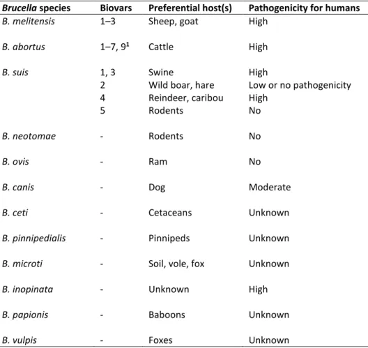
Brucella strains have been isolated from diverse animal sources such as wild rodents (Tiller et al., 2010), frogs (Eisenberg et al., 2012) and fish (Eisenberg et al., 2016) that will likely to be proposed as new species in the future. Despite of Brucella spp. difference in host affinity, they display very similar pathogenic behavior, while varying in virulence. The preferential hosts and the pathogenicity for humans of the 12 recognized Brucella species are described in Table 1.1.
Table 1.1. Brucella species, preferred host(s) and pathogenicity for humans. Brucella species Biovars Preferential host(s) Pathogenicity for humans
B. melitensis 1–3 Sheep, goat High
B. abortus 1–7, 91 Cattle High
B. suis 1, 3 Swine High
2 Wild boar, hare Low or no pathogenicity
4 Reindeer, caribou High
5 Rodents No
B. neotomae - Rodents No
B. ovis - Ram No
B. canis - Dog Moderate
B. ceti - Cetaceans Unknown
B. pinnipedialis - Pinnipeds Unknown
B. microti - Soil, vole, fox Unknown
B. inopinata - Unknown High
B. papionis - Baboons Unknown
B. vulpis - Foxes Unknown
1 B. abortus biovar 8 was deleted by the Brucella Subcommittee in 1978 (Osterman and Moriyón, 2006)
All Brucella species have identical 16S ribosomal (r)RNA- and recA gene sequences, and are almost identical in the majority of housekeeping genes (Whatmore et al., 2009, 2007; Scholz et al., 2008b; Gee et al., 2004). The current understanding of Brucella spp. phylogeny has been elucidated by several methods such as Multilocus Variable Number Tandem Repeat Analysis (MLVA, Scholz & Vergnaud, 2013; Al Dahouk et al., 2012; Le Flèche et al., 2006), Multilocus Sequencing Typing (MLST) and MLS Analysis (MLSA) (Whatmore et al., 2016, 2009, 2007), microarray studies (Foster et al., 2012; Bohlin et al., 2010; Rajashekara et al., 2004), and Single Nucleotide Polymorphisms (SNP, Wattam et
al., 2014, 2012, 2009; Foster et al., 2009, 2008; Chain et al., 2005). The phylogenetic analysis showed
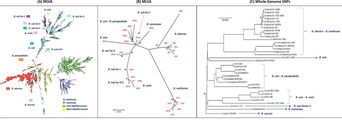
that all Brucella species are monophyletic and distinct from Ochrobactrum. Moreover, most of the analysis referred in the literature distinguish at least six lineages within Brucella spp.: B. abortus-B.
melitensis; B. canis-B. suis, B. ovis, B. ceti-B. pinnipedialis, B. neotomae, and B. microti (Figure 1.2).
From these, B. abortus and B. melitensis are the most closely related, and a close relationship has also been detected between B. canis and B. suis biovars 3 and 4, while B. suis biovars 1, 2 and 5 were allocated to different branches. B. neotomae and B. ovis demonstrate greater divergence levels from
other Brucella species (Olsen & Palmer, 2014; Wattan et al., 2014; Scholz & Vergnaud, 2013). Few studies have been made that include the novel species B. inopinata, B. papionis and B. vulpis. Nevertheless, a recent work confirms the abovementioned lineages and place B. papionis more closely related to B. ovis. The “atypical” B. inopinata and B. vulpis are placed in two separate branches (Whatmore et al., 2016).
1.2. The Brucella species: approaches on diversity and epidemiology
The accessibility of whole-genome sequence data opened the way for comprehensive molecular analyses and subsequent development of molecular typing tools that allow identification and differentiation of Brucella at the species, biovar and individual strain level (Scholz & Vergnaud, 2013). Since the 90’s, several PCR-based methods have been developed and implemented in diagnostic laboratories to confirm pure cultures of brucellae and differentiate among Brucella species and biovars and vaccine strains (Whatmore et al., 2016; Scholz & Vergnaud, 2013; López-Goñi et al., 2011; Yu & Nielsen, 2010; Mayer-Scholl et al., 2010). Moreover, considering that distinguishing individual bacterial lineages within a species is the basis of infectious disease epidemiology, several PCR-based genotyping tools have been developed, including enterobacterial repetitive intergenic consensus sequence PCR (ERIC-PCR), repetitive intergenic palindromic sequence PCR (REP-PCR), randomly amplified polymorphic DNA PCR (RAPD-PCR) or arbitrarily primed PCR (AP-PCR), and restriction fragment length polymorphism PCR (RFLP-PCR) of the omp2 (including omp2a and omp2b genes) and omp31 loci. From these, only RFLP-PCR assay targeting the omp2 (Cloeckaert et al., 1995) or omp31 (Vizcaíno et al., 1995)
loci are more commonly used in laboratories. In fact, PCR-RFLP analysis of omp2a, omp2b and omp31
genes define five different restriction patterns (haplotypes) for each B. suis biovar (1 to 5) and four additional haplotypes are further disclosed within biovar 2 isolates. This method have been useful to differentiate among Portuguese and Spanish isolates from those isolated in other European countries (Muñoz et al., 2010; Garcia-Yoldi et al., 2007; Ferrão-Beck et al., 2006).
As already mentioned, current approaches to study Brucella spp. diversity and phylogeny include methods such as MLVA, MLST/MLSA, SNPs analysis as well as whole-genome sequencing, which serves as a robust and unbiased method to resolve intraspecies relationships for closely related species such as Brucella spp. (Tan et al., 2015). Nevertheless, the highly discriminatory MLVA or MLSA represent a perfect first-line tools for molecular epidemiological studies within outbreak investigations, and MLSA is also appropriate for phylogenetic reconstructions, owing to the highly clonal evolution of the different species (Whatmore et al., 2016; Allen et al., 2015; Scholz & Vergnaud, 2013).
Figure 1.2. Phylogenetic analysis of the Brucella species. (A) Minimum spanning tree based on multilocus variable number of tandem repeats analysis (MLVA) data from 1,925 isolates. Branch lengths up to three are shown. Adapted from Scholz & Vergnaud, 2013. (B) Unrooted phylogenetic reconstruction of the relationships between sequence types based on multilocus sequencing analysis (MLSA). The tree was constructed with the concatenated sequence data of the nine loci (4,396 bp) using the neighbour joining approach. Adapted from Whatmore et al., 2007; (C) Phylogenetic tree based on maximum parsimony of the core Brucella genomes. The tree was rooted with B. microti. All branches have 100% support unless otherwise noted. Adapted from Wattan et al., 2014.
The epidemiological surveillance of animal and human brucellosis has clearly benefited from the appearance and improvement of molecular typing (Valdezate et al., 2010). To date one of the best approaches to obtain valuable discrimination in brucellae is MLVA-16 (using 16 VNTR markers), as it is an easy and cost effective methodology. Since brucellosis has a worldwide distribution, laboratories of different countries try to apply the same genotyping techniques, thus facilitating the exchange of information. In this way, a collaborative public online database based on a MLVA-16 scheme has been built up with the aim of promoting the creation of a global epidemiological map of Brucella spp. (http://mlva.u-psud.fr/), where data can be submitted and compared to other published results. MLVA-16 exhibits an intraspecies discriminatory power and is extremely discriminant and highly efficient in differentiating strains within a local outbreak. Most of the epidemiological studies were applied to B. melitensis or B. abortus due to its important economic and medical concerns. The epidemiological relationship between B. melitensis isolates using MLVA-16 assay has shown that three separate clusters are observed when geographical origin is considered: the American, the Eastern Mediterranean, and the Western Mediterranean groups (Valdezate et al., 2010). For instance, in a study using 126 Portuguese isolates, the majority of the isolates (83%) were included in the Eastern Mediterranean group, which also include isolates from Spain and Turkey, and the remainder (17%) was included in the American group (Ferreira et al., 2012). In contrast, most isolates from Italy belong to the Western Mediterranean lineage (Garafolo et al., 2013). Although less variability is found within B.
abortus populations, MLVA is still a useful tool in ongoing disease surveillance of B. abortus outbreaks,
especially when combined with accurate epidemiological information on disease tracings, geographical clustering of cases and chronology of infection (Allen et al., 2015). Furthermore, although the validity of B. abortus or B. melitensis biovars established by classical microbiological methods cannot be confirmed by MLVA clustering, this method can significantly contribute to epidemiological trace-back analysis of human Brucella infections and may advance surveillance and control of human brucellosis (Ferreira et al., 2012; Al Dahouk et al., 2007). In addition, it was also showed that MLVA-16 can be an essential assay to guarantee the quality and stability of live anti-bacterial vaccines being produced worldwide, such as the B. melitensis REV1 vaccine (Garcia-Yoldi et al., 2007).
Regarding B. suis species, recent studies using both MLVA and MLSA have also shown that considerable intraspecies genetic diversity is observed even when considering isolates sharing the same biovar (Whatmore et al., 2016; Kreizinger et al., 2014; Muñoz et al., 2010; Garcia-Yoldi et al., 2007). However, less work have been made in order to better understand the phylogeny and the epidemiologic relationships of this pathogen.
1.3. Brucella suis biovar 2 and brucellosis
Within the species B. suis, biovars 1, 2 and 3 are the most relevant constituting the main etiological agents involved in swine brucellosis. B. suis infection due to biovars 1 and 3 have been reported in several non-natural domestic and wild host animal species, such as cattle, dogs, horses, sheep, reindeer, caribou, hares, red foxes and various murine species. These biovars 1 and 3 are important human pathogens mainly prevalent in South America and South-East Asia (EFSA, 2009). In Europe they have only been reported in Croatia, indicating that may be restricted to this geographic region (Cvetnic et al., 2009; Garcia-Yoldi et al., 2007; Cvetnic et al., 2005).
B. suis biovar 2 has a very specific pathogenicity for suidae and hares but does not infect
humans (Olsen & Palmer, 2014). It is usually the causative agent of swine brucellosis in Europe (Godfroid et al., 2011; Muñoz et al., 2010; EFSA, 2009; Garcia-Yoldi et al., 2007) and is widely spread amongst Eurasian wild boar (Sus scrofa) and European brown hare (Lepus europaeus) populations, which are identified as the potential source of transmission of biovar 2 to outdoor or extensively reared pigs (Olsen & Palmer, 2014; Munoz et al., 2010; Galindo et al., 2010; Cvetnic et al., 2009; Bergagna et
al., 2009; Leuenberger et al., 2007). Moreover, it is known that biovar 1, 2 and 3 can be transmitted
from swine to cattle, inducing transient seroconversion, which can confound B. abortus diagnostic assays (Olsen & Palmer, 2014; Musser et al., 2013).
Brucellae produce abortion and infertility in infected pigs and is easily transmitted in extensive production systems where animals share the same environment and have contacts with wild animals, namely wild boars and hares. The control and eradication of the disease is only possible with well-planned strategies, adapted to the local reality and globally integrated in the production cycle, serving the interest of public and animal health and also of the production economy. Although the infections in wildlife reservoirs are a lesser threat for causing human infection, they can be a source for the reintroduction of infection into domestic livestock, which poses a new challenge to eradication of the disease worldwide (Musser et al., 2013; Plotkin, 2009). The pathogenesis and pathobiology of brucellosis in domestic pigs are likely to be identical to those in wild boar. In a primary infection the bacteria can spread within a few months from one infected animal to more than 50% of animals on the farm. The infection can often reach 70% to 80% of infected animals at the start of the outbreak, and the infected herds manifest a high percentage of abortions, increased neonatal mortality and infertility, with adverse economic consequences (Aparício, 2013). Nevertheless, as there tend to be few -or mild clinical signs, the disease can go unnoticed in infected groups (EFSA, 2009) and abortion may be the only clinical sign observed, usually occurring during the second or third month of gestation (Poester et al., 2013).
Swine brucellosis is considered to be inexistent or of low prevalence in domestic pigs, therefore it is usually excluded from National Surveillance Programs in the European Union. Consequently, diagnosis is only performed on pigs for International Trading purposes, in selected Artificial Insemination Centers or for investigation of suspected cases. However, the increasing number of outbreaks in pig farms all over Europe, and the high prevalence of B. suis biovar 2 in wild boars and hares, suggest that swine brucellosis could be an emergent but still unrecognized problem in many countries (Olsen & Palmer, 2014; Godfroid et al., 2013; Munoz et al., 2010; Godfroid & Käsbohrer, 2002).
Occurrence of B. suis biovar 2 infection in Portugal
The existence of B. suis biovar 2 infection has been confirmed by isolation of the agent from animals belonging to different pig farms in diverse regions of Portugal where sporadic outbreaks occurred. However, no systematic surveillance is carried out and few studies have been performed to evaluate the real status of the infection in Portugal.
There are three recognized autochthonous pig breeds in Portugal reared in extensive production systems, and therefore more susceptible to B. suis biovar 2 infection: the Bísaro (produced in Trás-os-Montes), the Alcobaça Spotted (“Malhado de Alcobaça”, created in the Centre region), and the Iberian pig (“Porco Alentejano”, raised in an unique integrated agricultural-forestry-livestock system in Alentejo, called "montado"). The first documented B. suis biovar 2 outbreak in domestic pigs occurred in 1999, where a farm in Alentejo (rearing “Porco Alentejano” breed) was traced back as the source of infection of two other farms (one in the North and other in the Centre, with an intensive production system) due to the introduction of infected males. As the majority of the animals in each farm were serologically positive (near 90%) and there was a high number of abortions, the Veterinary Services decided to slaughter the totality of the animals. After that, between 1999 and 2000, about 12 new outbreaks were identified in Alentejo by the veterinary services. The occurrence of abortions and the increase of serologically positive animals have major economic implications as it prevents the free movement of animals and products. For that reason, the surveillance of brucellosis in domestic pigs as well as in wild boars is an important issue and, when feasible, control and eradication programs should be implemented in the affected areas.
1.4. From the outside-in: the cell envelope and virulence of brucellae
All of the various surface components of a bacterial cell are important in its ecology since they mediate the contact of the bacterium with its environment and consequently supports its own existence and survival in that environment.
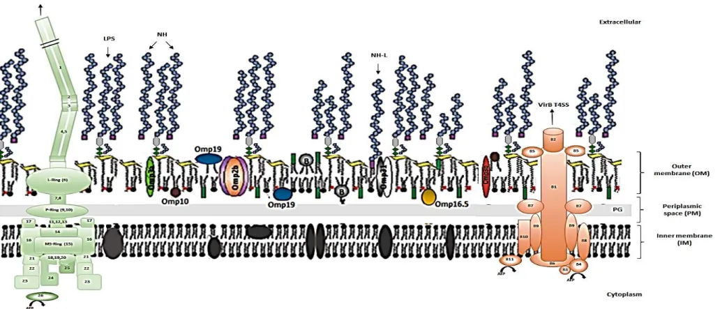
Electron microscopy of Brucella spp. cells shows the classical structure of the Gram-negative cell envelope with an outer-membrane (OM) of 6.5–8.0 nm and an inner membrane (IM) of similar thickness, both separated by a periplasmic space (PS) (Moreno & Moriyón, 2006). The IM is a phospholipid bilayer that contains proteins involved in subtract transport and other metabolic processes. The PS encloses a peptidoglycan (PG) mesh layer and some periplasmic soluble components, such as cyclic β-glucans, and proteins. The OM is the most external layer of the cell constituting an important barrier for survival in hostile environments and an accessible target for the interaction of bacterial pathogens with the host and defense mechanisms of the immune system (Vizcaíno & Cloeckaert, 2012). Structurally, it is an asymmetrical lipid bilayer composed of lipopolysaccharide (LPS) and other haptenic polysaccharides, such as hapten native (NH), proteins and phospholipids (PL), with the LPS molecules and PL located in the outer and inner leaflet, respectively (Figure 1.3). Most of Gram-negative bacteria share this basic structure with OM molecules bearing pathogen-associated molecular patterns (PAMPs) easily recognized by innate immunity. However, brucellae OM has an atypical composition giving distinctive traits to the bacterium when compared with other Proteobacteria (Vizcaíno & Cloeckaert, 2012), and it has been proposed that some of its OM molecules, such as LPS, lipoproteins, flagellin and ornithine lipids, display a reduced PAMP (Gil-Ramírez et al., 2014; Palacios-Chaves et al., 2011; Barquero-Calvo et al., 2007).
The different surface molecules are involved in various important processes, such as bacterial growth, sensing of and protection from environmental stresses, adhesion, and invasion of host cells, signaling, and interaction with the immune system (Bierne & Cossart, 2007). Likewise, a comprehensive characterization of the OM repertoire is required to better understand the factors that contribute to the success of a bacterial pathogen in colonizing different environmental niches and its mammalian host. Smooth brucellae such as B. abortus, B. melitensis and B. suis have OMs that are unusually resistant to the disrupting action of bactericidal peptides and complement (Palacios-Chaves
et al., 2011). It is believed that this stability is related to several specific characteristics, namely: (1)
brucellae OMs contain large amounts of phosphatidylcholine (PC) and blockage of the synthesis of PC with the subsequent replacement by phosphatidylethanolamine (PE, which is the major PL in Gram-negative bacteria) generates attenuation, suggesting that PC is essential for brucellae virulence (Conde-Álvarez et al., 2006); (2) the presence of positively charged ornithine lipids (OL), locate in the
outer leaflet of the OM (Moriyón & López-Goñi, 1998), although recent studies indicate that OL have become dispensable in the existing brucellae. This is consistent within the trend observed in α-Proteobacteria animal pathogens to reduce and eventually eliminate the envelope components susceptible of recognition by innate immunity (Palacios-Chaves et al., 2011); (3) the strong association of some outer membrane proteins (OMPs) to LPS and PG (Moriyón & López-Goñi, 1998), and (4) the presence of very long chain fatty acids (VLCFAs) in the lipid A of LPS that have the potential to span the OM displaying the terminal hydroxyl group in the periplasmic space, probably favoring a strong anchorage of the LPS and the integrity of the OM (Vizcaíno & Cloeckaert, 2012; Barquero-Calvo et al., 2009).
In addition, it was confirmed that the B. abortus LPS core has a branched structure. Based on the observation that the mutation of glycosyltransferase WadC, results in a lipopolysaccharide that, while keeping the O‐polysaccharide essential for optimal protection, shows a truncated core that is more readily recognized by elements of innate immunity, it was proposed that the Brucella LPS core branch is a virulence-related structure accounting in part for the stealthy behavior of these bacteria (Gil-Ramírez et al., 2014; Kubler-Kielb & Vinogradov, 2013; Conde-Álvarez et al., 2012). The characteristic surface properties make brucellae furtive pathogens and more resistant to several host defense compounds, contributing to the intracellular survival and its ability to establish chronic infection (Ruiz-Ranwez et al., 2015).
The brucellae LPS possess unusual immunological properties such as low toxicity, high resistance to macrophage degradation and protection against immune responses, being a major virulence factor in Brucella (Lapaque et al., 2005). Since LPS is the most relevant antigen during infection and vaccination, LPS and LPS-related molecules are extensively used in immunological studies and in the diagnosis of brucellosis (Moreno & Moriyón, 2006; Cardozo et al., 2006; Aragón et al., 1996). Among Gram-negative bacteria, the genus Brucella is the unique in which some species express the smooth (S)-type LPS (B. abortus, B. melitensis, B. suis, B. microti, B. neotomae, B. ceti, B. pinnipedialis,
B. inopinata, B. papionis and B. vulpis) and others have naturally rough (R)-type LPS (B. canis and B. ovis). The S-LPS and R-LPS differ mostly in the most external LPS moiety (the O-polysaccharide), which
is not synthetized in rough Brucella species and is also missing in the surface of the rough mutants of the smooth species that appear spontaneously during culture dissociation (Moreno & Moriyón, 2006). It is known that the production of a complete LPS is necessary for virulence of smooth Brucella strains (González et al., 2008; Rittig et al., 2003). However, the rough species B. canis and B. ovis are virulent in their respective preferred natural hosts and in animal models, and it was shown that rough brucellae attract and infect monocytes more effectively than smooth brucellae although only S-LPS phenotypes establish a specific host cell compartment permitting successful parasitism (Rittig et al., 2003).
13
Figure 1.3. Schematic representation of Brucellae cellular membrane. Adapted and modified from Moreno & Moriyón (2006), Fabienne et al. (2008), Feroozand Letesson (2010), Ferooz et al. (2011), Vizcaíno and Cloeckaert (2012), Gíl-Ramírez et al. (2014), Sankarasubramanian et al. (2016). The cellular membrane is constituted by the inner membrane (IM), the periplasmic space (PM) with the peptidoglycan (PG, light grey), and the outer membrane (OM). IM proteins are represented in grey; the OM contains the LPS (constituted by the lipid-A diaminoglucose disaccharide backbone represented by yellow trapezoids, the core oligosaccharide with its branch, and the O‐polysaccharide chain), several outer membrane proteins (OMPs), ornithine lipids (green rectangles), phospholipids, mainly phosphatidylcholine (dark circles) and lipoproteins, namely the Braun’s lipoprotein (B, grey circles) that can be linked to peptidoglycan or free in the OM. The purples squares mark the reducing ends of the native haptens (NH), an unknown sugar that in some cases may be linked to a lipid (NH-L). NH of various sizes are intertwined with the O-polysaccharides of the LPS in the OM, forming a dense layer. It is also shown the hypothetical model of the brucellae VirB Type IV secretion system (T4SS) and flagellum. B1 to B11 represent the 11 proteins that constitute the VirB T4SS. Numbers 1 to 26 symbolize proteins that compose the flagellum: 1.FliC (filament); 2.FlgL and 3.FlgK (Hook-filament junction); 4.FlgE and 5.FlgD (Hook); 6.FlgH, 7.FliL, 8.FlgG, 9. FlgL, 10.FlgA, 11.FlgB, 12.FlgC, 13.FlgF, 14.FliE and 15.FliF (Basal body); 16.MotA and 17.MotB (Motor); 18.FliP, 19.FliQ, 20.FliR, 21.FliG, 22.FliM, 23.FliN, 24.FlhA and 25.FlhB (Export apparatus); 26.FliI (ATP synthetase).
The most virulent species with higher zoonotic spectrum are those from domesticated animals, such as B. melitensis, B. suis (with the exception of biovar 2), B. abortus and B. canis; while those displaying lower pathogenicity and zoonotic potential are B. ovis and those from wildlife animals, like
B. suis biovar 2 and biovar 5, B. neotomae, B. microti, B. papionis, B. vulpis, B. pinnipedialis and B. ceti. Brucella spp. are able to infect multiple hosts but some species are highly adapted to a single-host
species, such B. ovis which is a pathogen for rams but does not infect other hosts (Figure 1.4) (Moreno, 2014; Olsen, 2014; Godfroid et al., 2011; Tsolis et al., 2009). In order to understand the pathomechanisms of infectious diseases with clinical significance in animals and humans we must first understand the biology of these agents, and their hosts, in order to unravel the interactions that occur at the host-agent interface. A comprehensive understanding of the basis of host specificity can provide insights into molecular pathogenesis, the evolution of pathogenic Brucella species, and the potential for these pathogens to cross the species barrier to infect new hosts.
Figure 1.4. Zoonotic and non-zoonotic Brucella species, modified from Moreno, 2014. Hosts in clockwise direction: (A) cattle, camel, sheep, goat, reindeer, swine, dog, unknown; (B) rams, wild boar, hare, wild rodent, wild rodent, common vole, baboon, red fox, pinnipeds, porpoise, dolphin. Regarding
B. microti, B. papionis, B. vulpis, B. pinnipedialis and B. ceti, pathogenicity for humans is still unknown.
One of the striking features that distinguish brucellae organisms from other pathogenic bacteria is that they do not display obvious virulence factors such as exotoxins, cytolysins, capsules, fimbria, plasmids, lysogenic phages, resistant forms, antigenic variation, endotoxic LPS or apoptosis inducers. Also unlike other pathogenic bacteria, brucellae virulence does not appear to be the result of relatively few virulence genes that can be transferred horizontally via plasmids, phages, or
assembled in pathogenicity islands. Instead, the true virulence elements of brucellae are those molecular determinants that allow to control their intracellular trafficking and adapt to the intracellular niche as well as on the extremely efficient adaptation to shield itself from the immune recognition and to manipulate key aspects of host cell physiology (apoptosis, vacuolar trafficking) (Ruiz-Ranwez et al., 2015; Gomez et al., 2013; von Bargen et al., 2012; Lamontagne et al., 2010; Rambow-Larsen et al., 2009; Gorvel, 2008; Seleem et al., 2008; Barquero-Calvo et al., 2007; Moreno & Moriyón, 2006; Lapaque et al., 2005; Gorvel and Moreno, 2002; Letesson et al., 2002). However, in recent years, various virulence factors have been identified as essential for infection, including LPS (Cardozo et al., 2006; Lapaque et al., 2005; Ugalde et al., 2000), β-cyclic glucan (Martirosyan et al., 2012; Arellano-Reynoso et al., 2005), BvrR/BvrS two component system (TCS) (Martín-Martín et al., 2012; Lamontagne et al., 2010; Viadas et al., 2010; Guzman-Verri et al., 2002), some Omps (Lim et al., 2012; Vizcaíno and Cloeckaert, 2012), and the VirB Type IV secretion system (T4SS) (Seleem et al., 2007; Celli et al., 2003; Boschiroli et al., 2002). Quorum sensing (QS) is also known to be involved in the regulation of brucellae virulence determinants mostly linked to the cell surface (T4SS, flagellum, Omps and exopolysaccharide), contributing to the adaptation of the metabolic network during the nutrient shift faced by brucellae all along its intracellular trafficking (Gorvel, 2014; Weeks et al., 2010; Rambow-Larsen et al., 2009; Letesson et al., 2002).
In both humans and animals, brucellae first target the respiratory epithelium, the conjunctiva, and sexual organs. However, even nowadays, the mechanisms involved in brucellae entry into host cells still remain to be characterized (Gorvel, 2014). The ability of Brucella spp. to successfully survive and replicate within different host cells explains their pathogenicity. Extensive replication of Brucella spp. in placental trophoblasts is associated with abortion in their preferential animal hosts, and persistence in macrophages leads to chronic infections that are a hall mark of brucellosis in both natural animal hosts and humans (Kim, 2015; Gorvel, 2014; Grilló et al., 2012; Roop et al., 2009).
In vitro studies were used as models to understand adhesion, internalization, intracellular
trafficking, survival, and replication of brucellae in susceptible hosts. After attachment to the epithelial cell surface receptors that contain sialic acid and sulfated residues, brucellae induces a zipper-like mechanism for internalization. Binding promotes activation of small GTPases that trigger a signaling cascade that reorganizes the actin cytoskeleton to induce a host cell membrane rearrangement along the surface of the pathogen that enhances invasion, and entry occurs within a few minutes after interaction (Rossetti et al., 2012). Brucella spp. organisms are capable of colonizing macrophages, monocytes, and dendritic cells as well as trophoblasts, fibroblasts, endothelial cells, and epithelial cells (Gorvel, 2014; Hamer et al., 2014; Martirosyan et al., 2011; Starr et al., 2008). Brucellae enters into host cells through lipid rafts (Barquero-Calve et al., 2007; Porte et al., 2003; Watarai et al., 2002), and
its entry depends on the expression of BvrR/BvrS TCS (Manterola et al., 2005; Guzman-Verri et al., 2002). In fact, it has been shown that mutants lacking LPS O-chain do not use lipid rafts and are killed by the host cell suggesting that O-chain plays an important role in early events of host infection (Porte
et al., 2003). Brucellae survive and replicate inside nonprofessional phagocytic cells up to 72 hours in
vitro and move across the epithelium in vivo by subverting the mucosal epithelial barrier function to
facilitate brucellae migration. At the same time, this interaction initiates a minimal innate immune response with weak proinflammatory activity (Rossetti et al., 2012; Barquero-Calvo et al., 2007). Brucellae survival strategies have been elucidated from analysis of intracellular trafficking in either macrophage or epithelial cell models. Once internalized (Figure 1.5), Brucella spp. cells resides within the brucellae-containing vacuole (BCV), a modified phagosome in which the bacterium survives and ultimately proliferates (Kim et al., 2015; Gomez et al., 2013; Lee et al., 2013; Starr et al., 2008; Celli et
al., 2003; Chaves-Olarte et al., 2002). The BCVs then fuse rapidly with the lysosome in a controlled
manner, as suggested by the presence of the lysosomal markers, lysosomal-associated membrane protein (LAMP), and CD63, on the surface of bacteria (Starr et al., 2008; Celli et al., 2003; Pizarro-Cerdá
et al., 1998). In this transient stage, most of the contents of the BCVs are subjected to phagolysosomal
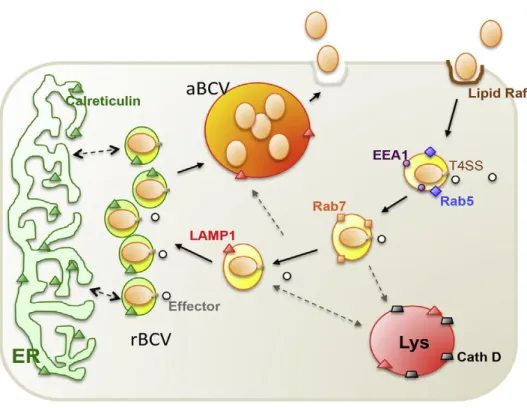
degradation, and 90% of internalized brucellae cells are killed by the action of hydrolyzing enzymes (Celli et al., 2003). However, the remaining 10% evade the host killing mechanisms through an unknown mechanism that probably involves the acidification of the BCVs, subsequent triggering of the virB operon and release of a large variety of effectors into the host cells' cytosol (Boschiroli et al., 2002). The bacteria then traffic and arrive at the endoplasmic reticulum (ER). Within the ER, the bacteria survive and establish their replicative niche, and multiply to large numbers (Celli et al., 2003). Recent studies have shown that autophagy-like vacuoles (i.e. autophagic “brucellae-replicating organelle”, aBCV) provide a replication-permissive compartment following the ER stage, that is essential for the completion of the intracellular lifecycle of brucellae and for cell-to-cell spreading (Starr et al., 2008).
Figure 1.5. Working model of brucellae intracellular trafficking in macrophage cells, extracted from Kim, 2015. Plasma membrane-associated lipid rafts mediate the internalization of smooth brucellae into macrophage cells. As the BCV matures, it sequentially associates with markers for early (EEA1, purple circle; Rab5, blue diamond) and late (Rab7, orange square) endosomes. The biogenesis and trafficking of BCVs is regulated by bacterial effector proteins (white circles), which are secreted through the brucellae T4SS. BCVs that contain virulent organisms do not fuse with lysosomes (cathepsin D, gray trapezoid), although transient association with LAMP1-positive membranes (orange triangles) is observed. The pathogen replicates in tight rBCVs that are decorated with calreticulin (green triangle), a marker for the ER. At a later point after infection (48 to 72 hours), the pathogen is observed in LAMP1-positive aBCVs that also contain LAMP1. Finally, the pathogen is released from the cell through lytic or nonlytic (shown) mechanisms. aBCV, autophagic brucellae-containing vacuole; BCV, brucellae-containing vacuole; Beclin1, coiled-coil myosin-like BCL2-interacting protein; EEA1, early endosome antigen 1; ER, endoplasmic reticulum; LAMP1, lysosome-associated membrane protein 1; rBCV, replicative brucellae-containing vacuole; T4SS, type IV secretion system; ULK1, Unc-51-like kinase 1.
1.5. Comparative genomics as a tool to understand evolution in brucellae
The way in which we perceive the taxonomic relationships among different bacteria influences our understanding of their basic biological and ecological features (Moreno & Moriyón, 2002). Distinguishing individual bacterial lineages within a species, initially by phenotypic and subsequently by genotypic typing techniques, has been the cornerstone of infectious disease epidemiology, allowing the identification and tracking of the organisms responsible for infection and disease (Parkhill and Wren, 2011). During the past decade, the understanding of evolution at the genomic level has been