S H O R T R E P O R T
Open Access
Molecular detection of tick-borne bacteria
and protozoa in cervids and wild boars
from Portugal
André Pereira
1, Ricardo Parreira
2,3, Mónica Nunes
2,3, Afonso Casadinho
1, Maria Luísa Vieira
2,3, Lenea Campino
2,4,5and Carla Maia
1,2,4*Abstract
Background:Wildlife can act as reservoir of different tick-borne pathogens, such as bacteria, parasites and viruses. The aim of the present study was to assess the presence of tick-borne bacteria and protozoa with veterinary and zoonotic importance in cervids and wild boars from the Centre and South of Portugal.
Methods:One hundred and forty one blood samples from free-ranging ungulates including 73 red deer
(Cervus elaphus), 65 wild boars (Sus scrofa) and three fallow deer (Dama dama) were tested for the presence of
Anaplasma marginale/A. ovis,A. phagocytophilum,Anaplasma/Ehrlichiaspp.,Babesia/Theileriaspp.,Borrelia burgdorferi
(sensu lato) (s.l.), andRickettsiaspp. DNA by PCR.
Results:Anaplasmaspp. DNA was detected in 33 (43.4 %) cervids (31 red deer and two fallow deer) and in two (3.1 %)
wild boars whileTheileriaspp. were found in 34 (44.7 %) cervids (32 red deer and two fallow deer) and in three (4.6 %)
wild boar blood samples. Sequence analysis ofmsp4sequences identifiedA. marginale,A. ovis, while the analysis of
rDNAsequence data disclosed the presence ofA. platysandA. phagocytophilumandT. capreoliandTheileriasp. OT3.
Anaplasmaspp./Theileriaspp. mixed infections were found in 17 cervids (22.4 %) and in two wild boars (3.1 %). All
samples were negative forBabesiasp.,B. burgdorferi(s.l.),Ehrlichiasp. orRickettsiasp.
Conclusions:This is the first detection ofAnaplasma marginale,A. ovis,A. phagocytophilum, A. platys, Theileria capreoli
andTheileriasp. OT3 in cervids and wild boars from Portugal. Further studies concerning the potential pathogenicity of
the different species of AnaplasmaandTheileriainfecting wild ungulates, the identification of their vector range, and
their putative infectivity to domestic livestock and humans should be undertaken.
Keywords:Anaplasmaspp., Fallow deer, PCR, Portugal, Red deer,Theileriaspp., Tick-borne pathogens, Wild boar
Background
Wildlife can harbor a high density of ticks that can transmit several pathogens, such as bacteria, parasites and viruses. In addition to their veterinary importance, many of these tick-borne pathogens can also affect the human population as a result of their zoonotic potential. Therefore, the management of such situation calls for a One Health approach, including the increased awareness
for their presence especially in sylvatic environments and areas associated with animal husbandry among veterinarians, physicians and general public [1].
Piroplasmoses in cattle is caused by tick-borne
protozoan parasites comprising several Theileria and
Babesia species. These diseases are a serious health
problem, being responsible for important economic losses to the cattle industry. In Europe, infections with different
Theileria spp. [Theileria sp. OT3,T. capreoli (formerly
Theileria sp. 3185/02), Theileria sp. ZS OT4,T. ovis] and
Babesia spp. [including, among others, B. bigemina, B.
capreoli,B. divergensandB. venatorum(formerlyBabesia
sp. EU1)] have been reported in cervids. These include fallow deer (Dama dama),red deer (Cervus elaphus) and
* Correspondence:[email protected]
1Faculty of Veterinary Medicine, Universidade Lusófona de Humanidades e
Tecnologias, Lisbon, Portugal
2Global Health and Tropical Medicine (GHTM), Instituto de Higiene e
Medicina Tropical (IHMT), Universidade Nova de Lisboa (UNL), Lisbon, Portugal
Full list of author information is available at the end of the article
roe deer (Capreolus capreolus) [2–5], whileTheileria sp.
and B. bigeminawere detected in wild boars (Sus scrofa)
[5]. In Portugal, the main pathogenic piroplasm species re-ported in cattle isTheileria annulata, although others, in-cluding T. buffeli and T. orientalis, considered as being moderately pathogenic or benign, are also present [6, 7].
In addition, several pathogenic species of Babesia (B.
bovis, B. divergens and B. bigemina) have also been
reported in cattle from central and southern Portugal
[6, 7]. Further, human babesiosis caused by B.
diver-gens, B. microti or B. venatorum have been reported
in several European countries [8], including one fatal case due to B. divergens in Portugal [9].
Anaplasmoses, caused by bacteria of the genus
Anaplasma, known for a long time in veterinary
medicine, are also considered as emerging human diseases, and are frequently associated with infection
with Anaplasma phagocytophilum [10]. This bacterium,
which is the causative agent of tick-borne fever, a disease of important negative economic impact to European animal husbandry (involving domestic ruminants), also causes human granulocytic anaplasmosis. Wild ruminants are one of its main reservoirs [11] while the role of wild boars in its natural cycle is still contradictory [12]. Other
Anaplasma spp. such as A. marginale and A. ovis have
also been detected in European cervids [13]. In Portugal,
antibodies reactive to A. phagocytophilum antigens were
detected in humans and other mammals [14], while A.
marginaleandA. ovis were detected in cattle [15] and in
sheep [16], respectively.
Among the diseases caused by tick-borne pathogens,
Lyme borreliosis caused by spirochetes of the Borrelia
burgdorferi (sensu lato) (s.l.) complex is currently the
most common tick-borne disease in Europe [17]. In Portugal, its notification in humans is mandatory, but the disease is clearly underdiagnosed and underreported [17]. Wild large vertebrates seem to be frequently exposed to these bacteria, as indicated by the detection of eitherBorrelia-specific antibodies in these animals or
Borrelia DNA in engorged ticks collected from them
[18, 19]. Finally, several tick-borneRickettsiaspp. associ-ated with human infections such asRickettsia conorii, R.
slovaca and R. raoultii have also been described in
several European countries, including Portugal [20].
Rickettsia spp. (e.g. R. helvetica, R. slovaca) DNA has
previously been detected in the peripheral blood [21] or in ticks removed from cervids and wild boars [4, 22], but the role these wild mammals play in the natural main-tenance of these bacteria has not yet been clarified.
No information about tick-borne pathogens circulating in wild ungulates from Portugal is available, with the
single exception of the recent detection of Borrelia
burgdorferi (s.l.) in wild boars from northern Portugal
[17]. Thus, the aim of the present study was to assess
the presence of tick-transmitted bacteria and protozoa with veterinary and zoonotic importance in cervids and wild boars from the Centre and South of the country.
Methods
Animals and samples
During the hunting seasons, from December 2013 to March 2015, a total of 141 free-ranging ungulates
including 73 red deer (Cervus elaphus), 65 wild boars
(Sus scrofa) and 3 fallow deer (Dama dama) from
both sexes were sampled in the districts of Castelo Branco (n= 31), Portalegre (n= 16), Lisboa (n= 19), Évora (n= 15) and Beja (n= 60). Animals were classified in two age cat-egories: young (1–3 years) and adults (> 3 years). Blood samples were collected from each animal by cardiac or thoracic punctures in EDTA tubes and stored at -20 °C until DNA extraction.
Ethical approval
This study was ethically approved by the board of the Faculty of Veterinary Medicine (ULHT).
PCR amplification
A commercial kit (PCR-template Preparation kit, Roche Diagnostics GmbH, Germany) was used to extract DNA from the collected blood samples, following the manu-facturer’s instructions.
In order to avoid false negative results due to PCR inhibition, and so as to validate the efficiency of the
DNA extraction, the modified vertebrate-universal cyt-b
specific primers (cytB1-F and cytB2-R) were used to amplify a 350 bp segment of the host mitochondrial
cytochrome b gene (cyt-b) [23]. PCR amplifications were
performed in a 25 μl final volume containing 12.5 μl of
NZYTaq 2x Green Master Mix (Nyztech, Portugal), 1μl
of each primer (10 pmol) and 2μl of template DNA.
Detection ofAnaplasma/Ehrlichiaspp.,A. marginale/A.
ovis,A. phagocytophilum,Babesia/Theileriaspp.,B.
burg-dorferi(s.l.) andRickettsiaspp. DNA in blood samples was
assessed by PCR, according to previously described proto-cols (Table 1).
PCR amplifications were performed in a final volume of
25 μl using NZYTaq 2× Green Master Mix, 3 μl of the
prepared DNA extracts and 10 pmol of each primer. In all amplifications, positive (containing genomic DNA of the targeted microrganism) and negative (without DNA) con-trols, were included. PCR amplifications were carried out in a Thermo Electron Corporation® Px2 Termal Cycler (VWR, USA) and the obtained PCR products visual-ized under UV illumination after electrophoresis on 1.5 % agarose gels stained with Greensafe premium® (Nzytech, Portugal) using a 100 bp DNA ladder as a molecular-weight size marker (Nzytech, Portugal).
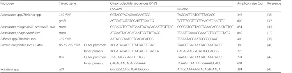
Table 1Sequences of the oligonucleotide primers used
Pathogen Target gene Oligonucleotide sequences (5′-3′) Amplicon size (bp) Reference
Forward Reverse
Anaplasmaspp./Ehrlichiaspp. 16S rRNA GGTACCYACAGAAGAAGTCC TAGCACTCATCGTTTACAGC 345 [38]
groEL ACTGATGGTATGCARTTTGAYCG TCTTTRCGTTCYTTMACYTCAACTTC 600 [39]
Anaplasma marginale/A. centrale/A. ovis msp4 GGGAGCTCCTATGAATTACAGAGAATTGTTTAC CCGGATCCTTAGCTGAACAGGAATCTTGC 851 [40]
Anaplasma phagocytophilum msp4 ATGAATTACAGAGAATTGCTTGTAGG TTAATTGAAAGCAAATCTTGCTCCTATG 849 [13]
Babesiaspp./Theileriaspp. 18S rRNA AATACCCAATCCTGACACAGGG TTAAATACGAATGCCCCCAAC 400 [38]
Borrelia burgdorferi(sensu lato) ITS 5S-23S rRNA Outer primmers ACCATAGACTCTTATTACTTTGAC TAAGCTGACTAATACTAATTACCC 380 [41]
Inner primers ACCATAGACTCTTATTACTTTGACCA GAGAGTAGGTTATTGCCAGGG 225
flaB Outer primmers TGGTATGGGAGTTTCTGG TAAGCTGACTAATACTAATTACCC 774 [42]
Inner primers CAGACAACAGAGGGAAAT TCAAGTCTATTTTGGAAAGCACC 604
Rickettsiaspp. gtlA GGGGGCCTGCTCACGGCGG ATTGCAAAAAGTACAGTGAACA 381 [43]
Parasites
&
Vectors
(2016) 9:251
Page
3
of
PCR products were purified from agarose gel slices with NZYGelpure® (Nzytech, Portugal) according to the manufacturer’s instructions. Purified products were sent
to LIGHTrun™ Sequencing Service (GATC-biotech,
Germany) for direct sequencing of the obtained ampli-cons by Sanger’s method with the same primers used for DNA amplification.
DNA sequence analyses
Species identity of the obtained sequences was assessed on the basis of the closest BLASTn match (identity≥98 % using the MegaBLAST and a query cover no smaller than 96 %) with homologous sequences deposited in the Gen-Bank database. The sequences obtained in the course of this work were deposited at DNA Data Bank of Japan (DDBJ) (http://www.DDBJ.nig.ac.jp).
Phylogenetic relationships were inferred from nucleo-tide sequence alignments produced with the MAFFT multiple alignment program using a combination of the G-INS-i alignment option [24]. Phylogenetic tree con-struction was carried out using a Maximum Likelihood (ML) approach, using the Kimura’s 2-P (K2P) evolutionary
model, and assuming a Γ distributed substitution rates
among sites, as indicated by Mega6 [25] on the basis of the Akaike information criterion. Alternatively, an
empir-ically defined model (GTR +Γ+ I) was also used. The
topological robustness of the obtained trees was assessed by bootstrapping, using 1000 resampling of the original alignment data. The final trees were manipulated for
display using FigTree v.1.2.2. (available at http://tree.-bio.ed.ac.uk/software/figtree/).
Statistical analysis
Percentages of positive samples forAnaplasma spp. and
Theileria spp. regarding the independent variables and
categories were compared by the Chi-square or Fisher’s exact tests. AP-value≤0.05 was considered as statistically significant. Exact binomial 95 % confidence intervals (CI) were defined for the proportions. Analyses were
performed with Epi Info™ 7.1.5.2 software for Centers
for Disease and Prevention.
Results
A 350 bp fragment of the host mitochondrial cyt-bgene
was amplified in all DNA blood samples.
Anaplasma spp. DNA was detected in 33 (43.4 % CI:
32.1–55.3 %) cervids (31 red deer and two fallow deer) and in two (3.1 % CI: 0.4–10.7 %) wild boars using a set
of general primers that target 16S rDNA. Seventeen
sequences obtained from red deer (accession numbers:
LC126854, LC126858-9, LC126863-5, LC126867-9,
LC126871, LC126873, LC126875, LC126878, LC126879 and LC126881-3) and two from wild boars (accession
numbers: LC126885-6) showed 99–100 % identity with
A. platys previously described in dogs from Portugal
(LC018182-3; [26]), Argentina (JX261979; [27]) and in a goat from Cyprus (EU090182; [28]). Further, eight
se-quences obtained from red deer (LC126855-6,
Anaplasma marginale(Red deer) LC126846
Anaplasma marginale(Red deer) LC126851
Anaplasma marginaleKJ512166
Anaplasma marginaleKF739428
Anaplasma marginale(Red deer) LC126847
Anaplasma marginale(Red deer) LC126850
Anaplasma marginale(Red deer) LC126848
Anaplasma marginaleEF190508
Anaplasma ovis KJ782397
Anaplasma ovis (Red deer) LC126849
Anaplasma ovis FJ460446
Anaplasma ovis AY702924
Anaplasma centraleAY054383
Anaplasma centraleAF428090
Anaplasma centraleCP001759
Anaplasma phagocytophilumHQ661156
Anaplasma phagocytophilumKM205442
Anaplasma phagocytophilumAY706390
100 90
89
97 86
89
0.1
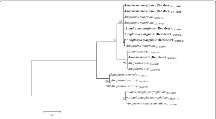
Fig. 1Phylogenetic tree ofAnaplasma spp.based on the analysis ofmsp4sequences
LC126860-2, LC126870, LC126876 and LC126884)
showed 99 % identity withA. phagocytophilumdescribed
in Swedish moose (KC800983; [29]) and in black-striped
field mice (Apodemus agrarius) in South Korea
(KR611719). Six sequences isolated from red deer
(LC126857, LC126866, LC126872, LC126874 and
LC126880) and two isolated from fallow deer (LC126852-3)
showed 100 % identity with A. marginale (KC335218,
KC335223) described in cows and withA. ovisdescribed in sheep (KC335231) and goats (KC335225) from Italy [30] as
well as with A. centrale described in goats from China
(KP062964, KP062966; [31]).
Sequencing of the msp4 gene amplified from the
samples where the presence of A. centrale/A.marginale/
A. ovis DNA had been detected with the 16S rDNA
primers, confirmed the presence of A. marginale in five
(6.6 % CI: 2.2–14.7 %) red deer from the Castelo Branco,
Portalegre and Beja districts and A. ovis in one (1.3 %
CI: 0–7.1 %) red deer from Beja. Attempts to amplify
msp4sequences from the two fallow deer failed. The
ob-tainedmsp4sequence data, along with related sequences
obtained from Genbank, were subjected to phylogenetic analyses (Fig. 1).
PCR reactions prepared using either groEL or A.
phagocytophilum species-specific primers revealed
repro-ducibly negative amplification results. On the contrary,
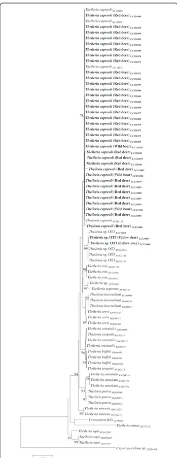
Theileria spp. were found in 34 (44.7 % CI: 33.3–56.6 %)
cervids (32 red deer and two fallow deer) and in three (4.6 % CI: 1.0–12.9 %) wild boar samples, using primers
targeting the 18S rRNA gene. Blast analysis showed that
the sequences obtained from red deer (accession numbers: LC131069-100) and wild boars (LC131101-3) presented 98–99 % identity toT. capreoli(KJ188207-8) described in Sika deer from China [32] while the two sequences obtained from fallow deer (LC131067-8) showed a high
identity (98–99 %) to the Theileria sp. OT3 (Genbank:
KF470868) described in sheep from China [33]. The
phy-logenetical analysis of the obtained 18S rDNAsequences
along with the related sequences from GenBank corrobo-rated the Blast identification (Fig. 2).
Anaplasmaspp./Theileriaspp. co-infections were found
in 17 cervids (22.4 % CI: 13.6–33.4 %). Of these, eight red deer were co-infected withA. platysandT. capreoli, four
withA. marginaleandT. capreoli, two withA.
phagocyto-philum andT. capreoli, one withA. ovisand T. capreoli,
while two fallow deer were co-infected with Anaplasma
sp. andTheileriasp. OT3. Two wild boars (3.1 % CI: 0.4– 10.7 %) were co-infected withA. platysandT. capreoli.
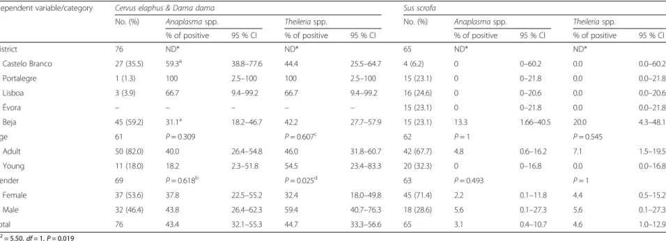
The frequency of Anaplasma infection was
signifi-cantly higher (P= 0.019) in red deer from the Castelo
Branco district than from Beja (Table 2). All the blood samples taken from wild boars with positive PCR ampli-fication results toT. capreoliorA. platyswere from the Beja district.
Theileria capreoliKJ188208 Theileria capreoli(Red deer) LC131084
Theileria capreoliKJ188207 Theileria capreoli(Red deer) LC131095
Theileria capreoli(Red deer) LC131094
Theileria capreoli(Red deer) LC131085
Theileria capreoli(Red deer) LC131096
Theileria capreoli(Red deer) LC131088
Theileria capreoli(Red deer) LC131074
Theileria capreoli(Red deer) LC131073
Theileria capreoliJX134575 Theileria capreoli(Red deer) LC131071
Theileria capreoli(Red deer) LC131083
Theileria capreoli(Red deer) LC131091
Theileria capreoli(Red deer) LC131086
Theileria capreoli(Red deer)LC131069
Theileria capreoli(Red deer) LC131089
Theileria capreoli(Red deer) LC131090
Theileria capreoli(Red deer) LC131077
Theileria capreoli(Red deer) LC131082
Theileria capreoli(Red deer) LC131078
Theileria capreoli(Red deer) LC131072
Theileria capreoli(Red deer) LC131075
Theileria capreoli(Red deer) LC131087
Theileria capreoli(Wild boar) LC131103
Theileria capreoli(Red deer) LC131099
Theileria capreoli(Red deer) LC131070
Theileria capreoli(Red deer) LC131100
Theileria capreoli(Red deer) LC131081
Theileria capreoli(Wild boar) LC131102
Theileria capreoli(Red deer) LC131079
Theileria capreoli(Red deer) LC131092
Theileria capreoli(Red deer) LC131098
Theileria capreoli(Red deer) LC131097
Theileria capreoli(Red deer) LC131076
Theileria capreoli(Wild boar) LC131101
Theileria capreoli(Red deer) LC131093
Theileria capreoliKJ188215 Theileria capreoli(Red deer) LC131080
Theileriasp. OT3 KF470868 Theileriasp. OT3 (Fallow deer) LC131067
Theileriasp. OT3 (Fallow deer) LC131068
Theileriasp. OT3 DQ866840
Theileriasp. OT3 AY533145
Theileriasp. OT3 EF092455
Theileria ovis KF697193
Theileria ovis GU726904
Theileria ovis KJ850942
Theileriasp. AY748463
Theileria separata AY260175
Theileria luwenshuniKC769995
Theileria luwenshuniJX469510
Theileria luwenshuniKJ850935
Theileria cerviKP407020
Theileria cerviHQ184411
Theileria cerviHQ184407
Theileria orientalisAB520956
Theileria sergentiKJ850938
Theileria orientalisHM538221
Theileria orientalisKJ850937
Theileria buffeliKJ806987
Theileria buffeliKJ806986
Theileria buffeliHQ840962
Theileria sergentiAY661515
Theileria annulataKM288518
Theileria annulataKF429799
Theileria annulataKT367872
Theileria parva HQ895968
Theileria parva HQ895973
Theileria parva HQ895972
Theileria sinensisHM538203
Theileria sinensisEU274472
Cytauxzoon felisGU903911
Theileria annaeKF773741
Theileria equiEU642509
Theileria equiHM229407
Theileria equiJX457819
Cryptosporidiumsp. JX294363 99 99 97 83 99 98 93 96 99 95 98 94 99 94 78 0.2
Fig. 2Phylogenetic tree ofTheileriaspp. based on18S rRNA
Table 2Prevalence of tick-borne pathogens as detected by PCR in 76 cervids and 65 wild boars from Centre and southern Portugal
Idependent variable/category Cervus elaphus & Dama dama Sus scrofa
No. (%) Anaplasmaspp. Theileriaspp. No. (%) Anaplasmaspp. Theileriaspp.
% of positive 95 % CI % of positive 95 % CI % of positive 95 % CI % of positive 95 % CI
District 76 ND* ND* 65 ND* ND*
Castelo Branco 27 (35.5) 59.3a 38.8
–77.6 44.4 25.5–64.7 4 (6.2) 0 0–60.2 0.0 0.0–60.2
Portalegre 1 (1.3) 100 2.5–100 100 2.5–100 15 (23.1) 0 0–21.8 0.0 0.0–21.8
Lisboa 3 (3.9) 66.7 9.4–99.2 66.7 9.4–99.2 16 (24.6) 0 0–20.6 0.0 0.0–20.6
Évora – – – – – 15 (23.1) 0 0–21.8 0.0 0.0–21.8
Beja 45 (59.2) 31.1a 18.2
–46.7 42.2 27.7–57.9 15 (23.1) 13.3 1.66–40.5 20.0 4.3–48.1
Age 61 P= 0.309 P= 0.607c 62 P= 1 P= 0.545
Adult 50 (82.0) 40.0 26.4–54.8 46.0 31.8–60.7 42 (67.7) 4.8 0.6–16.2 7.1 1.5–19.5
Young 11 (18.0) 18.2 2.3–51.8 54.5 23.4–83.3 20 (32.3) 0 0–16.8 0.0 0.0–16.8
Gender 69 P= 0.618b P= 0.025d 63 P= 0.493 P= 1
Female 37 (53.6) 37.8 22.5–55.2 32.4 18.0–49.8 45 (71.4) 2.2 0.1–11.8 4.4 0.5–15.2
Male 32 (46.4) 43.8 26.4–62.3 59.4 40.7–76.3 18 (28.6) 5.6 0.1–27.3 5.6 0.1–27.3
Total 76 43.4 32.1–55.3 44.7 33.3–56.6 65 3.1 0.4–10.7 4.6 1.0–12.9
a
χ2= 5.50,df= 1,P= 0.019
b
χ2= 0.25,df= 1
c
χ2= 0.26,df= 1
dχ2= 5.03,df= 1
ND* Statistically significant difference(s) not confirmed after pairwise comparisons
Pereira
et
al.
Parasites
&
Vectors
(2016) 9:251
Page
6
of
None of the samples analysed revealed the presence of
Babesiasp., B. burgdorferi(s.l.),Ehrlichiasp. or
Rickett-siasp.
Discussion
Concern about the role of wildlife in the natural maintenance transmission of tick-borne pathogens is increasing, especially in areas where free-ranging animals regularly interact with domestic livestock and humans [5, 34].
This study, which aimed at the detection of tick-borne bacteria and protozoa of veterinary and zoonotic import-ance in cervids and wild boars, disclosed, to our know-ledge, the first evidence for the circulation of Anaplasma
spp. and Theileria spp. among red deer, fallow deer and
wild boars in central/southern Portugal. In this study,
Anaplasmaspp. infections were detected in the three wild
ungulate species analysed as revealed by the amplification
of 16S rDNA sequences using genus-specific primers.
Anaplasma platyscauses canine cyclic thrombocytopenia
and is presumably transmitted by ticks of the
Rhipicepha-lus sanguineus group. As A. platys DNA has previously
been reported in dogs [26], ticks [35] and red foxes [36] from Portugal, its detection in the red deer and wild boars sampled herein indicates that these animals are also ex-posed to the bacterium. However, the ability ofA. platysto cause disease in these animals has not been established yet. As wild cervids are considered one of the main reservoirs
ofA. phagocytophilum[11], the detection of this bacterium
in eight red deer blood samples using16S rDNAprimers it is not entirely surprising, especially when it is known that the pathogen is circulating in different Portuguese verte-brate hosts, as well as inIxodes ricinus,its most frequently associated vector [14]. However, the absence of detection ofmsp4specific sequences may indicate the circulation of
divergent A. phagocytophilumvariants among Portuguese
red deer different from the ones previously reported [37]. In any case, this issue deserves future clarification. Further-more, the presence ofA. ovisandA. marginalein red deer
was confirmed by msp4phylogenetic analysis, confirming
the susceptibility of this cervid to the agents responsible for bovine and ovine anaplasmoses [13]. Both pathogens have been reported in cattle from the Alentejo region (which includes the Évora and Beja districts [15]), and in sheep raised throughout the country [16].
The occurrence of Theileria spp. infections in
Euro-pean cervids is well documented [2–5]. In the present
study, and for the first time in Portugal,T. capreoli and
Theileria sp. OT3 msp4 sequences were amplified from
red deer and fallow deer samples, respectively, corrobor-ating previous data from northern Spain [3]. The overall prevalence ofBabesia spp. andTheileria spp. infections previously reported in cattle from the central and south-ern regions of Portugal ranged from 23.1 % [7] to 74.7 %
[6], respectively, with T. annulata and T. buffeli being
the most commonly detected species and B. bigemina,
B. bovis and B. divergens being the least frequently
found. As none of the Theileria and Babesia species
known to circulate in the Portuguese cattle were de-tected in the present study, it seems that the tested wild ungulate species might not play a significant role in their transmission, at least in the regions where samples were collected. Furthermore, although deer have been previ-ously appointed as the source for ovine infection with
Theileria sp. OT3 [3], no data is yet available regarding
the circulation of piroplasmids in small ruminants from Portugal.
Despite the fact thatB. burgdorferi(s.l.) andRickettsia
spp. have already been detected in ticks and/or in the blood collected from cervids and wild boars [4, 17, 19, 22], their presence was not revealed in any of the sam-ples analysed in the present study. This observation sup-ports the hypothesis that wild ungulates, at least in the studied areas, are not pivotal players in the natural maintenance cycles of these bacteria, as previously re-ported [4, 21].
As large wildlife are important to maintain tick popula-tions, and since ticks may become infected with several pathogens during their life cycle, the detection of
Ana-plasma spp./Theileria spp. co-infections in the present
study is not surprising, and falls in line with previously published observations in wild ungulates [4]. The inter-action of different pathogens within the vertebrate host might lead to increased susceptibility to other infections as well as a modification of the pathogenesis of each microorganism with profound consequences for disease management programs and wildlife conservation [4].
Conclusions
The present study provides information regarding the presence ofAnaplasma marginale,A. ovis,A.
phagocyto-philum, A. platys, Theileria capreoli and Theileria sp.
OT3 in cervids and wild boars from central and south-ern Portugal. Further studies concsouth-erning the potential
pathogenicity of the different Anaplasma and Theileria
species infecting wild ungulates, the identification of their vector range, and their infectivity to domestic livestock and humans should be undertaken.
Competing interests
The authors declare that they have no competing interests.
Authors’contributions
Acknowledgments
The authors wish to acknowledge FCT for funds to GHTM–UID/Multi/04413/ 2013 and the hunters for their assistance in obtaining the blood samples and B. Almeida and G. Acto for technical assistance. M. Nunes was supported by the Ministry of Education and Science of Portugal, Fundação para a Ciência e a Tecnologia, through a PhD grant (SFRH/BD/78325/2011). The work of C. Maia was done under the frame of EurNegVec COST Action TD1303.
Publication of this paper has been sponsored by Bayer HealthCare–Animal Health Division in the framework of the 11th CVBD World Forum Symposium.
Author details
1Faculty of Veterinary Medicine, Universidade Lusófona de Humanidades e
Tecnologias, Lisbon, Portugal.2Global Health and Tropical Medicine (GHTM),
Instituto de Higiene e Medicina Tropical (IHMT), Universidade Nova de Lisboa (UNL), Lisbon, Portugal.3Medical Microbiology Unit, IHMT, UNL, Lisbon,
Portugal.4Medical Parasitology Unit, IHMT-UNL, Lisbon, Portugal. 5Department of Biomdical Scienecs and Medicine, Universidade do Algarve,
Lisbon, Portugal.
Received: 6 April 2016 Accepted: 25 April 2016
References
1. Dantas-Torres F, Chomel BB, Otranto D. Ticks and tick-borne diseases: a One Health perspective. Trends Parasitol. 2012;28:437–46.
2. Duh D, Petrovec M, Bidovec A, Avsic-Zupanc T. Cervids as babesiae hosts, Slovenia. Emerg Infect Dis. 2005;11:1121–3.
3. García-Sanmartín J, Aurtenetxe O, Barral M, Marco I, Lavin S, García-Pérez AL, et al. Molecular detection and characterization of piroplasms infecting cervids and chamois in Northern Spain. Parasitology. 2007;134:391–8. 4. Overzier E, Pfister K, Herb I, Mahling M, Böck G, Silaghi C. Detection of
tick-borne pathogens in roe deer (Capreolus capreolus), in questing ticks
(Ixodes ricinus), and in ticks infesting roe deer in southern Germany. Ticks
Tick Borne Dis. 2013;4:320–8.
5. Zanet S, Trisciuoglio A, Bottero E, de Mera IG, Gortazar C, Carpignano MG, et al. Piroplasmosis in wildlife:BabesiaandTheileriaaffecting free-ranging ungulates and carnivores in the Italian Alps. Parasit Vectors. 2014;7:70. 6. Silva MG, Marques PX, Oliva A. Detection ofBabesiaandTheileriaspecies
infection in cattle from Portugal using a reverse line blotting method. Vet Parasitol. 2010;174:199–205.
7. Gomes J, Soares R, Santos M, Santos-Gomes G, Botelho A, Amaro A, et al. Detection ofTheileriaandBabesiainfections amongst asymptomatic cattle in Portugal. Ticks Tick Borne Dis. 2013;4:148–51.
8. Yabsley MJ, Shock BC. Natural history of ZoonoticBabesia: Role of wildlife reservoirs. Int J Parasitol Parasites Wildl. 2013;2:18–31.
9. Centeno-Lima S, do Rosário V, Parreira R, Maia AJ, Freudenthal AM, Nijhof AM, et al. A fatal case of human babesiosis in Portugal: molecular and phylogenetic analysis. Trop Med Int Health. 2003;8:760–4.
10. Doudier B, Olano J, Parola P, Brouqui P. Factors contributing to emergence
ofEhrlichiaandAnaplasmaspp. as human pathogens. Vet Parasitol.
2010;167:149–54.
11. Stuen S, Granquist EG, Silaghi C.Anaplasma phagocytophilum-a widespread multi-host pathogen with highly adaptive strategies. Front Cell Infect Microbiol. 2013;3:31.
12. de la Fuente J, Gortazar C. Wild boars as hosts of human-pathogenic
Anaplasma phagocytophilumvariants. Emerg Infect Dis. 2012;18:2094–5.
13. de la Fuente J, Massung RF, Wong SJ, Chu FK, Lutz H, Meli M, et al. Sequence analysis of themsp4gene ofAnaplasma phagocytophilumstrains. J Clin Microbiol. 2005;43:1309–17.
14. Santos AS, Bacellar F, Dumler JS. A 4-year study ofAnaplasma
phagocytophilumin Portugal. Clin Microbiol Infect. 2008;15:46–7.
15. Gomes J, Rebêlo E. Seroprevalência da anaplasmose bovina em Portugal. In: Abstract book–IV Veterinary Sciences Congress. Portugal: Portuguese Society of Veterinary Sciences; 2008. p. 211.
16. Renneker S, Abdo J, Salih DE, Karagenç T, Bilgiç H, Torina A, et al. Can
Anaplasma ovisin small ruminants be neglected any longer? Transbound
Emerg Dis. 2013;60:105–12.
17. Faria AS, Paiva-Cardoso MD, Nunes M, Carreira T, Vale-Gonçalves HM, Veloso O, et al. First Detection ofBorrelia burgdorferisensu lato DNA in Serum of the Wild Boar (Sus scrofa) in Northern Portugal by Nested-PCR. Ecohealth. 2015;12:183–7.
18. Doby JM, Betremieux C, Rolland C, Barrat J. The large forest mammals reservoirs forBorrelia burgdorferi, agent of the Lyme disease? Serological examination of 543 deers and wild-boars. Rec Med Vet Ec Alfort. 1991;167:55–61.
19. Wodecka B, Rymaszewska A, Skotarczak B. Host and pathogen DNA identification in blood meals of nymphalIxodes ricinusticks from forest parks and rural forests of Poland. Exp Appl Acarol. 2014;62:543–55. 20. Oteo JA, Portillo A. Tick-borne rickettsioses in Europe. Ticks Tick Borne Dis.
2012;3:271–8.
21. Stefanidesova K, Kocianova E, Boldis V, Kostanova Z, Kanka P, Nemethova D, et al. Evidence ofAnaplasma phagocytophilumandRickettsia helvetica infection in free-ranging ungulates in central Slovakia. Eur J Wildl Res. 2008;54:519–24.
22. Sprong H, Wielinga PR, Fonville M, Reusken C, Brandenburg AH, Borgsteede F, et al.Ixodes ricinusticks are reservoir hosts forRickettsia helveticaand potentially carry flea-borneRickettsiaspecies. Parasit Vectors. 2009;2:41. 23. Maia C, Parreira R, Cristóvão JM, Freitas FB, Afonso MO, Campino L.
Molecular detection ofLeishmaniaDNA and identification of blood meals in wild caught phlebotomine sand flies (Diptera: Psychodidae) from southern Portugal. Parasit Vectors. 2015;8:173.
24. Katoh K, Toh H. Recent developments in the MAFFT multiple sequence alignment program. Brief Bioinform. 2008;9:286–98.
25. Tamura K, Stecher G, Peterson D, Filipski A, Kumar S. MEGA6: Molecular Evolutionary Genetics Analysis version 6.0. Mol Biol Evol. 2013;30:2725–9. 26. Maia C, Almeida B, Coimbra M, Fernandes MC, Cristóvão JM, Ramos C, et al.
Bacterial and protozoal agents of canine vector-borne diseases in the blood of domestic and stray dogs from southern Portugal. Parasit Vectors. 2015;8:759.
27. Eiras DF, Craviotto MB, Vezzani D, Eyal O, Baneth G. First description of naturalEhrlichia canisandAnaplasma platysinfections in dogs from Argentina. Comp Immunol Microbiol Infect Dis. 2013;36:169–73. 28. Chochlakis D, Ioannou I, Sharif L, Kokkini S, Hristophi N, Dimitriou T, et al.
Prevalence ofAnaplasmasp. in goats and sheep in Cyprus. Vector Borne Zoonotic Dis. 2009;9:457–63.
29. Malmsten J, Widén DG, Rydevik G, Yon L, Hutchings MR, Thulin CG, et al. Temporal and spatial variation inAnaplasma phagocytophiluminfection in Swedish moose (Alces alces). Epidemiol Infect. 2014;142:1205–13. 30. Zobba R, Anfossi AG, Pinna Parpaglia ML, Dore GM, Chessa B, Spezzigu A,
et al. Molecular Investigation and Phylogeny ofAnaplasmaspp. Mediterranean Ruminants Reveal the Presence of Neutrophil-Tropic Strains Closely Related toA. platys. Appl Environ Microbiol. 2014;80:271–80. 31. Yan G, Hongmei Y, Yasuko R, Weiqing P, Hong Y. Molecular detection of
tick-borne rickettsiales in goats and sheep from southeastern China. Vector Borne Zoonotic Dis. 2016. doi:10.1089/vbz.2015.1884.
32. Li Y, Chen Z, Liu Z, Liu J, Yang J, Li Q, et al. Molecular identification of
Theileriaparasites of northwestern Chinese Cervidae. Parasit Vectors.
2014;7:225.
33. Tian Z, Liu G, Yin H, Xie J, Wang S, Yuan X, et al. First report on the occurrence ofTheileriasp. OT3 in China. Parasitol Int. 2014;63:403–7. 34. de la Fuente J, Estrada-Pena A, Venzal JM, Kocan KM, Sonenshine DE.
Overview: Ticks as vectors of pathogens that cause disease in humans and animals. Front Biosci. 2008;13:6938–46.
35. Maia C, Ferreira A, Nunes M, Vieira ML, Campino L, Cardoso L. Molecular detection of bacterial and parasitic pathogens in hard ticks from Portugal. Ticks Tick Borne Dis. 2014;5:409–14.
36. Cardoso L, Gilad M, Cortes HC, Nachum-Biala Y, Lopes AP, Vila-Viçosa MJ, et al. First report ofAnaplasma platysinfection in red foxes (Vulpes vulpes) and molecular detection ofEhrlichia canisandLeishmania infantumin foxes from Portugal. Parasit Vectors. 2015;8:144.
37. Stuen S, Pettersen KS, Granquist EG, Bergström K, Bown KJ, Birtles RJ.
Anaplasma phagocytophilumvariants in sympatric red deer (Cervus elaphus)
and sheep in southern Norway. Ticks Tick Borne Dis. 2013;4:197–201. 38. Harrus S, Perlman-Avrahami A, Mumcuoglu KY, Morick D, Eyal O, Baneth G.
Molecular detection ofEhrlichia canis,Anaplasma bovis,Anaplasma platys,
CandidatusMidichloria mitochondrii andBabesia canis vogeliin ticks from
Israel. Clin Microbiol Infect. 2011;17:459–63.
39. Barber R, Li Q, Diniz P, Porter B, Breitschwerdt E, Claiborne M, et al. Evaluation of brain tissue or cerebrospinal fluid with broadly reactive polymerase chain reaction forEhrlichia,Anaplasma, spotted fever group
Rickettsia,Bartonella, andBorreliaspecies in canine neurological diseases
(109 cases). J Vet Intern Med. 2010;24:372–8.
40. de la Fuente J, Van Den Bussche RA, Prado TM, Kocan KM.Anaplasma
marginale msp1alphagenotypes evolved under positive selection pressure but
are not markers for geographic isolates. J Clin Microbiol. 2003;41:1609–16. 41. Schwartz JJ, Gazumyan A, Schwartz I. rRNA gene organization in the Lyme
disease spirochete,Borrelia burgdorferi. J Bacteriol. 1992;174:3757–65. 42. Wodecka B, Leońska A, Skotarczak B. A comparative analysis of molecular
markers for the detection and identification ofBorreliaspirochaetes in
Ixodes ricinus. J Med Microbiol. 2010;59:309–14.
43. Regnery RL, Spruill CL, Plikaytis BD. Genotypic identification of rickettsiae and estimation of intraspecies sequence divergence for portions of two rickettsial genes. J Bacteriol. 1991;173:1576–89.
• We accept pre-submission inquiries
• Our selector tool helps you to find the most relevant journal
• We provide round the clock customer support
• Convenient online submission
• Thorough peer review
• Inclusion in PubMed and all major indexing services
• Maximum visibility for your research
Submit your manuscript at www.biomedcentral.com/submit