Frequency and antimicrobial susceptibility of
Staphylococcus pseudintermedius in dogs with otitis externa
Frequência e sensibilidade antimicrobiana de Staphylococcus
pseudintermedius isolados de cães com otite externa
Carolina Boesel Scherer
1Larissa Silveira Botoni
1Fernanda Morcatti Coura
2Rodrigo Otávio Silva
2Rafael Dantas dos Santos
3Marcos Bryan Heinemann
4Adriane Pimenta Costa-Val
1*ISSNe 1678-4596
INTRODUCTION
Staphylococcus pseudintermedius is the
leading cause of pyoderma and otitis externa in
dogs, and is ultimately associated with urinary tract
infections (VAN DUIJKEREN et al., 2011). Otitis
disease affects nearly 20% of dogs admitted to
veterinary clinics (COLE et al., 2006; SANCHEZ
1Departamento de Clínica e Cirurgia Veterinárias (DCCV), Escola de Veterinária (EV), Universidade Federal de Minas Gerais (UFMG), Belo Horizonte, MG, Brasil. E-mail: adriane@ufmg.br. *Corresponding author.2Departamento de Medicina Veterinária Preventiva (DMVP), Escola de Veterinária (EV), Universidade Federal de Minas Gerais (UFMG), Belo Horizonte, MG, Brasil.
3Empresa Brasileira de Pesquisa Agropecuária (EMBRAPA), Semiárido, Aracaju, SE, Brasil.
4Departamento de Medicina Veterinária Preventiva e Saúde Animal, Faculdade de Medicina Veterinária (FMV), Universidade de São Paulo (USP), São Paulo, SP, Brasil.
ABSTRACT: Infections by Staphyloccocus pseudintermedius in the skin of dogs have been object of studies, since such microorganisms
often present multiple resistance to antibiotics. This study aimed to identify and evaluate the antimicrobial susceptibility of Staphylococcus pseudintermedius (SP) strains isolated from dogs with otitis. Swabs from both ears of 52 dogs with otitis (n=104) were included. Bacteria were cultured using Muller-Hinton agar (supplemented with 5% equine blood and incubated at 37°C for 24 to 48 hours. All colonies underwent biochemical evaluation for identification of staphylococci. The identity of colonies as SP was confirmed by polymerase chain reaction. The antimicrobial susceptibility of SP strains was evaluated by disk diffusion. The presence of the gene mecA was evaluated in all SP isolates by PCR. Forty-four SP strains were isolated from swabs of 31 dogs (31/52, 59.6%). Seventy-five percent of the strains were susceptible to cephalexin and 93.2% to amoxicillin plus clavulanic acid. Less than 23% of the strains were susceptible to penicillin. For non-beta-lactam antimicrobials, 63.6% of the strains showed resistance to sulfamethoxazole-trimethoprim, 61.4% to tetracycline, and 38, 64% to enrofloxacin. Aminoglycoside resistance rate was 27.3% for gentamicin. Resistance to oxacillin in vitro was detected in 13 of the 44 strains (29, 55%). A total of 12 strains (27.3%) were positive for mecA gene and five of these 12 strains were susceptible to in vitro oxacillin. Twenty-six (59, 1%) strains were resistant to three or more classes of antimicrobials, and classified as multi resistant. Our results showed high frequency of SP and multi resistant isolates to antimicrobials commonly used in veterinary.
Key words: dogs, MRSP, methicillin-resistant staphylococcus, otitis.
RESUMO: Infecções por Staphyloccocus pseudintermedius (SP) na pele de cães tem sido objeto de estudos, uma vez que esses microrganismos
geralmente apresentam resistência múltipla à antibióticos. Este estudo teve como objetivo identificar e avaliar a susceptibilidade antimicrobiana das cepas de SP isoladas de cães com otite. Amostras de ambas orelhas de 52 cães com otite (n=104) foram incluídas. As bactérias foram cultivadas em ágar Muller-Hinton suplementado com 5% de sangue equino e incubadas a 37ºC por 24 a 48h. Todas as colônias foram submetidas à avaliação bioquímica para identificação de estafilococos. A identificação das colônias como SP foi confirmada pela reação em cadeia da polimerase. A susceptibilidade antimicrobiana das cepas SP foi avaliada pela técnica de difusão em disco. A presença do gene mecA foi avaliada em todos os isolados de SP por PCR. 44 estirpes de SP foram isoladas de 31 cães (31/52, 59,6%). 75% das cepas foram suscetíveis a cefalexina e 93,2% à amoxicilina mais ácido clavulânico. Menos de 23% das estirpes eram suscetíveis à penicilina. Para antimicrobianos não beta-lactâmicos, 63,6% apresentaram resistência ao sulfametoxazol-trimetoprim, 61,4% à tetraciclina e 38,64% à enrofloxacina. A frequência de resistência a aminoglicosídeos foi de 27,3% para gentamicina. A resistência a oxacilina in vitro foi detectada em 13 das 44 estirpes (29,55%). Um total de 12 amostras (27,3%) foram positivas para mecA, cinco das quais foram suscetíveis à oxacilina na difusão em disco. 26 amostras (59,1%) foram resistentes a três ou mais classes de antimicrobianos e classificadas como multirresistentes. Nossos resultados mostram elevada frequência de isolados SP e multiresistentes para os antimicrobianos comumente usados em veterinária.
Palavras-chave: cães,MRSP, otite, Staphylococcus resistente a meticilina.
et al., 2011). Multiple antibiotic resistance is rapidly
emerging in S. pseudintermedius, limiting the
treatment options available (ROBERTS et al., 2014).
Methicillin resistance in staphylococci
is commonly associated with the presence of the
mecA gene. mecA encodes an altered
penicillin-binding protein 2a (PBP2a) (FRANK & LOEFFLER,
2012) and confers resistance to all beta-lactam
antibiotics, including penicillins, cephalosporins,
and carbapenems (KANIA et al., 2004; VAN
DUIJKEREN et al., 2011; PRIYANTHAet al., 2016).
In addition, methicillin-resistant S. pseudintermedius
(MRSP) strains are commonly resistant to several
other classes of non-beta-lactam antibiotics, including
aminoglycosides, quinolones, macrolides, phenicols,
sulfonamides, and tetracyclines (PAPICH, 2012).
Recent studies have shown that the
incidence of MRSP is increasing in dogs (KASAI et
al., 2016, PRIYANTHA et al., 2016). In Brazil, a high
incidence of MRSP in dogs with pyoderma has been
reported over the last few years (LOPES et al., 2015;
BOTONI et al., 2016; BOURGUIGNON et al., 2016).
Moreover, this microorganism has been increasingly
implicated in human infections (SOMAYAJI et al.,
2016; ROBB et al., 2017).
Despite the known importance of MRSP
in companion animals and humans in other countries,
few studies have been performed in Brazil. Moreover,
studies regarding the prevalence of this microorganism
in otitis and its antimicrobial resistance profile
are even less common in the literature. Therefore,
the purpose of this research was to determine the
frequency of S. pseudintermedius in dogs diagnosed
with otitis, and the antimicrobial susceptibility of the
strains to several antibiotics, including methicillin,
and to screen for the presence of the mecA gene.
MATERIALS AND METHODS
Fifty-two dogs of different breeds were
selected from the Department of Dermatology at
the Veterinary Hospital of the Universidade Federal
de Minas Gerais (UFMG). Dogs were examined
by veterinarians for the clinical diagnosis of otitis
externa. All dogs used in the study had two or more
clinical signs of otitis externa: pain; head shaking;
itching; erythema; excoriations; hyperkeratosis;
hyperpigmentation; partial or total ear canal stenosis;
changes in color, odor, appearance and/or quantity
of ear discharge; and positive coccoid bacteria by
cytology. This experiment was approved by the Ethics
Committee on Animal Use of UFMG under protocol
number 246/2013.
Swabs from the horizontal channel of the
external ear canal were collected from both ears, totaling
104 swabs. Samples were plated onto Muller-Hinton agar
(Difco Laboratories, EUA) supplemented with 5% equine
blood and incubated at 37°C for 24 to 48h. All colonies
underwent biochemical evaluation for identification
of staphylococci, as previously described by QUINN
et al. (2011) and BANNOEHR & GUARDABASSI
(2012). Biochemical examinations analyzed catalase,
oxidase, coagulase, and urease production, carbohydrate
fermentation (mannitol, sucrose, and trehalose), arginine
production, Voges Proskauer (for acetoin production),
and response to polymyxin B.
Strains identified as belonging to the
Staphylococcus genus were subjected to polymerase chain
reaction (PCR) to amplify a 430-bp portion of the nuc
gene locus to confirm their identity as S. pseudintermedius
(SASAKI et al., 2010). Primers used for this analysis were:
inF 5’CATGTCATATTATTGCGAATGA3’, and inR3
5’ AGGACCATCACCATTGACATATTGAAACC3’.
Amplicons were separated by electrophoresis on
a 1.5% agarose gel with a 1-kb molecular marker
(ThermoScientific
®Waltham, Massachusetts, USA).
S. pseudintermedius MRSP 3279, a reference strain
previously used in other studies (BANNOEHR et al,
2009; BOURGUIGNON et al., 2016) were used as
positive controls and amplification of sterile water served
as a negative control.
The antimicrobial susceptibility of S.
pseudintermedius strains was evaluated by disk
diffusion method according to the Clinical and
Laboratory Standards Institute document VET
01-A4 (CLSI, 2013a). Criteria for interpretation of
disk diffusion method described in CLSI document
VET01-A4 were performed according to CLSI
document VET 01-S2 (CLSI, 2013b). After isolation
and identification, three to five colonies were transferred
to a tube containing 3mL of Mueller-Hinton broth and
incubated at 37°C until cultures reached a standard
turbidity of 0.5, based on the McFarland scale. Using
a sterile swab, contents of the tube were distributed on
a 150-mm plate containing Mueller-Hinton agar, and
10 equidistant paper discs containing antibiotics: 20µg
amoxicillin + 10µg clavulanic acid, 30µg cephalexin,
5µg enrofloxacin, 10µg gentamicin, 1µg oxacillin,
10µg penicillin, 30µg tetracycline, and 1.25g/23.75µg
trimethoprim-sulfamethoxazole (DME
®Laboratory,
halo size following the CLSI standardization (CLSI,
2013b). The interpretation criteria are shown in table 1.
Additionally, all strains were subjected to PCR analysis to
screen for the presence of the mecA gene (MEHROTRA
et al., 2000).
RESULTS AND DISCUSSION
In the present study, 44 of the isolated
strains were confirmed as S. pseudintermedius. These
strains were isolated from 31 dogs (31/52 – 59.6%). Of
the 44 strains, 13 were obtained from same dogs but
different ears, that means, only one strain was obtained
from each ear of the dogs. Six strains were susceptible
to all antimicrobials tested. The percentage of S.
pseudintermedius strain antimicrobial susceptibility is
shown in table 2. Table 3 shows the resistance pattern
of the strains.
S. pseudintermedius was detected in almost
60% of dogs with otitis externa. This rate is higher than
that reported in previous studies (LILENBAUM et al.,
2000; HOEKSTRA & PAULTON, 2002; PENNA et al.,
2010; SAPUTRA et al., 2017). This variation in results
might be due to geographical differences, and clinical
conditions (with and/or without otitis), age, and breed
of the dogs. In Brazil, studies examining otitis caused
by
S. pseudintermedius in dogs are scarce, making
any comparison difficult. The isolation rate obtained
in this study was greater than that reported by the two
previously published studies in Brazil (LILENBAUM
et al., 2000; PENNA et al., 2010). Taken together,
our results indicated that S. pseudintermediu
s
is an
important bacterium causing otitis in dogs and should
be considered when treating this disease.
High beta-lactam susceptibility rates
were detected for some antibiotics, with 75% of
the strains susceptible to cephalexin and 93.2%
to amoxicillin plus clavulanic acid. In contrast,
less than 23% of the strains were susceptible to
penicillin. High susceptibility rates to amoxicillin
plus clavulanic have been reported previously
(PENNA et al., 2010;KROEMER et al., 2014;
DE MARTINO et al., 2016). However, resistance
to penicillin has not been previously reported (
LILENBAUM et al., 2000, KROEMER et al.,
2014;; DE MARTINO et al., 2016). Cephalexin is
used more commonly to treat pyoderma, a common
infection caused by S. pseudintermedius. Since only
first-generation cephalosporins, such as cephalexin,
are suitable for oral usage, it is a common choice
for most clinicians. Moreover, in veterinary
dermatology, studies suggested that pathogenic
staphylococci slowly acquired resistance to
cephalosporins (MASON & KIETZMANN, 1999),
which may account for the high susceptibility to
cephalexin detected in this study.
Owing to improve in vitro stability, oxacillin,
a penicillinase-resistant semi-synthetic penicillin, is
commonly used in veterinary microbiology laboratories
to detect methicillin resistant staphylococci (COLE et
al., 2006). While not routinely used for therapy, the
inclusion of oxacillin in antibiogram tests is useful
as methicillin-resistant staphylococci are commonly
resistant to all beta-lactams, and to aminoglycosides,
quinolones, macrolides, phenicols, sulfonamides,
and tetracyclines (PAPICH, 2012). This pattern was
observed in our study (Table 3). Here, resistance to
oxacillin by disk diffusion was detected in 13 of 44
strains (29,55%). However, PCR analysis for mecA
revealed that seven out of the 13 strains classified as
resistant to oxacillin by disk diffusion harbored the
mecA gene and five strains that were positive for mecA
Table 1 - Susceptibility test interpretive criteria for antibiotic against Staphylococcus spp. according to the Clinical and Laboratory Standards Institute document VET 01-S2 (CLSI, 2013).
Antibiotic ---Disk diffusion zone (mm)---
Susceptible Intermediate Resistant
Amoxicillin-clavulanic acid >20 - ≤19
Cephalexin* ≥18 15-17 ≤14
Enrofloxacin ≥23 17-22 ≤16
Gentamicin** ≥15 13-14 ≤12
Oxacillin*** ≥17 - ≤18
Penicillin ≥29 - ≤28
Tetracycline ≥19 15-18 ≤14
Trimethoprim-sulfamethoxazole** ≥16 11-15 ≤10
gene in the PCR were susceptible to oxacillin by disk
diffusion. That means that a total of 12 (27.3%) strains
tested positive for mecA by PCR. If we consider both
in vitro and PCR results, a total of 18 (40, 9%) strains
were classified as MRSP. Discrepancies in the results
obtained by the disk diffusion test and PCR detection
of mecA has been previously reported (ELHASSAN et
al., 2015; BOURGUIGNON et al., 2016). Although,
mecA seems to be the most common gene associated
with meticillin resistance in S. pseudintermedius, some
studies have shown that another gene (mecC) could also
be responsible for this resistance profile (KJELLMAN
et al., 2015). This could explain the absence of mecA
in some of the strains that were resistant to oxacillin in
the present study.
For non-beta-lactam antimicrobials, 63.6%
of the strains showed resistance to
sulfamethoxazole-trimethoprim, 61.4% to tetracycline and 38, 64% to
enrofloxacin. Aminoglycoside resistance was observed
in 27.3 of strains for gentamicin. Previous studies have
shown that resistance to aminoglycosides is commonly
high (PENNA et al., 2010; DE MARTINO et al., 2016).
Resistance to sulfamethoxazole-trimethoprim
was also reported in other studies (LILENBAUM et al.,
2000; DE MARTINO et al., 2016). However, a survey
in Europe revealed that sensitivity to sulfonamides
was very high (>90.0%). Similar resistance rates were
observed for tetracycline in Italy (DE MARTINO et al.,
2016). One study in Brazil reported that more than 90%
of staphylococci strains were sensitive to tetracycline
(LILENBAUM et al., 2000). Reports of enrofloxacin
resistance described similar results (PENNA et al., 2010;
DE MARTINO et al., 2016), while in Europe the level
of susceptibility to fluoroquinolones has been reported as
over 90% (KROEMER et al., 2014).
Recent studies have shown that MRSP
is responsible for healthcare-associated infections in
companion animals (WALTHER et al., 2017). In addition
to canine health problems, MRSP infection is increasing
in humans, especially those with close relationships
with dogs (SOMAYAJI et al., 2016; ROBB et al., 2017)
Furthermore, antimicrobial resistance surveillance to
inform evidence-based empiric therapeutic selection
suggested that a new clonal population of MRSP,
resistant to most therapeutically antibiotics, is emerging
(PRIYANTHA; GAUNT & RUBIN, 2016; VIDELA,
2017). According to KASAI et al. (2016), the incidence
of MRSP has been increasing worldwide, limiting
treatment options.
S u l f a m e t h o x a z o l e - t r i m e t h o p r i m ,
tetracycline, and penicillin had the highest resistance
rates in this study. While statistics concerning
the veterinary antibiotic market in Brazil are not
available, data from the USA and all European Union
countries showed that tetracycline is the most sold
antimicrobial followed by penicillin and sulfonamides
(FDA, 2014; EMA, 2015). Therefore, resistance to
these compounds is of great clinical importance, as
Table 2 - Percentages of antimicrobial resistance in S. pseudintermedius (SP) isolated from dogs (n = 44) with external otitis in Brazil, asdetermined by disk diffusion method.
AMO CFE GEN OXA ENO PEN SUT TET
6.8% 25% 27.3% 29.6% 38.6% 77.3% 63.6% 61.4%
AMO, Amoxicillin+clavulanic acid; CFE, cephalexin; ENO, enrofloxacin; GEN, gentamicin; OXA, oxacillin; PEN, penicillin; SUT, trimethoprim-sulfamethoxazole; TET, tetracycline.
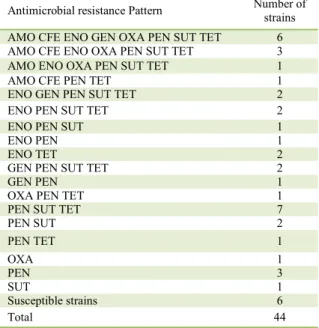
Table 3 - Resistance pattern of Staphylococcus pseudintermedius strains isolated from otitis externa in dogs.
Antimicrobial resistance Pattern Number of strains
AMO CFE ENO GEN OXA PEN SUT TET 6
AMO CFE ENO OXA PEN SUT TET 3
AMO ENO OXA PEN SUT TET 1
AMO CFE PEN TET 1
ENO GEN PEN SUT TET 2
ENO PEN SUT TET 2
ENO PEN SUT 1
ENO PEN 1
ENO TET 2
GEN PEN SUT TET 2
GEN PEN 1
OXA PEN TET 1
PEN SUT TET 7
PEN SUT 2
PEN TET 1
OXA 1
PEN 3
SUT 1
Susceptible strains 6
Total 44
they are frequently used in the treatment of human
infections (GUPTA et al., 2011).
It is also interesting to note that 26 (59,
1%) strains were resistant to three or more classes
of antimicrobials, classifying them as multi resistant
isolates. This rate is higher than that in a previous
report examining staphylococci isolated from otitis in
Brazil (LILENBAUM et al., 2000). However, recently
studies in Brazil, and other countries, have reported
high multi resistance rates of staphylococci group
isolates (PENNA et al., 2009; DE MARTINO et al.,
2016), emphasizing the emergency of SP resistance.
Antimicrobial agents are indispensable
for decreasing mortality and morbidity associated
with infectious diseases in animals and humans. In
veterinary medicine, they have been used for therapy,
metaphylaxis, prophylaxis, and growth promotion
(SCHWARZ et al, 2001). Recent studies on S.
pseudintermedius have shown a significant increasing
temporal trend in resistance to several antibiotics
(PRIVANTHA et al., 2016; QEKWANA 2017a,b).
Our results are consistent with those of these reports
and highlighted the emergence of antimicrobial
resistance in S. pseudintermedius, imposing a great
challenge for antimicrobial therapy in animals
(PRIYANTHA et al., 2016).
Despite the increasing importance of
MRSP in veterinary medicine, and for humans,
there is a lack of studies concerning MRSP
in Brazil. Here, we observed a high isolation
frequency of S. pseudintermedius from dogs
with otitis. Additionally, half of the isolates were
classified as MRSP, and almost 60% were multi
resistant. This study highlighted the need for
increased surveillance of antibiotic resistance in
veterinary settings in Brazil.
BIOETHICS AND BIOSSECURITY
COMMITTEE APPROVAL
This experiment was approved by the Comitê de Ética em Uso Animal of Universidade Federal de Minas Gerais (UFMG) under protocol number 246/2013.
ACKNOWLEDGEMENTS
The authors would like to thank the Pró Reitoria de Pesquisa da Universidade Federal de Minas Gerais (PRPq/UFMG) for the support.
DECLARATION OF CONFLICTING INTERESTS
The authors declared no potential conflicts of interest with respect to the research, authorship, and/or publication of this article.
REFERENCES
BANNOEHR, J. et al. Molecular diagnostic identification of
Staphylococcus pseudintermedius. Journal of Clinical Microbiology, v. 47, n. 2, p. 469-471, 2009. Available from: <http://jcm.asm.org/ content/47/2/469.full>. Acessed: Mar. 12, 2018.
BANNOEHR, J.; GUARDABASSI, L. Staphylococcus pseudintermedius
in the dog: Taxonomy, diagnostics, ecology, epidemiology and pathogenicity. Veterinary Dermatology, v. 23, n. 4, p. 1–16, 2012. Available from: <http://onlinelibrary.wiley.com/doi/10.1111/j.1365-3164.2012.01046.x/epdf>. Accessed: Aug. 11, 2017.
BOTONI, L. S. et al. Prevalence and in vitro susceptibility of methicillin-resistant Staphylococcus pseudintermedius (MRSP)
from skin and nostrils of dogs with superficial pyoderma.
Pesquisa Veterinaria Brasileira, v. 36, n. 12, p. 1178–1180, 2016. Available from: <http://www.scielo.br/pdf/pvb/v36n12/1678-5150-pvb-36-12-01178.pdf>. Accessed: Aug. 11, 2017.
BOURGUIGNON, E. et al. Description of methicillin-resistant
Staphylococcus pseudintermediusfrom canine pyoderma in Minas Gerais state, Brazil. Arquivos Brasileiros de Medicina Veterinária e Zootecnia, v. 68, n. 2, p. 299–306, 2016. Available from: <http:// www.scielo.br/pdf/abmvz/v68n2/0102-0935-abmvz-68-02-00299. pdf>. Accessed: Aug. 11, 2017.
CLSI. Performance standards for antimicrobial disk and dilution suscetibility tests for bacteria isolated from animals; Aproved standard. Fourth edition. CLSI document VET01-A4. Clinical and Laboratory Standards Institute. 33 (7), VET01-A4, 2013a.
CLSI. Performance standards for antimicrobial disk and dilution suscetibility tests for bacteria isolated from animals. Second informational supplement. CLSI document VET01-S2. Clinical and Laboratory Standards Institute. 33 (8), VET01-S2, 2013b.
COLE, L. K. et al. Short communications staphylococci in dogs with end-stage otitis. Veterinary Record, v. 159, p. 418–419, 2006. Available from: <http://veterinaryrecord.bmj.com/content/ vetrec/159/13/418.full.pdf>. Accessed: Aug. 11, 2017.
DE MARTINO, L. et al. An update on microbiological causes of canine otitis externa in Campania Region, Italy. Asian Pacific Journal of Tropical Biomedicine, v. 6, n. 5, p. 384–389, 2016. Available from: <http://veterinaryrecord.bmj.com/content/vetrec/159/13/418.full. pdf>. Accessed: Aug. 11, 2017.
ELHASSAN et al. Absence of the mecA Gene in methicillin resistant
Staphylococcus aureus isolated from different clinical specimens in Shendi City, Sudan. BioMed Research International, v. 2015, Article ID 895860, 5 pages, 2015. Available from: <https://www. hindawi.com/journals/bmri/2015/895860/cta/>. Accessed: Aug. 11, 2017.
EMA. European Medicines Agency, European Surveillance of Veterinary Antimicrobial Consumption, 2015. Sales of veterinary antimicrobial agents in 26 EU/EEA countries in 2013. (EMA/387934/2015). Available from: <http://www. ema.europa.eu/docs/en_GB/document_library/Report/2015/10/ WC500195687.pdf>. Accessed: Aug. 11, 2017.
administration. 2014. Available from: <https://www.fda.gov/ downloads/ForIndustry/UserFees/AnimalDrugUserFeeActADUFA/ ucm277657.pdf>. Accessed: Aug. 11, 2017.
FRANK, L. A.; LOEFFLER, A. Meticillin-resistant Staphylococcus pseudintermedius: Clinical challenge and treatment options. Veterinary Dermatology, v. 23, n. 4, 2012. Available from: <http:// onlinelibrary.wiley.com/doi/10.1111/j.1365-3164.2012.01047.x/ epdf>. Accessed: Aug. 11, 2017.
GUPTA, K. et al. Soper. International clinical practice guidelines for the treatment of acute uncomplicated cystitis and pyelonephritis in women: A 2010. Update by the Infectious Diseases Society of America and the European Society for Microbiology and Infectious Diseases. Clinical Practice Guidelines d CID. Clinical Infectious Diseases, v.52, 2011. Available from: <https://academic.oup.com/cid/ article-lookup/doi/10.1093/cid/ciq257>. Accessed: Aug. 11, 2017. HOEKSTRA, K.A., PAULTON, R.J. Clinical prevalence and antimicrobial susceptibility of Staphylococcus aureus and Staph. intermedius in dogs. Journal Applied Microbiology, v.93, p. 406-13, 2002. Available from: <http://onlinelibrary.wiley.com/doi/10.1046/ j.1365-2672.2002.01708.x/epdf>. Accessed: Aug. 11, 2017. KANIA, S. A. et al. Methicillin resistance of staphylococci isolated from the skin of dogs with pyoderma. American Journal of Veterinary Research, v. 65, n. 9, p. 1265–1268, 2004. Available from: <https://www.ncbi.nlm.nih.gov/pubmed/15478775>. Accessed: Aug. 11, 2017.
KASAI, T. et al. New categories designated as healthcare-associated and community-associated methicillin-resistant Staphylococcus Pseudintermedius in Dogs. Microbiology and Immunology, v.60, p. 540-551, 2016. Available from: <http://onlinelibrary.wiley.com/ doi/10.1111/1348-0421.12401/pdf>. Accessed: Aug. 11, 2017. KROEMER, S. et al. Comparative immunology, microbiology and infectious diseases antibiotic susceptibility of bacteria isolated from infections in cats and dogs throughout Europe (2002 – 2009). Comparative Immunology, Microbiology and Infectious Diseases, v. 37, n. 2, p. 97–108, 2014. Available from: <http:// www.sciencedirect.com/science/article/pii/S0147957113000775/ pdfft?md5=c73ed48b947f410b5531dbcde9817363&pid=1-s2.0-S0147957113000775-main.pdf>. Accessed: Aug. 11, 2017. LILENBAUM, W. et al. Antimicrobial susceptibility of staphylococci isolated from otitis externa in dogs. Letters in applied microbiology, v. 31, n. 1, p. 42–45, 2000. Available from: <http:// onlinelibrary.wiley.com/doi/10.1046/j.1472-765x.2000.00759.x/ epdf>. Accessed: Aug. 11, 2017.
LOPES, G.V. et al. Methicillin-resistant Staphylococcus pseudintermediusclonal groups isolated from canine pyoderma in Brazil. Acta Scientiae Veterinariae, v.43, p.1138, 2015. Available from: <http://revistas.bvs-vet.org.br/actascivet/article/ view/35417/39822>. Accessed: Aug. 11, 2017.
MASON, I. S.; KIETZMANN, M. Cephalosporins - pharmacological basis of clinical use in veterinary dermatology. Veterinary Dermatology, v. 10, p. 187–192, 1999. Available from: <http:// onlinelibrary.wiley.com/doi/10.1046/j.1365-3164.1999.00183.x/ epdf>. Accessed: Aug. 11, 2017.
MEHROTRA, M.; WANG, G.; JOHNSON, W. M. Multiplex PCR for detection of genes for Staphylococcus aureus
enterotoxins, exfoliative toxins, toxic shock syndrome toxin 1,
and methicillin resistance. Journal of Clinical Microbiology, v. 38, n. 3, p. 1032–1035, 2000. Available from: <https://www. ncbi.nlm.nih.gov/pmc/articles/PMC86330/pdf/jm001032.pdf>. Accessed: Aug. 11, 2017.
PAPICH, M. G. Selection of antibiotics for meticillin-resistant
Staphylococcus pseudintermedius: Time to revisit some old drugs. Veterinary Dermatology, v. 23, n. 4, p. 1–10, 2012. Available from: <http://onlinelibrary.wiley.com/doi/10.1111/j.1365-3164.2011.01030.x/ epdf>. Accessed: Aug. 11, 2017.
PENNA, B. et al. Species distribution and antimicrobial susceptibility of staphylococci isolated from canine otitis externa. Veterinary Dermatology, v. 21, n. 3, p. 292–296, 2010. Available from: <http:// onlinelibrary.wiley.com/doi/10.1111/j.1365-3164.2009.00842.x/ epdf>. Accessed: Aug. 11, 2017.
PRIYANTHA, R.; GAUNT, M. C.; RUBIN, J. E. Antimicrobial susceptibility of Staphylococcus pseudintermedius colonizing healthy dogs in Saskatoon, Canada. Canadian Veterinary Journal, v. 57, n. 1, p. 65–69, 2016. Available from: <https://www.ncbi.nlm. nih.gov/pmc/articles/PMC4677612/ >. Accessed: Aug. 11, 2017. QEKWANA, D.N. et al. Burden and predictors of Staphylococcus aureus and S. pseudintermedius infections among dogs presented at an academic veterinary hospital in South Africa (2007-2012). PeerJ, v.5, e3198, 2017a. Available from: <https://www.ncbi. nlm.nih.gov/pmc/articles/PMC5392248/pdf/peerj-05-3198.pdf>. Accessed: Aug. 11, 2017.
QEKWANA, D.N. et al. Patterns and predictors of antimicrobial resistance among Staphylococcus spp. from canine clinical cases presented at a veterinary academic hospital in South Africa. BMC Veterinary Research, v.13, p.116, 2017b. Available from: <https://www.ncbi.nlm.nih.gov/pmc/articles/ PMC5410067/pdf/12917_2017_Article_1034.pdf>. Accessed: Aug. 11, 2017.
ROBB, A.R. et al. Skin infection caused by a novel strain of
Staphylococcus pseudintermediusin a Siberian husky dog owner. JMM Case Reports, v.20, jmmcr005087, 2017. Available from: <https://www.ncbi.nlm.nih.gov/pmc/articles/PMC5382809/pdf/ jmmcr-4-5087.pdf>. Accessed: Aug. 11, 2017.
ROBERTS, A.L. Identification of Staphylococcus epidermidis in the
clinical microbiology laboratory by molecular methods. Methods in Molecular Biology, v.33, p.53, 2014. Available from: <https:// link.springer.com/protocol/10.1007%2F978-1-62703-736-5_3>. Accessed: Aug. 11, 2017.
SANCHEZ, R. et al. Aislamiento bacteriano en casos de otitis canina y su susceptibilidad antibiotica. Revista de Investigaciones Veterinarias del Peru, v. 22, n. 2, p. 161–166, 2011. Available from: <http://www.scielo.org.pe/pdf/rivep/v22n2/a13v22n2.pdf>. Accessed: Aug. 11, 2017.
SAPUTRA, S. et al. Antimicrobial resistance in coagulase-positive staphylococci isolated from companion animals in Australia: A one year study. PLoS One, v.12, e0176379, 2017. Available from:
<http://journals.plos.org/plosone/article/file?id=10.1371/journal.
pone.0176379&type=printable>. Accessed: Aug. 11, 2017.
SASAKI, T. et al. Multiplex-PCR method for species identification
SCHWARZ, S.; KEHRENBERG, C.; WALSH, T. R. Use of antimicrobial agents in veterinary medicine and food animal production. International Journal of Antimicrobial Agents, v. 17, p. 431–437, 2001. Available from: <http://www. sciencedirect.com/science/article/pii/S0924857901002977/ pdfft?md5=89f7d132a8e7da1745f82848857a0510&pid=1-s2.0-S0924857901002977-main.pdf>. Accessed: Aug. 11, 2017.
SOMAYAJI, R. et al. Human infections due to Staphylococcus pseudintermedius, an emerging zoonosis of canine origin: report of 24 cases. Diagnostic Microbiology and Infectious Disease, v. 85, n. 4, p. 471–476, 2016. Available from: <http://www.sciencedirect.com/science/article/pii/ S0732889316301304/pdfft?md5=aa10d78e16a4afc985586 dcb5fef071f&pid=1-s2.0-S0732889316301304-main.pdf>. Accessed: Aug. 11, 2017.
VIDELA, R. et al. Clonal complexes and antimicrobial susceptibility
profiles of Staphylococcus pseudintermediusisolates from dogs in
the United States. Microbial Drug Resistance. Ahead of print, 2017. doi: 10.1089/mdr.2016.0250. Available from: <https://www. ncbi.nlm.nih.gov/pubmed/28504897>. Accessed: Aug. 11, 2017. VAN DUIJKEREN, E. et al. Review on methicillin-resistant Staphylococcus pseudintermedius. Journal of Antimicrobial Chemotherapy, v. 66, n. 12, p. 2705–2714, 2011. Available from: <https://academic.oup.com/jac/article-lookup/doi/10.1093/jac/dkr367>. Accessed: Aug. 11, 2017.