Leaf Morphoanatomy of Diploon Cronquist (Sapotaceae Juss.)
Renata Gabriela Vila Nova de Lima1* , Liliane Ferreira Lima1, Angélica Cândida Ferreira1,
Josiane Silva Araújo2 & Carmen Silvia Zickel1
1Universidade Federal Rural de Pernambuco, Departamento de Biologia, Rua Dom Manuel de Medeiros, s/n, Dois Irmãos, 52171-900, Recife, PE, Brasil.
2Universidade Estadual do Piauí, Departamento de Biologia, BR 343, s/n, Bairro Campo Velho, 64800-000, Floriano, PI, Brasil
*Corresponding author: Renata Gabriela Vila Nova de Lima, e-mail: [email protected]
LIMA, R.G. V. N.; LIMA, L.F.; FERREIRA, A. C.; ARAÚJO, J. S.; ZICKEL, C. S. Leaf Morphoanatomy of
Diploon Cronquist (Sapotaceae Juss.) Biota Neotropica. 19(1): e20180600. http://dx.doi.org/10.1590/1676-0611-BN-2018-0600
Abstract: Diploon is a monospecific genus represented by Diploon cuspidatum, an arboreal species that has
morphological characteristics distinct from those of other Sapotaceae species. In this study, Diploon cuspidatum leaves were characterized morphoanatomically in order to reveal additional diagnostic characters of their external morphology of the genus. The Diploon petiole presents shape and arrangement of the vascular system flat-convex, occasionally with one or two accessory bundles, many laticifers, and many prismatic crystals. The midrib is biconvex with a U-shaped cuticle on the abaxial side, and laticifers are associated with the vascular tissues. Mesophyll is dorsiventral, palisade parenchyma has two cell layers, T- and Y-shaped malpighiaceous trichomes are on the abaxial epidermis with a small stalk cell and long arm. The venation pattern is brochidodromous. Intersecondary veins run parallel to the secondary veins, and quaternary veins branch freely. Higher order veins are not present. Morphoanatomical analysis revealed important characteristics that reveal a set of structures common to Sapotaceae, in addition to characters that are important for the recognition and identification of D. cuspidatum.
Keywords: Leaf anatomy, leaf architecture, micromorphology, taxonomy, Chrysophylloideae.
Morfoanatomia Foliar de Diploon Cronquist (Sapotaceae Juss.)
Resumo:Diploon é um gênero monoespecífico representado por Diploon cuspidatum, espécie arbórea com
características morfológicas peculiares em relação a outros gêneros de Sapotaceae. A espécie teve suas folhas caracterizadas morfoanatomicamente, a fim de fornecer caracteres diagnósticos adicionais à morfologia externa, subsidiar pesquisas no âmbito da anatomia vegetal, dendrologia e filogenia. D. cuspidatum evidenciou pecíolo plano-convexo, com feixe vascular plano-convexo, presença ocasional de até dois feixes acessórios, presença de muitos laticíferos e cristais prismáticos. A nervura central é biconvexa, com cutícula em forma de U no lado abaxial, laticíferos associados aos elementos vasculares. Mesofilo dorsiventral, parênquima paliçádico com duas camadas descontínuas, tricomas malpighiáceos do tipo T e Y na epiderme abaxial com pedúnculo pequeno e braço longo. O padrão de venação é do tipo broquidódroma. Veias intersecundárias paralelas as veias secundárias, veias quaternárias em ramos livres. Ausência de veias de ordem superior. A análise morfoanatômica realizada evidenciou caracteres importantes que retratam um conjunto de estruturas comuns a Sapotaceae e também importantes para reconhecimento e identificação D. cuspidatum.
Palavras-chave: Anatomia foliar, Arquitetura foliar, micromorfologia, taxonomia, Chrysophylloideae.
Introduction
Sapotaceae is a family with a pantropical distribution that includes about 60 genera and 1300 species (Pennington 1991; Govaerts et al.
2001, The PlantList 2013). It is one of the largest families of eudicots. It is known for having species of great morphological diversity that are especially important in lowland wet tropical forests because they provide essential resources to native fauna and humans (Lawrence 1951, Barroso 1978, Pennington 1991, Felippi et al. 2008, Gomes
et al. 2008, Reis et al. 2013). In Brazil, there are approximately 234
species distributed in 12 genera (Carneiro et al. 2015). Among these is Diploon Cronquist, which is of particular importance because of its
peculiar reproductive morphology.
Diploon is a monospecific genus represented by Diploon cuspidatum (Hoehne) Cronquist, which has as its basionym Chrysophyllum cuspidatum Hoehne. This arboreal species has alternating leaves, axillary flowers, basally connate petals, and it lacks staminodes. Its ovary is glabrous and unilocular with two ovules, and it has basal placentation, in addition to succulent fruits containing a single seed that has a small, basilateral scar and no endosperm (Pennington 1990, 1991). Together, these characters contribute to a good generic delimitation because some features of the genus Diploon are not common in other
Sapotaceae, which facilitates its identification. Despite having a distinct reproductive morphology (unilocular ovary and biovular with basal
placentation), Diploon shares morphological, anatomical, chemical, and
phylogenetic characteristics with other Sapotaceae genera (Cronquist 1946, Kukachka 1979, Pennington 1990, Swenson & Andeberg 2005).
Diploon cuspidatum, popularly known as preta” or “bapeba-roxa,” is native to Brazil, but it can also be found in Bolivia, Guyana, and Peru (Pennington 1990). In Brazil, it is distributed throughout two phytogeographic domains: the Amazon and the Atlantic Forest, occurring in the North, Northeast, Southeast, and South regions of the country (Carneiro et al. 2015). Although it is a relatively well-distributed
species, D. cuspidatum is present in biomes regarded as biodiversity hotspots, which face serious environmental threats caused by the most diverse anthropic disturbances. Because of the richness in flora and fauna, these areas require more studies to expand our knowledge of the biology of their species. Concerning D. cuspidatum, the literature has been largely restricted to references to local flora and floristic surveys (Kurtz & Araújo 2000, Alves-Araújo & Alves 2010, Palazzo et al. 2010,
Barreto & Catharino 2015) that gathered data on geographic distributions, identification keys, or morphological descriptions. Anatomical studies are even scarcer, even though they are relevant to our understanding of a species’ biology and they provide data to support taxonomic, dendrological, micromorphological, and phylogenetic studies.
In the last proposals regarding the taxonomic classification of
Sapotaceae (Pennington 1991, Swenson & Andeberg 2005), the positioning of the genus Diploon in the family was mainly based on its floral morphology and the presence of chemical components (flavonoids). Kukachka (1979) showed common anatomical characters between Diploon and the other genera of the family after analyzing woods of neotropical Sapotaceae. Regarding leaf architecture, Pennington (1990) used different terminology to describe the foliar venation pattern in Diploon and other genera, revealing useful features to identify D. cuspidatum,
which, in the vegetative state, may be similar to some Micropholis (Griseb.) Pierre species. These data, along with current research on
other Sapotaceae genera (Monteiro et al. 2007a, Almeida-Jr et al. 2012),
show that morphoanatomical studies can contribute significantly to plant systematics at different hierarchical levels. Therefore, the present study aimed to characterize and discuss the leaf morphoanatomy of D. cuspidatum from a taxonomic perspective in order to identify diagnostic characters that contribute to our knowledge of this genus.
Material and Methods
This study was based on the collection of Diploon specimen
branches found in the Atlantic and Amazon Forest (in Brazil’s North and Northeast areas), in addition to the analysis of material deposited in the national herbarium (BHCB, ESA, HEPH, HRBC, IAC, IAN, INPA, MBML, PEUF, SPSF, UB, and VIC).
The material used to perform the anatomical analysis was obtained during field expeditions and from herbarium samples (voucher: IAC43362, IAN123562, PEUFR53995, PEUFR53996 and SPSF3507).
Samples were selected according to the following criteria: 1) herbal
material in good preservation state, 2) reliable taxonomic identification, and 3) presence of fully expanded leaves.
The botanical material was submitted to a process of reverse herborization according to Smith & Smith (1942). The process consisted of treating the samples with 2% sodium hydroxide for 2 h, washing them in distilled water, and dehydrating them in graded ethanol solutions. Subsequently, samples were stored in 70% ethanol until the anatomical
slides were prepared.
Free-hand transverse sections were cut from the petiole (apex,
middle, and base regions) and the middle region of the leaf blade.
Sections were clarified in 50% sodium hypochlorite and stained with Astra blue and basic fuchsin. Samples were mounted on slides using glycerinated gelatin and sealed with colorless nail polish (Kraus & Arduin 1997).
For the analysis of leaf architecture, diaphanization was carried out according to the protocol developed by Shobe & Lersten (1967 apud Kraus & Arduin 1997). The clarified samples were then washed in distilled water, dehydrated in an ethanol series, and stained with basic fuchsin in 50% ethanol. Samples were mounted with glycerinated
gelatin (Kaiser 1880), and slides were sealed with colorless nail polish
(Kraus & Arduin 1997).
Morphoanatomical analysis was performed using an optical microscope (L2000i, Bioval), and images were obtained using a digital camera (Digital Microscope Imager, 44421, Celestron) attached to the
microscope.
Leaf blades, petioles, and trichome characters were described in
accordance with the terminology of Metcalfe & Chalk (1983), Howard (1979), and Theobald et al. (1979), respectively. The description of leaf venation patterns was based on the terminology of Ellis et al. (2009).
Results
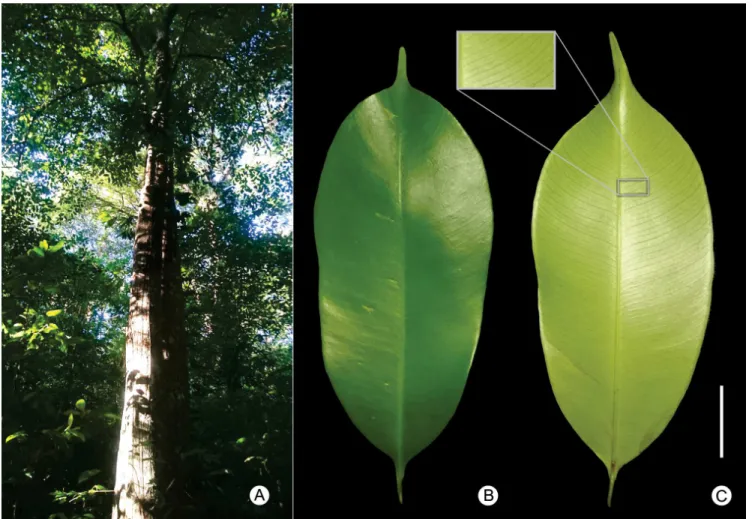
Figure 1. Diploon cuspidatum (Hoehne) Cronquist. A. Individual located at the Parque Estadual Dois Irmãos, PE, Brazil; B. Leaf blade showing the adaxial side; C. Leaf blade showing the abaxial side and detailing the venation pattern. * Scale bar: 2.00 cm.
The analysis of leaf architecture allowed the identification of a brochidodromous venation pattern with a pinnate midrib (Figure 2a). Secondary veins are excurrent from the primary vein, and they branch out without reaching the margin, which is formed by submarginal veins (Figure 2a). The spaces between adjacent secondary veins are regular, and the angle between the primary vein and secondary veins increases smoothly as these approach the leaf base. Intersecondary veins are 50% greater in length than lower secondary veins, and they run parallel to secondary veins. It is possible to observe one or more per intercostal area (Figure 2a). Intercostal tertiary veins display an alternate percurrent course, crossing the midrib in a reticulate pattern (Figure 2b). External tertiary veins are also observed, ending at the leaf margin parallel to the secondary veins. Quaternary veins end freely in a single branch (Figure 2b). Higher order veins were not observed.
In cross-section, the petiole has a flat-convex shape (Figure 2c), and when it is close to the leaf base, it presents two lateral projections in the adaxial portion, resulting in the canaliculate morphology of the petiole observed as part of the external morphology. The epidermis is unstratified with cells that are usually quadrangular and covered by a thin layer of cuticle. It is possible to observe scars left by trichomes. Below the epidermis, there is a continuous layer of collenchyma tissue followed by parenchyma. This region has a higher concentration of solitary prismatic crystals and laticifers, but few druses. Prismatic crystals and laticifers, however, can be found over the entire petiole at
a lower frequency. The vascular bundle, which is present in the middle and the petiole, is flat-convex (Figure 2c). One or two accessory bundles surrounded by a sclerenchymatous sheath are observed. Laticifers and prismatic crystals are present in the medullar region.
The leaf blade has a thicker cuticle on the adaxial side compared to the abaxial side. On both sides, the epidermis is unstratified and formed mainly by quadrangular cells. The abaxial side, however, displays simple, unicellular, T- or Y-shaped malpighiaceous trichomes with a short peduncle and long arms, which are concentrated close to the midrib (Figure 2g). Leaves are hypostomatic and have anisocytic stomata. In cross-section, guard cells are on the same level as other epidermal cells. The mesophyll is dorsiventral and consists of two discontinuous layers of palisade parenchyma followed by 10 layers of spongy parenchyma (Figure 2d). The palisade parenchyma of the mesophyll sometimes extends into the region of the midrib that is interrupted by annular collenchyma. Laticifers and collateral bundles occur throughout the mesophyll, and sclereids are present in the abaxial side of the leaf blade (Figure 2d).
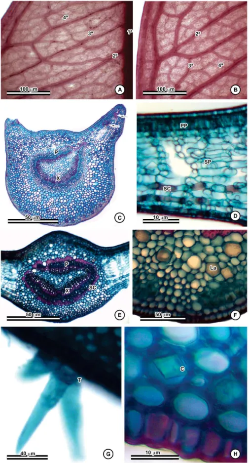
Figure 2. Morphoanatomic characters of D. cuspidatum. A–B. Venation pattern showing primary, secondary, intersecondary, tertiary, and quaternary veins; C.
Cross-section of the petiole with accessory bundles (arrows); D. Cross-section of the leaf blade; E. Cross-section of the mesophyll; F. Laticifers in the midrib; G.
Malpighiaceous trichomes on the abaxial face; H. Detail of prismatic crystals in the midrib. Abbreviations. C: prismatic crystals; La: laticifers; P: phloem; Pp:
a plano-convex shape. It is also surrounded by a sclerenchymatous sheath. Laticifers are present around every vascular bundle. The rest of the structure is filled with parenchyma.
Discussion
Some morphoanatomical characters described by previous authors (Solereder 1908, Metcalfe & Chalk 1950, Pennington 1990, Monteiro et al. 2007a, Almeida-Jr et al. 2012) as characteristic of the
Sapotaceae family were corroborated by the taxonomic analysis in the present study. The literature shows that leaf venation patterns can provide a set of characters that are variable among different Sapotaceae genera, sections, and species. Pennington (1990) described leaf venation using characters of primary to quaternary veins and by analyzing shape, organization, course, branching, and areolation. In this family, secondary veins can be craspedodromous, eucamptodromous, or brochidodromous, and they can have intersecondary veins. The course of tertiary and quaternary veins is variable with or without areolation.
The leaf vein system may vary in response to the environmental conditions of the different biomes. (Boeger et al. 2009, Sack & Scoffoni 2013). Despite this, the leaf venation pattern is an important character used for taxonomic distinction (Ellis et al. 2009, Borrero et al. 2016,
Coutinho 2016). We highlight some conservative characters that can help in the identification of D. cuspidatum in the vegetative stage: both
in the Atlantic forest biome and in the Amazon forest. These characters include the course of the midrib, the types of spaces between secondary veins, the angle between primary and secondary veins, and the pattern of intersecondary veins.
Laticifers and malpighiaceous trichomes are notable anatomical characters, which together allow any member of the Sapotaceae family to be easily recognized, even in sterile stages (Solereder 1908, Metcalfe & Chalk 1950, Monteiro 2007b, Almeida-Jr et al. 2012).
Additionally, the U-shaped cuticle on the abaxial side (which resembles smooth ondulations), hypostomatic leaves, anisocytic stomata, the predominance of a dorsiventral mesophyll, and the occurrence of sclerenchymatous elements and solitary or clustered crystals are also potentially diagnostic characters of Sapotaceae (Solereder 1908, Metcalfe & Chalk 1950, Monteiro 2007a, Almeida-Jr et al. 2012).
The shape and arrangement of the vascular system of the petiole and midrib have been deemed useful in plant systematics (Araújo et al. 2010, Leandro et al. 2015, Coutinho et al. 2016, Feio et al. 2018,
Guesdon et al. 2018, Pereira et al. 2018). Metcalfe & Chalk (1950)
described eight shapes for the petiole of Sapotaceae. Of these, Diploon is classified as “adaxially flattened closed cylinder” or flat-convex. The vascular bundle of the petiole is also flat-convex, occasionally displaying one or two accessory bundles. In the midrib, the contour and conformation of the vascular bundle is biconvex. For Diploon, as
a monospecific genus, these characters are also taxonomically relevant.
In other genera of Sapotaceae, the aforementioned characteristics were also present; however, the shape of the petiole, the midrib, and the
conformation of the vascular bundles may vary among species, which
was observed for Manilkara Adans. and Pouteria Aubl. (Monteiro et al.
2007a, Almeida-Jr et al. 2012).
The thickness and number of layers of the palisade parenchyma are usually related to the habitat of the individual species, and it is
mainly affected by the humidity and the extent of luminous intensity (Hlwatika & Bhat 2002, Rossatto & Kolb 2010; Schmidt et al. 2017;
Muniz et al. 2018). However, the number of layers is fixed for some species (Solereder 1908). Generally, for Sapotaceae, the palisade tissue consists of one or more cell layers, and it is present only on the adaxial side (dorsiventral mesophyll). In most Sapotaceae species, the spongy parenchyma has large intercellular spaces (Solereder 1908, Metcalfe & Chalk 1950, Monteiro 2007a, Almeida-Jr et al. 2012). Variations
in the number of layers, a dorsiventral mesophyll, two discontinuous layers of palisade parenchyma, and a spongy parenchyma with large intercellular spaces were observed in D. cuspidatum.
In Diploon, laticifers are present between the parenchyma cells of the midrib and the petiole, and they are easily distinguished from other epidermal and parenchymal elements. In Sapotaceae, laticifers are arranged in longitudinal rows and they have resinous substances that are sometimes accompanied by calcium oxalate crystal sand and starch (Solereder 1908). In the leaf blade, they are distributed predominantly in the midrib region, but are also found along the mesophyll and petiole (Solereder 1908, Metcalfe & Chalk 1950, Metcalfe & Chalk 1983, Monteiro et al. 2007a). In different genera of this family, the location of laticifers throughout these structures (leaf blade, mesophyll, and petiole) is variable, and they may occur along the vascular bundles of the veins or they can be immersed in the mesophyll (in Chrysophyllum L. and
Pouteria Aubl.) or distributed in the cortical and medullary regions
of the leaf blade (in Manilkara Adans.) (Solereder 1908, Metcalfe &
Chalk 1950, Monteiro et al. 2007a, Almeida-Jr et al. 2012). This study emphasizes the marked presence of laticifers in the family, which can be found in different parts of the leaf blade depending on the genera
and species.
According to Pennington (1990), D. cuspidatum displays glabrous leaves, but the anatomical analysis only allowed the visualization of trichomes along the midrib. Trichomes are unicellular malpighiaceous with long arms and a short peduncle. In Sapotaceae, most species are covered by an indumentum formed by persistent or deciduous adpressed trichomes with arms of different sizes and of variable density on the abaxial and adaxial side (Pennington 1990). The present study demonstrated the occurrence of trichomes in D. cuspidatum,which
may be deciduous. In many cases, it was possible to observe only the
scars of the trichomes.
For Solereder (1908) and Metcalfe & Chalk (1950), the occurrence of large numbers of cells rich in solitary or clustered prismatic crystals is a good generic diagnostic characteristic for Sapotaceae, especially when combined with other characters. We observed the presence of large prismatic crystals dispersed in the midrib and petiole regions in D. cuspidatum, which were more frequent in the first subepidermal layers of the petiole. Crystal idioblasts present in the palisade parenchyma
were important for the taxonomic delimitation of Pouteria Aubl. and Pradosia Liais (Solereder 1908, Monteiro et al. 2007a).
The description of D. cuspidatum leaf architecture and anatomy
has revealed a set of characters that have been reported for the
Sapotaceae family and characters that aid in the identification of the species. In addition, the findings of the present study contribute to
the morphoanatomic characterization of the Diploon and provide
Acknowledgements
We thank the Laboratório de Florística e Ecossistemas Costeiros (UFRPE) for providing the infrastructure necessary for this study and the Laboratório de Biologia Vegetal (UESPI) for the technical support in anatomical analyses. This study was supported by a research grant from the Conselho Nacional de Desenvolvimento Científico e Tecnológico (CNPq) and Coordernação de Aperfeiçoamento de Pessoal de Nível Superior – Brasil (CAPES) – Finance code 001.
Author Contributions
Renata Gabriela Vila Nova de Lima: Substantial contribution in the concept and design of the study; Contribution to data collection; Contribution to data analysis and interpretation; Contribution to manuscript preparation; Contribution to critical revision, adding intelectual content.
Liliane Ferreira Lima: Substantial contribution in the concept and design of the study; Contribution to data collection; Contribution to data analysis and interpretation; Contribution to manuscript preparation; Contribution to critical revision, adding intelectual content.
Angélica Cândida Ferreira: Contribution to data analysis and interpretation; Contribution to manuscript preparation; Contribution to critical revision, adding intelectual content.
Josiane Silva Araújo: Contribution to data analysis and interpretation; Contribution to manuscript preparation; Contribution to critical revision, adding intelectual content.
Carmen Silvia Zickel: Contribution to manuscript preparation; Contribution to critical revision, adding intelectual content.
Conflicts of interest
The authors declare that they have no conflict of interest related to the publication of this manuscript.
References
ALMEIDA-JR, E.B., ARAUJO, J.S., SANTOS-FILHO, F.S. & ZICKEL, C.S. 2012. Leaf morphology and anatomy of Manilkara Adans. (Sapotaceae)
from Northeastern Brazil. Plant Syst. Evol. 299(1):1-9.
ALVES-ARAÚJO, A. & ALVES, M. 2010. Flora da Usina São José, Igarassu, Pernambuco: Sapotaceae. Rodriguésia61(2):303-318.
ARAÚJO, J.S., AZEVEDO, A.A., SILVA, L.C. & MEIRA, R.M.S.A. 2010. Leaf anatomy as an additional taxonomy tool for 16 species of Malpighiaceae found in the Cerrado area (Brazil). Plant Syst. Evol. 286(1-2):117-131. BARRETO, E.H.P. & CATHARINO, E.L.M. 2015. Florestas maduras da região
metropolitana de São Paulo: diversidade, composição arbórea e variação florística ao longo de um gradiente litoral-interior, Estado de São Paulo, Brasil. Hoehnea 42(3):445-469.
BARROSO, G.M. 1978. Sistemática de angiospermas do Brasil: volume 1. Rio de Janeiro: Livros técnicos e científicos Editora, 255p.
BOEGER, M.R.T., BIU, C. & GOLDENBERG, R. 2009. Arquitetura foliar
comparativa de Miconia sellowiana (DC.) Naudin (Melastomataceae)
em diferentes fitofisionomias no Estado do Paraná, Brasil. Acta bot. bras. 23(3):657-665.
BORRERO, P.A.P, BOHREN, A.V., KELLER, H.A., GRANCE, L.A. & DUMMEL, C.J. 2016. La arquitectura foliar de las especies de Lauraceae Nativas de Misiones, Argentina. Bol. Soc. Argent. Bot. 51(1):37-57.
CARNEIRO CE, ALVES-ARAUJO A, ALMEIDA-JR EB, TERRA-ARAÚJO MH. 2015. Sapotaceae/Diploon. In: Lista de Espécies da Flora do Brasil,
2015 Jardim Botânico do Rio de Janeiro. http://floradobrasil.jbrj.gov. br/2012/FB000217 (último acesso em 09/04/2018).
COUTINHO, I.A.C., RANDO, J.G., CONCEIÇÃO, A.S. & MEIRA, R.M.S.A. 2016. A study of the morphoanatomical characters of the
leaves of Chamaecrista (L.) Moench sect. Apoucouita
(Leguminosae-Caesalpinioideae). Acta bot. bras. 30(2): 205-221.
CRONQUIST, A.J. 1946. Studies in the Sapotaceae-VI. Miscellaneous Notes. Bulletin of the Torrey Botanical Club 73(5):465-471.
ELLIS, B., DALY, D.C., HICKEY, L.J., JOHNSON, K.R., MITCHELL, J.D., WILF, P. & WING, S.L. 2009. Manual of leaf architecture, first printing. Published in Association with The New York Botanical Garden.
FEIO, A.C., MEIRA, R.M.S.A. & RIINA, R. 2018. Leaf anatomical features and their implications for the systematics of dragon’s blood, Croton section
Cyclostigma (Euphorbiaceae). Bot. J. Linn. Soc. 187:614-632.
FELIPPI, M., GROSSI, F., NOGUEIRA, A.C. & KUNIYOSHI, Y.S. 2008. Fenologia e germinação de sementes de Aguai, Chrysophyllum gonocarpum
(Mart. & Eichl.) Engl. Floresta 38(2):229-243.
GOMES, R., PINHEIRO, M.C.B. & LIMA, H.A. 2008. Fenologia reprodutiva de quatro espécies de Sapotaceae na restinga de Maricá, RJ. Revista Brasil. Bot. 31(4):679-687.
GOVAERTS, R., FRODIN, D.G. & PENNINGTON, T.D. 2001. World checklist and bibliography of Sapotaceae. R. Bot. Gard. Kew, UK.
GUESDON, I.R., AMORIM, A.M. & MEIRA, R.M.S.A.. 2018. The
hydrochorous Amazonian genus Glandonia (Malpighiaceae): new
records, morphoanatomy updates and taxonomic contributions. Phytotaxa 345(1):013–025.
HLWATIKA, C.N.M. & BHAT, R.B. 2002. An ecological interpretation of the difference in leaf anatomy and its plasticity in contrasting tree species in Orange Kloof, Table Mountain, South Africa. Ann. Bot. 89(1):109-114. HOWARD, R.A. 1979. The petiole. In: Metcalfe CR, Chalk L (eds) Anatomy of
the dicotyledons: systematic anatomy of the leaf and stem, Oxford Claredon, Oxford, v.1, p.88–96.
KAISER, E. 1880. Verfahren zur herstellung einer tadellosen glycerin-gelatine. Botanisch Zentralb, Stuttgart, v.180, p.25-26.
KRAUS, J.E. & ARDUIN, M. 1997. Manual básico de métodos em morfologia vegetal. Editora da Universidade Federal Rural do Rio de Janeiro, Rio de
Janeiro, v.1, 198p.
KUKACHKA, B.F. 1979. Wood anatomy of the neotropical Sapotaceae. VIII,
Diploon. Res. Pap. FPL-349. Madison, WI: U.S. Department of Agriculture,
Forest Service, Forest Products Laboratory. 6p.
KURTZ, B.C. & ARAÚJO, D.S.D. 2000. Composição florística e estrutura do componente arbóreo de um trecho de Mata Atlântica na Estação Ecológica Estadual do Paraíso, Cachoeiras de Macacu, Rio de Janeiro, Brasil. Rodriguésia 51(78/79):69-111.
LAWRENCE, G.H.M. 1951. Taxonomia das plantas vasculares. New York: Macmillan. 823p.
METCALFE, C.R. & CHALK, L. 1950. Anatomy of dicotyledons: leaves, stem, and wood in relation to taxonomy with notes on economic uses. Oxford: Clarendon Press, v.2, p.871-880.
METCALFE, C.R. & CHALK, L. 1983. Anatomy of the Dicotyledons. Volume
II: Wood Structure and Conclusion of the General Introduction. Second edition. Oxford science publications.
MONTEIRO, M.H.D.A., NEVES, L.J. & ANDREATA, R.H.P. 2007a. Taxonomia e anatomia das espécies de Pouteria Aublet (Sapotaceae) do
estado do Rio de Janeiro, Brasil. Pesquisas, Botânica 58:7-118.
MONTEIRO, M.H.D.A., ANDREATA, R.H.P. & NEVES, L.J. 2007b. Estruturas secretoras em Sapotaceae. Pesquisas, Botânica 58:253-262.
MUNIZ, L.F., BOMBOA, A.B., FILARTIGAA, A.L. & APPEZZATO-DA-GLÓRIA, B. 2018. Can climate and soil conditions change the morpho-anatomy among individuals from different localities? A case study
PALAZZO, F.M.A., NETO, A.O.D., MONTEIRO, M.H.D.A. & ANDREATA, R.H.P. 2010. Sinopse comentada de Sapotaceae no município de Rio das Ostras (RJ, Brasil). Pesquisas, Botânica 61:293-306.
PENNINGTON, T.D. 1990. Sapotaceae. In Flora Neotropica. The New York Botanical Garden, New York., v.52, 770p.
PENNINGTON, T.D. 1991. The genera of Sapotaceae. R. Bot. Gard. Kew,
UK. 295p.
PEREIRA, L.B.S., COSTA-SILVA, R., FELIX, L.P. & AGRA, M.F. 2018. Leaf morphoanatomy of “mororó” (Bauhinia and Schnella, Fabaceae). Rev. bras.
farmacogn. 28(4):383–392.
REIS, L.P., SILVA, J.N.M., REIS, P.C.M., CARVALHO, J.O.P., QUEIROZ, W.T. & RUSCHEL, A.R. 2013. Efeito da exploração de impacto reduzido em algumas espécies de Sapotaceae no leste da Amazônia. Floresta 43(3):395-406.
ROSSATTO, D.R. & KOLB, R.M. 2010. Gochnatia polymorpha (Less.) Cabrera
(Asteraceae) changes in leaf structure due to differences in light and edaphic conditions. Acta Bot. Bras. 24(3):605-612.
SACK, L. & SCOFFONI, C. 2013. Leaf venation: structure, function, development, evolution, ecology and applications in the past, present and future. New Phytol. 198(4):983-1000.
SCHMIDT, D., CARON, B.O., PILAU, J., MAICON NARDINO, M. & ELLI, E.F. 2017. Morfoanatomia foliar de azevém no sub-bosque de espécies arbóreas em sistemas agroflorestais. Rev. Ceres, Viçosa 64(4):368-375. SMITH, F.H. & SMITH, E.C. 1942. Anatomy of the inferior ovary of Darbya.
Am. J. Bot. 29(6):464–471.
SOLEREDER, H. 1908. Systematic Anatomy of the Dicotyledons. Oxford: Clarendon Press. v.1. p.512-515.
SWENSON, U. & ANDERBERG, A.A. 2005. Phylogeny, character evolution, and classification of Sapotaceae (Ericales). Cladistics 21(2):101-130. THEOBALD, W.L., KRAHULIK, J.L. & ROLLINS, R.C. 1979. Trichome
description and classification. In: METCALFE, C.R., CHALK, L. (eds) Anatomy of the dicotyledons: systematic anatomy of the leaf, stem, 2nd ed. Oxford Claredon, Oxford, 1:40–53.
THE PLANT LIST, 2013. Version 1.1. Published on the Internet; http://www. theplantlist.org/ (ultimo acesso em: 07/03/2018).