UMinho | 2014
Universidade do Minho
Escola de Ciencias da Saúde
Flávia Raquel Teixeira de Castro
Impact of IL-10 and SIRT2 in the
protective immune response to
and
Mycobacterium tuberculosis
Listeria monocytogenes infections
Maio de 2014
Mycobacterium tuberculosis
Flávia Raquel Teixeira de Castro
Impact of IL
-1
0 and SIR
T2 in t
he pro
tective immune response to
and Lis
teria monocytogenes infections
My
cobacterium tuberculosis
My
Flávia Raquel Teixeira de Castro
Impact of IL-10 and SIRT2 in the
protective immune response to
and
Mycobacterium tuberculosis
infections
Listeria monocytogenes
Maio de 2014Dissertação de Mestrado
Mestrado em Ciências da Saúde
Trabalho Efetuado sob a orientação do
Doutor António Gil Castro
e da
Doutora Lúcia Moreira Teixeira
Universidade do Minho
Escola de Ciencias da Saúde
Mycobacterium tuberculosis
Listeria monocytogenes
ii
DECLARAÇÃO
Nome: Flávia Raquel Teixeira de Castro
Endereço electrónico: flaviacastro@ecsaude.uminho.pt Telefone: +351 913 220 186
Número do Bilhete de Identidade: 13892586
Título da dissertação: Impact of IL-10 and SIRT2 in the protective immune response to Mycobacterium tuberculosis and Listeria monocytogenes infection
Orientador:
Doutor António Gil Castro Co-orientação:
Doutora Lúcia Moreira Teixeira
Ano de conclusão: 2014
Designação Ramo de Conhecimento do Mestrado: Ciências da Saúde
DE ACORDO COM A LEGISLAÇÃO EM VIGOR, NÃO É PERMITIDIDA A REPRODUÇÃO DE QUALQUER PARTE DESTA TESE.
Universidade do Minho, 21 de Maio de 2014
iii
The work presented in this dissertation was done in the Microbiology and Infection Research Domain of the Life and Health Sciences Research Institute (ICVS), School of Health Sciences, University of Minho, Braga, Portugal (ICVS/3B’s – PT Government Associate Laboratory, Braga/Guimarães, Portugal).
v
Para ser grande, sê inteiro: nada teu exagera ou exclui. Sê todo em cada coisa. Põe quanto és no mínimo que fazes.
Assim em cada lago a lua toda brilha, porque alta vive. Ricardo Reis
vii
ACKNOWLEDGEMENTS
Esta tese não teria sido possível sem o apoio profissional e pessoal de todos aqueles que estiveram comigo ao longo dos últimos 2 anos.
Assim começo por agradecer ao Doutor Gil Castro, meu orientador, por me ter dado a oportunidade de integrar a sua equipa, pela sua orientação, pelo apoio profissional e pessoal. Obrigada por ter acreditado em mim e por me ter dado a liberdade e a confiança de fazer crescer comigo este projeto. Comigo levo a sua admiração e dedicação à ciência e a vontade de fazer sempre mais e melhor. Também adotei os pMT-10!
Quero também expressar a minha gratidão à Doutora Margarida Saraiva por também me ter orientado. Agradeço-lho por acreditar em mim, por partilhar comigo os seus conhecimentos e os seus projetos. Agradeço-lhe pelas discussões científicas, levo comigo sem dúvida mais rigor e clareza científica.
É também com imensa gratidão que agradeço à Lúcia, que tanto me ensinou!!! Agradeço-te pelo apoio no laboratório, pelo acompanhamento profissional mas também pessoal. Obrigada por todos os ensinamentos, obrigada por me tornares capaz de conduzir um “mega” in vivo até ao fim, obrigada pelas discussões científicas e pela confiança que sempre me transmitiste. Obrigada pela amizade, pelos cafezinhos e “lab meetings” de quarta à tarde.
Egídio, nem sei por onde começar! Quantas vezes precisaria de agradecer? Obrigada pela ajuda imensa que deste no projeto, pela confiança e por me desafiares constantemente. Obrigada por partilhares comigo a tua visão crítica e as tuas ideias. Obrigada por me incentivares sempre a ser melhor. Obrigada pelo companheirismo e obrigada por simplesmente estares sempre que é preciso. Obrigada!!!
Quero também agradecer à Doutora Teresa Pais pela colaboração com os SIRT2 e pela disponibilidade para discutirmos os resultados. Obrigada!
Às meninas pequeninas mas Grandes do laboratório, Vânia e Joana Gaifem, pela ajuda nas grandes experiências, por estarem sempre dispostas a animarem-me! Obrigada pelo companheirismo, pela amizade, pelas horas intermináveis no p3 e pelos lanchinhos.
viii
Ao Diogo … Obrigada por partilhares comigo as culturas de microglia, por me ajudares a perder o medo dos ratinhos, pelas vezes sem conta que me incentivaste a ser melhor, pelo apoio profissional mas sobretudo pessoal. Um obrigado gigante por todos os discursos de motivação e por todas as conversas sérias.
Ao Jeremy, obrigada pela ajuda! Agradeço também à Filipa e ao Henrique por serem sempre tão prestáveis e companheiros. Obrigada meninos!
A todos aqueles que fazem parte do I3.02 e que me acolheram muito bem. E são eles: os sempre bem-dispostos Bruno, Alice e Bernardo; as companhias amigáveis das noites no
laboratório – a Palmira e o Nuno; a amiga secreta Cláudia; a polivalente e divertida Alex; a
inspiradora Isabel; as sossegadinhas Ana, Rita e Gabriela. Obrigada por tornarem o laboratório um lugar social muito apetecível.
À minha Joaninha Gouveia, special… Oh miúda, gosto tanto de ti! Obrigada por todo o apoio, por estares lá quando preciso e por me teres aberto as portas da tua casa. Obrigada pelos abracinhos e pelo carinho. Obrigada! Levo sem dúvida comigo a tua coragem e o teu sorriso sempre confiante.
Às minhas meninas, Vânia, Dias e Machado, há anos que me aturam! Obrigada pela vossa amizade, pelo vosso apoio e pelos nossos cafés intermináveis! Um especial agradecimento à Vânia, sempre te quis proteger, e agora sinto que és tu que me proteges. Um especial agradecimento também à Marisa, és fantástica sabes? És do melhor que levo de Braga!
À minha querida amiga Cris, um obrigado gigante por tudo, pelo companheirismo mesmo distante, pela amizade incondicional. Pelos abraços, por viveres comigo os meus sucessos mas também os meus fracassos. “Cris, Su, Mimi e Fla” tenho saudades!
Aos meus pais…um agradecimento mais do que especial! Obrigada pela confiança, pelo apoio incondicional, por serem quem são! Obrigada por aguentarem todas as minhas lágrimas e vibrarem com as minhas vitórias! Vocês são os meus pilares! À minha Marcinha por fazeres de
mim a irmã mais “nova” e por cuidares de mim! Ao meu Tiago, obrigada pelo carinho, pela
ix
ABSTRACT
The host immune system needs to respond to the pathogens with adequate intensity and duration to control and eradicate the infection, without compromise the host homeostasis. In this respect, several mechanisms are involved in the control and regulation of the immune response to prevent an exaggerated immune response to pathogens. Here, we focused on two key molecules and addressed their impact in the control of the intracellular pathogens Mycobacterium tuberculosis and Listeria monocytogenes: interleukin (IL)-10, an important inflammatory cytokine; and sirtuin2 (SIRT2), a histone deacetylase with important anti-inflammatory properties poorly explored during infection.
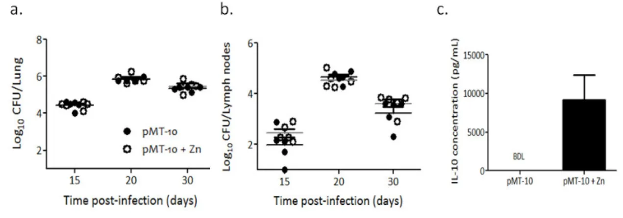
To address the role of IL-10 during different phases of the infection, we used the pMT-10 mice model, which over-expression of IL-10 is under the control of a zinc-inducible promoter.
We show that IL-10 over-expression during early and chronic phase of immune response to M.
tuberculosis did not significantly impact the immune response. When IL-10 was over-expressed during the period corresponding to T cell priming, the immune response was altered but without consequences in the control of infection. Specifically, we observe an increase in interferon -producing CD4 T cells and a decrease in IL-17--producing CD4 T cells. The myeloid response was also altered, with DC expressing low MHC class II and, surprisingly, inflammatory monocytes expressing more MHC class II. On the other hand, in L. monocytogenes infection high levels of
IL-10 increased susceptibility to infection. In sheer contrast with M. tuberculosis infection, the
susceptibility of pMT-10 mice to L. monocytogenes was associated with a significant decrease in number and activation of inflammatory monocytes in the spleen.
In what regards SIRT2, we observed that this deacetylase did not significantly impact the
inflammatory response of macrophages in vitro or in vivo the control of M. tuberculosis in the
lung. Interestingly however, we found higher bacterial burdens in the liver of SIRT2 deficient mice, suggesting that this deacetylase may have an important impact in the dissemination of mycobacteria.
Understanding the temporal and site-specific impact of immunomodulatory pathways during infection will be a significant step towards understanding the dynamics between the pathogen and host immune response. This will have an important impact in the development of more rational therapeutic approaches.
xi
RESUMO
A intensidade e duração da resposta imune são essenciais para garantir o controlo de agentes infeciosos sem comprometer a homeostasia do hospedeiro. Diferentes mecanismos, cujo papel é a regulação da intensidade da resposta imune, foram já identificados como sendo importantes para prevenir respostas exageradas do hospedeiro. Nesta tese, estudamos o papel de duas moléculas, a interleucina-10 (IL-10) e a sirtuina 2 (SIRT2), uma citocina e uma histona deacetilase com importantes propriedades imunossupressoras e anti-inflamatórias, na infeção por Mycobacterium tuberculosis e por Listeria monocytogenes.
Para avaliar o papel da IL-10 durante as diferentes fases da infeção por M. tuberculosis,
utilizamos ratinhos transgénicos (pMT-10) que sobre-expressam IL-10 sobre controlo de um promotor induzido por zinco. A sobre-expressão de IL-10 durante as fases inata e crónica da
resposta imune não influenciou a capacidade dos ratinhos para controlar a infeção por M.
tuberculosis, enquanto que a sobre-expressão da IL-10 no período de tempo correspondente à ativação das células T levou a alterações da resposta imune do hospedeiro, sem alterar o controlo da infeção. Nesta fase da infeção, após a indução de níveis elevados de IL-10, observou-se um aumento nas células CD4 produtoras de interferão (IFN)- e uma diminuição das células CD4 produtoras de IL-17, nos pulmões. A indução de níveis elevados de IL-10 também influenciou a ativação das células mieloides, observando-se uma menor expressão de MHC-II pelas células dendríticas mas curiosamente uma maior expressão de MHC-II pelos monócitos inflamatórios.
Por outro lado, na infeção por L. monocytogenes, a indução de níveis elevados de IL-10
aumentou a suscetibilidade dos ratinhos pMT-10 à infeção, que poderá estar relacionado com a diminuição do número de monócitos inflamatórios e da sua menor ativação no baço.
Relativamente ao papel da SIRT2, observamos que a ausência deste mediador nas células mieloides não influenciou a resposta dos macrófagos nem o controlo da infeção in vitro por M. tuberculosis. Curiosamente, in vivo observamos um aumento da carga bacteriana no fígado, o que sugere que esta deacetilase pode ter um papel importante na disseminação desta micobactéria.
A compreensão das vias imunomoduladoras durante a infeção será um passo importante para a compreensão da dinâmica entre o agente patogénico e a resposta imune do hospedeiro. Isto poderá ter um impacto importante no desenvolvimento de abordagens terapêuticas mais racionais.
xiii
TABLE OF CONTENTS
Abstract ... ix
Resumo ... xi
Table of contents ... xiii
Figure Index ... xv
List of Abbreviations ... xvii
Introduction ... 1
1.1 The immune system ... 3
1.2 The immune response to intracellular bacteria ... 5
1.3 Modulation of the immune response ... 12
1.3.1 IL-10 – an immunosuppressive cytokine... 12
1.3.2 Sirtuin 2 ... 16
Aims ... 19
Material and Methods ... 23
Results – Part I ... 31
1.1 Effect of IL-10 over-expression during the early stages of the immune response against M. tuberculosis infection ... 33
1.2 Effect of IL-10 over-expression during the chronic stage of the immune response against M. tuberculosis infection ... 36
1.3 Effect of IL-10 over-expression prior to T cell response to M. tuberculosis infection ... 38
1.4 Effect of IL-10 over-expression in the outcome of L. monocytogenes infection ... 52
Results – Part II ... 55
2.1 Impact of SIRT2 in in vitro M. tuberculosis infection ... 57
2.2 Impact of SIRT2 in in vivo M. tuberculosis infection ... 59
Discussion ... 61
xv
FIGURE INDEX
Cell-mediated immune response in M. tuberculosis infection. ... 8
Innate immune activation by L. monocytogenes. ... 10
Regulation of the immune response during M. tuberculosis infection. 3... 15
Gene activation and repression are regulated by acetylation/deacetylation of core histones. ... 17
Early IL-10 over-expression does not impact the control of aerosol M. tuberculosis infection. .... 34
Early IL-10 over-expression induces a delay in the accumulation of T cells in the lung during M. tuberculosis infection. ... 35
Late IL-10 over-expression does not impact the control of aerosol M. tuberculosis infection. .. 37
Late IL-10 over-expression does not impact the kinetics of T and B cells in the lung during M. tuberculosis infection.. ... 38
IL-10 over-expression between day 7 and day 14 does not impact the control of aerosol M. tuberculosis infection ... 39
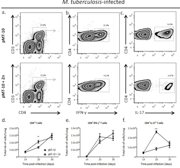
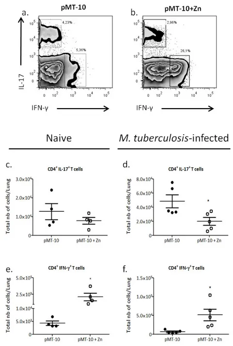
IL-10 over-expression for seven days differently impacts the CD4 T cell response ... 41
The low CD4+ IL-17+ T cells accumulation in the lungs in the presence of high IL-10 levels does not correlates with a decrease in IL-23p19 expression ... 42
The high CD4+ IFN-γ+ T cells accumulation in the lungs in the presence of high IL-10 levels correlates with a T-cell chemoattrancts expression.. ... 43
IL-10 over-expression between day 7 and 14 does not impact the accumulation and activation of alveolar macrophages in the lungs. ... 45
IL-10 over-expression between day 7 and 14 inhibits the up-regulation of MHC class II expression by DCs in the lungs.. ... 46
IL-10 over-expression between day 7 and 14 promotes the accumulation and activation of inflammatory monocytes in the lungs. ... 48
IL-10 over-expression between day 7 and 14 decreases the accumulation of neutophils in the lungs.. ... 49
Schematic representation of our hypothesis about the possible mechanisms involved in increase of CD11b+ Ly6G- SSC low MHC II high in the presence of high levels of IL-10, in the lungs. ... 50
Neutralization of IFN- does not impact the IL-10-induced accumulation of activated inflammatory monocytes in the lungs.. ... 51
xvi
IL-10 over-expression during L. monocytogenes infection impacts the accumulation and activation of CD11b+Ly6ChighMHC II+ cells.. ... 54 The activity of SIRT2 does not impact the control of M. tuberculosis infection by macrophages in vitro... 58 The absence of SIRT2 does not impact the control of M. tuberculosis infection but seems to favor the dissemination of bacilli to the liver.. ... 59 The absence of SIRT2 does not impact the development of tissue lesion in the liver at day 30 .. 60
xvii
LIST OF ABBREVIATIONS
APC Antigen presenting cell
BCG Bacillus Calmette-Guérin
BMDM Bone marrow-derived macrophages
BHI Brain heart infusion
CCL CC-chemokine ligand
CCR Chemokine receptor
CLR C-type lectin receptor
CXCL chemokine (C–X–C motif) ligand
CR Complement receptor
DCs Dendritic cells
CFU Colony-forming unit
FBS Fetal bovine serum
FoxP3 Forkhead box P3
GM-CSF Granulocyte-macrophage colony-stimulating factor
HDAC Histone deacetylase
HAT Histone acetyltransferases
HIV Human immunodeficiency virus
IFN Interferon
IL Interleukin
iNOS Inducible nitric oxide synthase
I.P Intraperitoneal
IRF Interferon regulatory factors
LLO Listeriolysin O
MAPK Mitogen activated protein kinases
MARCH1 Membrane associated ring-CH1
MHC Major histocompatibility complex
mRNA Messenger RNA
MyD88 Myeloid differentiation primary-response protein 88
xviii
NF-kβ Nuclear factor kappa beta
NK Natural killer
ON Overnight
PAMP Pathogen-associated molecular patterns
PBS Phosphate-buffered saline
PRR Pattern recognition receptors
RNI Reactive nitrogen intermediates
ROI Reactive oxygen intermediates
RT Room temperature
Sir2p Silent information regulator 2 protein
SIRT Sirtuin
TB Tuberculosis
Th T helper
TLR Toll like receptor
TGF-β Transforming growth factor
TNF Tumor necrosis factor
Treg T regulatory
TRIF TIR domain-containing adaptor inducing interferon
1
3
1.1 THE IMMUNE SYSTEM
The immune system, composed by two functional subsystems, the innate and the adaptive immune systems, that have evolved, to protect the host from infectious agents that exist in the environment, by reciprocal selective pressures and diverse invasion and evasion mechanisms on both participants [1, 2]. The innate immune system confers the first line of defense to infection, it is crucial for the early pathogen recognition which has a critical impact in the development of the protective response [1, 2]. On the other hand, the adaptive immune system triggers a specific
response often able to clear the pathogen and to generate immunological memory [2].
1.1.1 Innate Immunity
The innate immune response is orchestrated at different levels. It is composed by the physical barriers, as epidermis, ciliated epithelium, endothelium and mucosal surfaces that produce antimicrobial secretions, which prevent the entry and spread of microorganisms [1, 2]. Wherever physical barriers are unable to prevent infections, the innate immune response takes over. This response encompasses chemical components such as soluble receptors, complement proteins and enzymes that hydrolyze the pathogen; and cytokines and chemokines that drive the immune response [2-4]. The cellular components of the innate immune system includes phagocytic cells (macrophages, neutrophils, monocytes and dendritic cells (DCs)), innate lymphoid cells (natural killer (NK) cells, NKT cells and T cells), mast cells, basophils and eosinophils that are involved in the recognition of pathogens and in the triggering of the antimicrobial immune response [2-4]. The innate immune response starts with the recognition of evolutionarily conserved structures of pathogens, known as pathogen-associated molecular patterns (PAMPs) through a limited number of germ line-encoded pattern recognition receptors (PRRs), such as toll-like receptors (TLRs), c-type lectins receptors (CLRs) and others; and also complement receptors (CR) [2, 5-7]. Upon PAMPs recognition, different intracellular signalling pathways downstream PRRs can be triggered. These pathways are mediated by adaptor molecules, including the myeloid differentiation primary-response protein 88 (MyD88) and TIR domain-containing adaptor inducing interferon (IFN)-β (TRIF). The main signalling pathways downstream TLR-mediated responses are nuclear factor kappa B (NF-kβ) [2, 7-9] and mitogen-activated protein kinases (MAPKs) [7, 10]. These
cascades are the principal responsible for the induction of IL-10 and pro-inflammatory cytokines and,
4
addition to these antimicrobial responses, the innate immune system has other mechanisms that recognize and limit the growth of pathogens such as, the complement system activation, phagocytosis by macrophages and polymorphonuclear cells, and autophagy [1, 4]. Furthermore, the innate immune system is also able to recognize and clear host factors that appear in abnormal locations or in aberrant conformations such as, extracellular DNA or ATP resulting from cell death caused by infection or stress [11].
1.1.2 Adaptive Immunity
The adaptive immune response is initiated with the presentation of antigens to naive T cells by antigen-presenting cells (APCs), including DCs, macrophages and B cells [12, 13]. The generation of antigen-specific responses begins with the recognition and internalization of pathogens, or their antigens, normally by DCs in peripheral tissues. These cells then migrate to the draining lymph nodes where antigens are presented to naive T cells in the context of major histocompatibility complex (MHC) molecules [2, 12, 14]. Pathogen recognition by PRRs has a critical role in this process as it promotes DC maturation and enhances their migratory phenotype [15]. This phenotype is characterized by the upregulation of MHC class II, costimulatory molecules (such as CD40, CD80 and CD86), cytokines (such as interleukin (IL)-12, IL-6 and tumor necrosis factor (TNF)) [14, 16, 17], and lymphoid tissue homing chemokine receptors (such as CCR7) [18, 19]. Altogether, the recognition of the complex MHC-peptide by T cells, together with costimulatory signals and the cytokine microenvironment promote the differentiation of naive T cells towards specific populations [13].
DCs are able to induce the differentiation of naive CD4 T cells into different helper (Th) subsets, including Th1, Th2 and Th17 characterized by a specific cytokine signature [20-23]. This differentiation is initiated by PAMP recognition by the APC that ultimately determines the type of cytokines produced [21]. In this regard, IL-12 produced by APCs [24], together with IFN- from memory T cells and NK cells, plays a critical role in inducing the Th1 phenotype [20, 21, 25]. IL-4 is in turn involved in the induction of Th2 responses [20, 21, 25, 26]. Finally, IL-1, transforming growth factor (TGF)-β and 6 promote the differentiation of Th17 responses and IL-23 maintains their phenotype [27-30]. The Th1 subset, characterized by the expression of IFN- , IL-2, and TNF, is critical for protection against intracellular pathogens by promoting macrophage activation [20-22]. Th2 cells, essential in immunity to extracellular pathogens and involved in allergic responses, are characterized by the production of IL-4, IL-5, IL-13 and B cell growth and
5
differentiation factors that support humoral immunity [20-22]. Th17 cells, important in protection against several pathogens, including intracellular and extracellular pathogens, are characterized by IL-17, IL-21 and IL-22 production [22, 27]. The activity of these effector cells is strictly regulated between them [31] and by natural and inducible regulatory T (Tregs) cells, whose differentiation and maintenance are dependent on the transcription factor Foxp3 [23, 32]. An abnormal activity of Th1 or Th17 cell responses may culminate in autoimmune diseases [22, 33], whereas excessive Th2 cell responses may result in allergic inflammatory disease [22]. In all, the innate and adaptive immune responses are interactive subsystems of the immune system that act in concern to protect the host from infection but can also cause important immunopathological consequences. Therefore, it is essential to understand how the balance between pathogen clearance and immunopathology in several infections is regulated.
1.2 THE IMMUNE RESPONSE TO INTRACELLULAR BACTERIA
Intracellular pathogens have the capacity to enter and to explore survival mechanisms inside the cells. As a result, the host immune protective response is mainly mediated by T cells with little participation of the humoral response [34]. Furthermore, the response to intracellular bacteria culminates in tissue reactions typically characterized by granuloma structures [34]. Despite intracellular bacteria being contained within these granulomata, sometimes pathogen clearance fails and the disease becomes chronic. Two classical examples of intracellular pathogens are Mycobacterium tuberculosis and Listeria monocytogenes.
1.2.1 M. tuberculosis Infection
Tuberculosis (TB) is an infectious disease caused by the bacillus M. tuberculosis, which is spread by respiratory transmission [35]. Typically, it affects the lungs (pulmonary TB), however this
disease can be extrapulmonary [35, 36]. TB was declared by the World Health Organization
(WHO) a global public health emergency in 1993 [35]. Despite the many efforts to improve TB prevention and treatment, TB remains a major cause of morbidity and mortality worldwide [35]. There were 8.6 million new cases and 1.3 million deaths in 2013 and it is estimated that one-third of the world’s population is currently infected with M. tuberculosis but remain asymptomatic, defined as latent TB [35]. Only 5-10% of these latent individuals develop active TB disease in their lifetime [37, 38]. Control of this global epidemic disease has been impaired by
6
Calmette-Guérin (BCG), is only efficient against childhood TB [39-41]; by the appearance of
multidrug-resistant M. tuberculosis strains [42, 43]; by co-infections with the human
immunodeficiency virus (HIV) [44, 45]; and by the lack of sensitive and rapid TB diagnosis [46].
1.2.1.1 Innate immune response
Once the bacillus is delivered to the lungs, the immune response to M. tuberculosis is initiated
with the bacterial recognition by phagocytic cells, mainly alveolar macrophages but also
neutrophils, monocytes and DCs [47-51], through different PRRs expressed on the surface of
these cells [52-54].Among TLRs, TLR2, -4 and -9 are the best characterized in the recognition of
M. tuberculosis [54-56]. Briefly, TLR2 recognizes the largest array of mycobacterial PAMPs, including lipoproteins, phosphatidylinositol mannans and lipomannan; TLR4 recognizes phosphatidyl-myo-inositol mannosides (PIMs) [56, 57], and TLR9 senses mycobacterial DNA [55, 57]. M. tuberculosis recognition via different TLRs triggers various intracellular signaling cascades that culminate with the production of important pro- and anti-inflammatory cytokines involved in protective host immune response, including TNF, IL-12 (both pro-inflammatory) and IL-10 (anti-inflammatory). There is experimental evidence supporting the key role of multiple TLRs in the control of M. tuberculosis [58-61]. Moreover, different polymorphisms in the human TLR2 gene were reported to associate with increased susceptibility to TB [62-64].
Upon M. tuberculosis recognition, phagocytic cells upregulate different microbicidal mechanisms. Alveolar macrophages are the primary cell type involved in the uptake of mycobacteria [50, 65,
66]and the depletion of these activated cells in mice led to impaired resistance to infection [67].
In vivo and in vitro studies in mice have shown that M. tuberculosis is phagocyted mainly by alveolar macrophages [68] but DCs [69, 70] and neutrophils [71-74] can also phagocytose M. tuberculosis. These cells protect the host in a non specific manner through essential microbicidal mechanisms, such as the production of toxic effector molecules such as reactive oxygen or nitrogen intermediates (ROI or RNI, respectively), phagossome acidification, limiting iron availability and undergoing apoptosis [47, 50, 51, 75]. Indeed, mice lacking the mediators of microbicidal macrophage functions, such as expression of inducible nitric oxide synthase (iNOS) [48, 76] or LRG-47 [77] are unable to control M. tuberculosis infection.
7
1.2.1.2 Adaptive immune response Th1 response
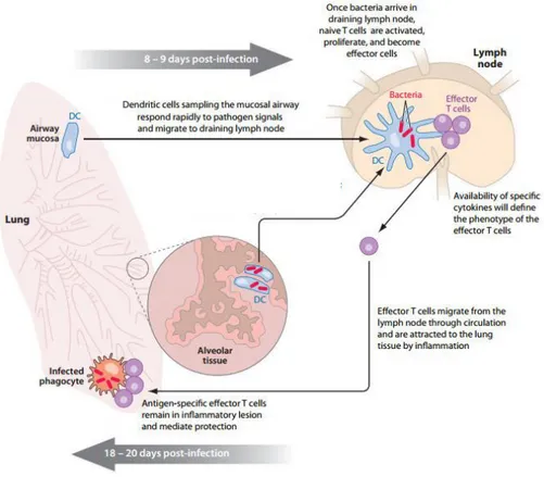
The innate immune response makes a critical contribution to the activation of the adaptive immunity. Indeed, inflammatory response mediated by phagocytic cells increase the flow of DCs, which around day 9 post-infection start to migrate to the draining lymph nodes, where they will drive naive T cell activation and differentiation (Figure 1) [78-81]. Some reports have shown that M. tuberculosis compromise DCs function, which may explain the slow dissemination of bacteria
to the lymph nodes [69, 82, 83].Upon T cells activation, after 2 weeks of infection, they begin
migrating to the lung where they mediate protective immunity by activating the infected phagocytes (Figure 1) [81, 84].
It is well established that CD4 T cells capable of making IFN- are required for protective
immunity [85, 86]and that loss of these cells increases the probability of mice to succumb to TB
[44]. Indeed, CD4 T cells-producing IFN- (Th1) are required for resistance to lethal infection, given that MHC class II [87] and IFN- [88-90] deficient mice rapidly succumb to infection. IFN- deficient mice fail to produce RNI and ROI and develop progressive tissue destruction, associated with uncontrolled bacterial growth [90]. These data highlight the important role of IFN- in the
control of M. tuberculosis infection; however, recent studies suggest IFN- -independent
mechanisms whereby CD4 T cells mediate protection [91].Althoughantigen specific CD8 T cells,
NK cells and T cells also produce IFN- during M. tuberculosis infection, they cannot
compensate for a lack of CD4 T cells [80, 81].
The induction of protective IFN- T cell response depends upon IL-12 [92, 93] which is mainly produced by activated DCs. Indeed, IL-12p40 deficient mice cannot control bacterial growth and this is associated with the absence the innate and adaptive sources of IFN- [92, 94]. Recently, IL-12p40 has been suggested to induce DCs migration to the lymph nodes [95], thus promoting T cell priming [95, 96]. Human studies have also shown a crucial role for the IL-12-CD4-IFN- axis in the control of mycobacterial infections [97]. Indeed, humans with mutations in gene encoding the IL-12 receptor are extremely susceptible to M. tuberculosis. Similarly, patients with mutations in IFN- receptor 1 (IFN- R1) and in gene encoding STAT1, an essential transducer of IFN- -mediated signals, are also extremely susceptible to pulmonary TB and BCG infection [97, 98]. Also in humans, CD4 T cells are critical in the control of M. tuberculosis as reported by the
8
In summary, the axis IL-12-CD4-IFN- is critical to the control of M. tuberculosis infection,
although these cells are unable to eradicate the pathogen or to prevent the chronic disease. Therefore, it is important to understand as the protective immune response to M. tuberculosis is regulated, for instance, by IL-10, in order to design novel immune therapies.
Figure 1. Cell-mediated immune response in M. tuberculosis infection. Upon M. tuberculosis aerosol
infection, the bacillus is deposited in the lower airways and the alveolar tissue. M. tuberculosis is recognized by
alveolar macrophages and DCs and the bacteria do not disseminate from the lung until day 9 post-infection. After, DCs migrate to the lymph nodes and present the antigen to naive T cells. The phenotype of T cells will depend on the availability of specific cytokines. After T cell activation and differentiation, these cells migrate to the lung in response to inflammation and mediate protection by macrophages further activation, stopping the bacterial growth. From Andrea M. Cooper, Cell-mediated Immune Response in Tuberculosis, Annual Reviews of Immunology, 2009
9
Th17 response
In addition to Th1 cells, Th17 cells are also involved in the immune response to mycobacteria [99, 100]. However, in the absence of IL-17, protection is not altered in response to a low dose
of M. tuberculosis infection [99]. On the other hand, when IL-17 is blocked during a high-dose
challenge, the recruitment of neutrophils, granuloma formation and protection are impaired [101, 102]. In the context of vaccination, repeated exposure to BCG results in a significant increase in IL-17 response, accompanied with an increase in influx of neutrophils/granulocytes and subsequently, pathological inflammation in the lungs [103]. Furthermore, Th17 cells seem to be able to anticipate the recruitment of Th1 cells to the lung, promoting the resistance to mycobacterial infection [101]. It has also been shown that IFN- can inhibit CD4 T cell-producing IL-17, impairing the recruitment and the accumulation of neutrophils, contributing to decreased lung inflammation and improved the outcome of the disease [104]. Thus, the IL-17 cellular response needs to be tightly regulated because the balance between protective and damaging immunity need to be achieved to control of M. tuberculosis proliferation and reduce transmission and mortality.
1.2.2 L. monocytogenes Infection
L. monocytogenes is the causative agent of human listeriosis, a potentially fatal foodborne infection [105]. The incidence of listeriosis varies between 0.1 and 11.3/1000000 in different
countries[106]. Despite adequate antimicrobial treatment, listeriosis has an average case-fatality
rate of 20-30% [106]. Pregnant woman and individuals who are immunocompromised or elderly
are particularly vulnerable to L. monocytogenes infection [107]. Clinical manifestations are
variable, ranging from febrile, gastroenteritis to more severe invasive forms, including sepsis,
meningitis, rhombencephalitis, perinatal infections, and abortions [105, 107]. Contrary to M.
tuberculosis, these bacteria cause an acute infection and the major targets organs of
experimental murine listeriosis are the spleen and liver [34] The murine model of L.
monocytogenes infection is an attractive tool to understand the interplay between the host and
pathogen [108]. Infection of mice with a sub-lethal dose of L. monocytogenes induces a robust
innate inflammatory response, that restricts bacterial growth and dissemination to the liver and spleen, preventing the spread into systemic and, consequently lethal infection [108].
10
1.2.2.1 Innate Immune Response
Upon intravenous infection of mice, the bacteria are rapidly taken up by resident myeloid cells of
spleen, including macrophages, neutrophils and DCs [108, 109]. Within the cell, L.
monocytogenes escapes the phagosome to cytosol by producing listeriolysin O (LLO), a virulence
factor that destroys the phagosomal membrane (Figure 2a) [110, 111]. In the cytosol, L.
monocytogenes induce NF-kβ pathway mediating the transcription of innate inflammatory cytokines and chemokines, as CC-chemokine ligand 2 (CCL2) and CCL7 (Figure 2b) [107]. These
chemokines are involved in the recruitment of Ly6CHigh inflammatory monocytes to infected tissues
[107, 109]. The recruitment of these inflammatory cells is essential for the control of L.
monocytogenes infection [112]. In response to microbial products, macrophages secrete TNF and IL-12 (Figure 2c). These cytokines drive NK cells to produce IFN- , which in turn increases the antimicrobial capacity of macrophages.
Figure 2. Innate immune activation by L. monocytogenes. (a) L. monocytogenes in the bloodstream is
rapidly recognized and internalized by macrophages in the spleen and other tissues. In the macrophages phagossome, bacteria secrete LLO, which lyses the phagossome membrane and activates NF-kβ pathway, mediating
the transcription of innate immune genes, as CCL2. (b) CCL2-producing infected cells induce the recruitment of
circulating monocytes that express CCR2, to the infected tissues. (c) Infected macrophages release microbial
products that activate recruited monocytes through TLRs in MyD88-dependent manner. (d) Monocytes differentiate
in DCs that release TNF and nitric oxide (Tip-DCs), which promote bacterial killing. From Eric G. Pamer, Immune Responses to Listeria monocytogenes, Nature Reviews Immunology, 2004
11
Accordingly, IFN- and TNF deficient mice are highly susceptible to L. monocytogenes infection [108, 109, 113]. Thus, the innate immune response is essential for host survival, as indicated by the impact in the outcome of infection on mice that lack the innate immune players [113]. Despite the importance of innate immunity for the initial control of L. monocytogenes, the T cell response is needed to bacteria clearance since SCID mice develop chronic infection [114].
1.2.2.2 Adaptive Immune Response
DCs play a central role in the recognition of L. monocytogenes, antigen presentation and
subsequent priming of T cells [107, 108]. Indeed, mice depleted of CD11c+ DCs are unable to generate a protective CD8 T cell response [115]. In this regard, CD8 T cells provide a more substantial contribution to a long-term protective response than CD4 T cells to L. monocytogenes. These cells can be separated into two populations: the first is restricted to classical MHC class I molecules, and the second is restricted to a non-classical MHC class I molecule (H2-M3-restricted T cells) [107]. During primary infection, H2-M3-(H2-M3-restricted CD8 T cells respond rapidly, whereas MHC class I-restricted CD8 T cells reach peaking frequencies only 7 to days following inoculation [107]. During a recall response however, MHC class I- restricted CD8 T cells are more important [107]. CD8 T cell response contributes to anti-listerial immunity by two synergistic mechanisms: lysing infected cells via perforin and granzymes, exposing intracellular bacteria; and secreting IFN- to activate macrophages [116].
The role of CD4 T cells during L. monocytogenes infection is still not well understood, but it is thought that CD4 T cells differentiate into Th1 cells [117] and facilitate granuloma formation
[118]. Furthermore, CD4 T cells were shown to contribute to CD8 T cells activation, but mice
lacking CD4 T cell responses mount a primary CD8 response similar to wild-type mice and rapidly clear the infection [108, 119]. In addition, in vivo depletion studies shown that memory
CD8 T cells are the most effective T cell subset able to confer protection [120].
In all, murine L. monocytogenes infection provides an interesting model to address innate and acquired mechanisms of protection during infection, including the role of cytokines and their function in different cell lineages.
12
1.3 MODULATION OF THE IMMUNE RESPONSE
During infection, the host immune system needs to respond to pathogens with adequate intensity and duration to control and eliminate the infection. However, exacerbated antimicrobial mechanisms can often cause collateral damage, which sometimes is more detrimental to the host than the infection itself. Thus, several mechanisms have been described to control and regulate the immune response to prevent immunopathological consequences. In the next section,
the importance of two key molecules in the inflammatory response, IL-10 – an important
mediator in immune response against several pathogens, and sirtuin2 – an immune mediator
unexplored in the context of M. tuberculosis infection are discussed.
1.3.1 IL-10 – an immunosuppressive cytokine
IL-10 Biology
IL-10 is an anti-inflammatory cytokine produced and recognized by many immune cells, from both myeloid and lymphoid lineages [121, 122]. Among innate immune cells, monocytes, macrophages and DCs (but not plasmocytoid DCs [123]) are the main producers of IL-10, but
NK cells, mast cells and granulocyte cells are also able to synthesize this cytokine [121, 122]. In
addition, many subsets of T cells can produce IL-10, such as Th1 cells, Th2 cells, CD8 T cells, and Treg cells; and also B cells. Recently, Th17 and Th22 were also found to produce IL-10 [121, 122].
10 acts through a surface receptor complex – 10R, which is composed by 10R1 and IL-10R2 subunits [121, 122]. This receptor transduces signals through the JAK-STAT signalling pathway and STAT transcription factors [122, 123]. IL-10 production is induced in response to the interaction between TLRs and PAMPs and it may also be induced through CLR, Card9 and DC-SIGN [122-124]. Moreover, IL-10 can be induced by pathogens as an evasion mechanism [125]. However, it is still not clear whether high concentrations of IL-10 during infections are a cause or a consequence of high pathogen loads [124].
The immunosuppressive action of IL-10 has a broad range of target cells in an autocrine/paracrine way, mainly on the innate cells, namely on macrophages and DCs. IL-10 produced early in the immune response can inhibit the release of pro-inflammatory mediators (TNF-α, IL-1β , IL-6, IL-12, granulocyte-macrophage colony-stimulating factor (GM-CSF) and others) by macrophages [121, 126, 127], as well as the expression of chemokines involved in
13
the recruitment of monocytes, DCs, neutrophils and T cells [121, 122, 124]; inhibit phagocytosis and suppress the production of ROI and RNI, such as nitric oxide, involved in bacterial killing [121, 125, 128]. In addition, IL-10 blocks antigen presentation by APCs via down-regulation of MHC class II and co-stimulating molecules (e.g. CD86 and CD80) [121, 122]. Moreover, IL-10 inhibits APCs migration to the lymph nodes by dampening IL-12 production [124]. As result, the effects of IL-10 in DCs and macrophages indirectly compromise the activation and differentiation of naive T cells and, subsequently, adaptive immunity [121, 129]. Moreover, the effect of IL-10 in Th1 cells decreases IFN- production and, consequently, amplifies the deactivation of APCs [121]. IL-10 also reduces IL-23 production by macrophages, which is essential for the proliferation and survival of Th17 cells [122]. In addition, IL-10 can also inhibit T cells proliferation and differentiation by impairing the production of cytokines important for these effects, namely, IFN- , IL-2, IL-4 and IL-5 [121, 124, 127].
Despite the inhibitory functions of IL-10, this cytokine is also important in the prevention of apoptosis of B cells, and in the recruitment, proliferation and cytotoxic activity of CD8 T cells and NK cells [121, 127]. Thus, IL-10 can directly regulate innate and adaptive responses by limiting T cell activation and differentiation in the lymph nodes, as well as suppressing proinflammatory responses in tissues, leading to impaired control of infections and/or reduced immunopathology.
IL-10 in Infections
Depending on the type of infection, the presence of IL-10 may either contribute for host protection or susceptibility [121, 127]. In certain cases, the presence of IL-10 prevents exacerbated inflammatory responses that frequently culminate in fatal immunopathology, such as those occurring upon infection with Toxoplasma gondii [130], Plasmodium chabaudi [131] and Trypanosoma cruzi [125]. This phenotype is confirmed in IL-10-deficient mice that develop lethal colitis in response to gut flora, as a sign of excessive immune reaction [132]. However, in other situations, the presence of IL-10 was shown to delay or impair protective immune responses. This is supported by studies showing that the absence of IL-10 results in improved clearance of L. monocytogenes [133] and Leishmania major [134] infections. Therefore, IL-10 is responsible for the fine balance between suppressing and activating immune responses to pathogens. The timing, as well as the level of IL-10 and the immune environment may play a critical role in these processes.
14
IL-10 in M. tuberculosis Infection
As discussed above, control of M. tuberculosis infection requires the activation of specific CD4 T cells in lymph nodes. These cells migrate to the site of infection where they activate macrophages [51], it is therefore expectable a detrimental impact of IL-10 in the course of the disease. Indeed, it has been shown that IL-10 can downregulate the M. tuberculosis-induced Th1 responses [135]. Moreover, IL-10 block phagocytosis and impair ROI and RNI production [128]; inhibit IL-12p40 dependent migration of DCs to the lymph nodes [136]; and down-regulate the expression of co-stimulatory molecules by DCs required by T cell differentiation (Figure 3) [137]. These findings suggest that IL-10 can suppress the development of the protective immune
response against M. tuberculosis [138, 139]. In spite of this, Jung et al. described that IL-10
deficient mice had a similar capacity to control M. tuberculosis infection when compared to wild-type mice [140]. On the other hand, Higgins et al., showed that IL-10 deficient mice infected with M. tuberculosis display exacerbated immunopathology and succumb to disease during late stages of infection [141]. More recently, Redford et al., reported reduced bacterial burden in the lungs and the spleen after M. tuberculosis infection in IL-10 deficient mice when compared with wild-type mice [142]. This decrease of bacterial loads is accompanied by an earlier and enhanced Th1 response in lung [142]. While these results are difficult to compare, due to the use of different mycobacterial strains, the dose of infection, the microbial flora of mice from different laboratories, and the time-points assessed they suggest a role for IL-10 in the protective immune response to M. tuberculosis.
A different approach to study the role of IL-10 in M. tuberculosis is transgenic mice that
over-express IL-10. These models show an increased susceptibility to M. tuberculosis infection and
other mycobacterial species [143]. While transgenic mice that over-express IL-10 in T–cell
compartment have enhanced susceptibility to M. tuberculosis [143] and BCG [144] infection
associated with impaired T cell responses, the increased susceptibility of transgenic mice that over-express IL-10 in the macrophage compartment is associated with macrophage deactivation, but similar T cell responses to that of wild-type mice [145]. These data suggest that, IL-10 may influence the immune response and susceptibility to reactivation disease. In support of these findings, it has been demonstrated that IL-10 is increased in patients with active pulmonary TB
[146]. Indeed, T cells co-expressing IFN-γ and IL-10 have been isolated from the bronchoalveolar
15
presence of human polymorphisms related with increased IL-10 responses. One example is SLC11 A1, a TB susceptibility locus, that has been associated with increased IL-10 production by monocytes [148]. A recent meta-analysis suggest, an important trend toward polymorphisms in the IL-10 gene and increased susceptibility [149] further supporting an important role for IL-10 during M. tuberculosis infection. Future studies will be important to further define its role.
Figure 3. Regulation of the immune response during M. tuberculosis infection. Following infection with
M. tuberculosis, several regulatory actions mediated by IL-10 and Tregs cells serve to limit host-induced immunopathology that may be detrimental to pathogen clearance. The induction of IL-10 during M. tuberculosis infection inhibits macrophage and DCs functions, blocking DCs migration to the lymph nodes and subsequently, antigen presentation. In the lymph nodes, IL-10 and Tregs cells can inhibit T cell differentiation and activation of Th1 response. Furthermore, IL-10 can block the migration of these cells to the lung through the downregulation of CXCL10 expression with consequences at macrophage activation levels. From Anne O’ Garra et al., The Immune Response in Tuberculosis. Annual Reviews of Immunology,2013
16
IL-10 in L. monocytogenes infection
As for M. tuberculosis infection, the role of IL-10 during L. monocytogenes infection is not yet
fully defined. Overall, IL-10 appears to have a detrimental role, since IL-10-deficient mice are
more resistant to L. monocytogenes infection, accompanied by an up-regulation of the Th1
response [133, 150]. However, some studies suggest that IL-10 may have a protective role, possibly associated with reduced immunopathology [151]. Future studies are required to address the role of IL-10 during L. monocytogenes infections, specifically in what respects to its impact in the protective T cell response and development of immunopathology.
1.3.2 Sirtuin 2
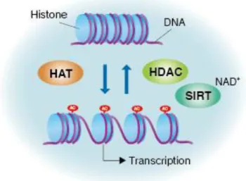
As discussed above, regulation of the immune response is critical for infection control and host survival. This regulation can be mediated by cytokines but recent data suggest that regulation of gene expression can have a significant impact in the immune response and outcome of the infection [152]. Gene regulation can occur at transcriptional and post-transcriptional levels, translational level and chromatin remodeling. One important modulator of gene expression is the specific modification of histones [153]. Histones are one of the most evolutionary conserved proteins and are essential for the homeostasis of eukaryotic organisms [154]. Each histone can be modified on different residues by phosphorilation, acetylation, metylation and others [154]. The cellular outcome of these modifications depends on the residue target and its cellular environment [154]. Acetylation and deacetylation of nucleosomal histones play an essential role in the modulation of chromatin structure, chromatin function and in the regulation of gene expression [155]. Acetylation is mediated by histone acetyltransferases (HATs), which use acetyl-coenzyme A as a cofactor to catalyze the transfer of an acetyl group to a lysine residue. This modification decreases the affinity between histones and DNA, allowing chromatin to adopt a more relaxed structure (Figure 4). As a consequence, chromatin becomes more permissive to transcription factor binding, activating gene expression [156]. Histones deacetylation, which is mediated by histone deacetylases (HDACs) counteracts the effects of HATs and it is associated with transcriptional repression (Figure 4). These two opposite classes of enzymes need to be tightly controlled to maintain a balanced gene transcription [156]. Indeed, discrepancies in the histone acetylation has been associated with carcinogenesis and cancer progression [155].
17
Sirtuins are a family of class III HDACs, which use nicotinamide adenine dinucleotide (NAD) as a cofactor (Figure 4) [157]. This family of proteins is highly conserved and is involved in regulation of ribosomal DNA recombination, gene silencing, DNA repair and chromosomal stability and longevity [158, 159]. In mammals, there are seven types of sirtuins described (SIRT1-7), which display diversity in subcellular localization that may account for differences in their biological function and substrate usage [160].
Figure 4 – Gene activation and repression are regulated by acetylation/deacetylation of core histones. Gene expression can be regulated at histones level. Histone acetylation is catalysed by HATs, whereas the
reverse reaction is performed by HDACs. When histones are acetylated, interaction of the N terminal of histones with the negatively charged phosphate groups of DNA is decreased. As a consequence, the condensed chromatin is transformed into a more relaxed structure that is associated with increase in gene transcription. In contrast, deacetylation performed by HDACs has the opposite effect. Deactelylation of histone tails become DNA more tightly wrapped around the histone cores, making it harder for transcription factors to bind to the DNA and, consequently,
gene transcription. Sirtuin family is HDACs and is involved in gene regulation. From
http://ruo.mbl.co.jp/e/product/cyclex/cellular-acetyle.html
Sirtuin type 2 (SIRT2) is the mammalian ortholog of silent information regulator 2 protein (Sir2p)
of Saccharomyces cerevisiae [160]. SIRT2 is a NAD+-dependent HDAC that plays an important
role in transcriptional silencing [158, 161]. This sirtuin is predominantly a cytoplasmatic protein that may shuttle to the nucleus, for instance during mitosis, where it functions as a mitotic checkpoint protein [162]. The role of SIRT2 has been mainly characterized in the cytoplasm, in regulation of microtubule dynamics once its localize with microtubules [161]. Moreover, SIRT2
18
has been involved in regulation of cell cycle, DNA repair, apoptosis, metabolism and aging by deacetylation of several transcriptions factors [162]. For example, SIRT2 has involved in regulation of NF-kβ gene expression through deacetylation of p65 [163], regulating the expression of a broad number of target genes involved in the immune and inflammatory response, apoptosis, cell proliferation, differentiation and survival [163].
Several studies have indicated that SIRT2 plays an important role in brain function, inhibiting inflammation and neurotoxicity mediated by microglia [164]. Interestingly, pharmacological or genetic inhibition of SIRT2 has been shown to be protective in Drosophila model of Parkinson’s disease [165, 166]. In some types of cancer, as melanoma, gliomas, and gastric carcinomas, levels of SIRT2 are decreased. SIRT2 resides in a genomic region frequently deleted in human gliomas and in vitro studies shown that expression of SIRT2 in glioma-derived cell lines markedly reduces their capacity to form colonies [160, 162]. These findings led to the hypothesis that the inactivation of SIRT2 may underlie the development of gliomas and that SIRT2 activation possibly protect against these diseases. Thus, SIRT2 may be a potential target for therapeutic treatments. There is evidence that histone modifications induced by pathogens can impact host immunity [167]. For instance, in vitro studies have shown that L. monocytogenes secret LLO that induce modifications in the host histones [167]. Similar effects were observed with other toxins of the
same family, such as perfringolysin of Clostridium perfringens and pneumolysin from
Streptococcus pneumonia [167]. In the mouse, a recent report suggest that L. monocytogenes induces SIRT2 translocation from cytosol to the nucleus, causing deacetylation of histone H3 on lysine 18 (H3K18), and impacting the control of infection [168]. These mechanisms of host subversion could be an important strategy used by other pathogens, including M. tuberculosis.
19
21
The immune response against pathogens needs to be tightly regulated to eliminate the pathogen without compromise the host homeostasis. Two important modulators of this balance are IL-10
and SIRT2. The role of IL-10 during M. tuberculosis infection or L. monocytogenes infection
remains controversial, and it is still not well understood whether this cytokine plays a role in host resistance or susceptibility. As for SIRT2, it has been recently suggested that the activation of this histone deacetylase is an active mechanism that L. monocytogenes exploits to establish infection.
We are interested in determining whether or not M. tuberculosis also uses this pathway to
promote infection.
To address the role of IL-10, we used a novel model of IL-10-over-expression, the pMT-10 mouse model that allows us to evaluate the impact of a transiently IL-10 over-expression during different phases of the immune response. To address the role of SIRT2 we used mice that are deficient in the gene encoding this enzyme in the myeloid lineage.
The main goals of this thesis are:
(i) Determine the impact of transient high levels of IL-10 in the immune response and the outcome of aerosol M. tuberculosis infection; specifically, we will focus on the impact of IL-10 at
the time of infection, during the acute phase and during the chronic phase of M. tuberculosis
infection.
(ii) Determine the role of IL-10 in the mechanisms of immune-mediated protection and outcome of L. monocytogenes infection.
(iii) Define the role of SIRT2 in the in vitro response of macrophages and the in vivo outcome of the M. tuberculosis infection.
23
25
Animals. pMT-10 mice on a C57BL/6 background were produced by Drs. António G. Castro and
Paulo Vieira at the Gulbenkian Institute of Science, Oeiras (IGC). A mouse IL-10 cDNA sequence
was cloned in the p169ZT vector containing the sheep metallothioneinc (MT) Ia promoter, a
β-globin splice site and the SV40 polyadenylation signal. The resulting vector (pMT-IL10) was injected in C57BL/6 eggs and the transgenic were identified by PCR using MT and IL-10 specific primers. The association of an MT promoter with the mouse IL-10 cDNA sequence allows for the over-expression of IL-10 in the presence of zinc. IL-10 over-expression was induced by administration of 2% sucrose solution containing 50 mM of zinc sulfate in the animals’ drinking water, ad libitum. A group of transgenic littermates were supplied with 2% sucrose solution as a control.
LysM-Cre+Sirt2 flox+/flox+ mice were obtained by Teresa F. Pais at IMM by crossing LysMcre mice (The Jackson Laboratory) with Sirt2 floxed mice, which were used through an MTA with Johan Auwerx & Kristina Schoonjans Laboratory of Integrative and Systems Physiology, NCEM, Ecole Polytechnique de Lausanne (EPFL), Switzerland. Experimental mice were matched for sex and age and were infected at between 8 to 12 weeks of age. All procedures involving live animals were carried out in accordance with the European Union Directive 86/609/EEC, and previously approved by the Portuguese National authority Direcção Geral de Veterinária.
Bacteria and Infection. M. tuberculosis H37Rv, originally from the Trudeau Institute
Mycobacterial Collection, was grown in Middlebrook 7H9 liquid media (BD Biosciences) for 7–10 days and then diluted into Proskauer Beck (PB) medium supplemented with 0.05% Tween 80
and 2% glicerol to the mid-log phase. Bacterial stocks were aliquoted and frozen at –80°C. To
determine the concentration of M. tuberculosis aliquots, 10-fold serial dilutions of 6 frozen vials were plated in Middlebrook 7H11 (BD Biosciences) agar plates supplemented with 10% oleic acid/albumin/dextrose/catalase (OADC) and 0.5% glycerol and viable bacteria were determined after 3 weeks post-incubation at 37°C.
Mice were infected with M. tuberculosis H37Rv via the aerosol route using an inhalation exposure
system (Glas-Col). Briefly, the mycobacterial inoculum was prepared to a concentration of 2x10⁶
CFUs from the frozen original stock, by passing it through a 26G needle 4-6 times to disrupt
bacterial clumps and diluted in water (Aqua B. Braun). Mice were exposed to the aerosol over a
26
infection dose was confirmed by counting of viable bacteria in the entire lung of 5 animals 3 days after the aerosol infection.
L. monocytogenes strain EGD was grown in brain heart infusion broth (BHIB) for 24h, washed twice with PBS, aliquoted and frozen in PBS containing 30% glycerol at -80ºC. To determine the
concentration of L. monocytogenes aliquots serial dilutions of 1 frozen vial were plated in BHI
medium plates and the viable bacteria were determined after 24 hours post-incubation at 37°C.
The 50% lethal dose (LD50) of this inoculum was approximately 1x10⁶ when administered
through the intraperitoneal (i.p.) route to wild-type C57BL/6 mice. For experimental infections,
mice were intraperitoneally infected with 5x10⁵ bacteria diluted in 200μl of phosphate-buffered
saline (PBS).
In vivo IFN-γ neutralization. In vivo IFN- blockade was carried out by i.p. injections of 0.5 mg
of anti-Mouse IFN- (clone XMG1.2, eBiosciense) twice a week, as described in the respective figure legends.
Preparation of Single Cell Suspensions. At selected time-points post-infection, mice were
killed by CO2 asphyxiation and the organs were aseptically excised. Lungs were first perfused with
PBS through the right ventricle of the heart to exclude blood cells, dissected with 2 sterile scalpels and incubated at 37ºC with collagenase IX (0.7mg/ml, from Sigma-Aldrich) for 30 minutes. Single cell suspensions from the digested lungs, liver, spleen or mediastinal lymph
nodes were homogenized and passing through a 40-μm-pore-size nylon cell strainer (BD
Biosciences). Lymph nodes cells were resuspended in cDMEM and used for bacterial load determination, flow cytometry analysis and RNA extraction. Liver cells were resuspended in cDMEM and used for bacterial burden determination. Lung cells were treated with erythrocyte
lyses solution (0.87% of NH4Cl solution and 5% of PBS in water) to lyse residual red blood cells
and resuspended in cDMEM and used for bacterial burden determination, flow cytometry analysis or RNA extraction. Single cell suspensions were counted using a Countess® Automated Cell Counter (Life Technologies).
Measurements of Survival. Mice infected with L. monocytogenes were monitored twice daily
and when the animals had a behavior unresponsive or recumbent were considered moribund and euthanized.
27
Bacterial Load Determination. To determine the M. tuberculosis bacterial burdens, lung,
liver or mediastinal lymph nodes single cell suspensions were incubated with 0.1% saponin
(Sigma) for 10 min to release intracellular bacteria. The number of CFU was determined by plating 10-fold serial dilutions of the disrupted cell suspensions in Middlebrook 7H11 agar plates
supplemented as described above. BBL™ MGIT™ PANTA™ antibiotic mixture (BD Bioscience)
was used to prevent contaminations. Viable mycobacteria colonies were counted after 3 weeks of incubation at 37ºC. For control of L. monocytogenes bacterial burden, spleen and liver were collected and placed in cold PBS. The liver and the remaining spleen were disrupted by high
pressure using a homogenizer releasing the bacteria to the solution. The number of L.
monocytogenes CFU was measured by plating of 10-fold serial dilutions of homogenate tissue onto BHI agar plates and incubating over-night (ON) at 37ºC.
Flow Cytometry Analysis. For intracellular cytokine staining, 2-3x10⁶ cells were stimulated with
a mixture of phorbol myristate acetate (PMA) (50ng/ml) and ionomycin calcium salt (4μg/ml), in
the presence of brefeldin A (10μg/ml) (all from Sigma-Aldrich) for 4 hours at 37ºC. After
stimulation, the cells were recovered and fixed ON with 10% neutral buffered formalin. Cells were pre-treated with Fc block (anti CD16/CD32) for 10 minutes to minimize a non-specific antibody binding and permeabilized with saponin FACS buffer (PBS containing 2% of FBS, 0.01% of azide and 0.5% saponin) for 10 minutes and stained for surface and intracellular antigens for 30 minutes at room temperature (RT). The following antibodies were used: CD3-PerCPcy5.5 (clone 145-2C11, eBioscience), CD4-APC-Cy7 (clone GK1.5, eBioscience), CD8-FITC (clone 5H10-1, Biolegend), CD11b-PE (clone M1/70, eBioscience); CD11c-BV421 (clone N418, Biolegend); CD19-APC (clone eBio1D3, eBioscience), Ly6G-APC (clone 1A8, Biolegend); MHC II-FITC (M5/114.15.2, Biolegend); Ly6C-PerCPCy5.5 (clone AL-21, Pharmingen); IL-17-APC (clone TC11-18H10.1, eBioscience ), IFN- -PECy7 (clone XMG1.2 from Biolegend), iNOS-FITC (6/iNOS/NOS Type II, eBioscience). Samples were acquired on a LSRII flow cytometry with Diva Software. All data were analysed using FlowJo version 7 software. The total number of cells in each gate was calculated using the total number of cells determined by Countess® Automated Cell Counter.
28
Differentiation and Infection of Bone Marrow-Derived Macrophages (BMDM). Bone
marrow cells were flushed from tibiae and femurs’ mice with complete Modified Eagle Medium (cDMEM, DMEM supplemented with 10% of heat-inactivated fetal bovine serum (FBS), 1% of HEPES 1M, 1% L-glutamine and 1% sodium pyruvate 100mM (all from GIBCO)). Bone marrow cells were counted and cultured in cDMEM supplemented with 20% L929-cell-conditioned
medium (LCCM) in 8-cm plastic petri dishes (Sterilin) in 8ml at a concentration of 0.5 x 10⁶
cells/ml and cultured during 7 days under 37ºC and 5% CO2 conditions. On day 4 of
differentiation, 10ml of cDMEM supplemented with 20% LCCM were added to the cultures. At day 7, adherent cells were scraped, harvested and counted using Neubauer chamber. After, BMDM
were plated at a concentration of 1x10⁶/200μl in 24-wells plates (Nunc) and incubated at 37°C.
The supernatant was recovered and the cells were infected with M. tuberculosis at a multiplicity of infection (MOI) of 2:1 (bacteria/macrophage ratio) and incubated at 37°C. Four hours post-infection, cells were washed 3 times with incomplete DMEM to remove the bacteria that were not internalized. Washed cells were ressuspended in 0.5 ml of cDMEM and incubated at 37ºC for 72 hours or used to determine bacterial internalization. For that, 0.1% saponin (Sigma) in PBS was added to the wells and the cells were incubated at RT for 10 minutes to release the intracellular bacteria. The number of viable bacteria was determined by plating 10-fold serial dilutions of the disrupted cell suspensions in supplemented Middlebrook 7H11, as described before. Viable mycobacteria colonies were determined after 3 weeks of incubation at 37ºC.
For evaluate cytokine production, cells were plated at a concentration of 0.5x10⁶/well in 24-wells
plates and infected with M. tuberculosis at MOI of 2:1 in a final volume of 0.5ml and incubated at 37°C. Twenty-four hours post-infection, supernatants were recovered, filtered with 0.2 µm filter and stored at -80°C.
Cytokine determination by ELISA. The concentration of IL-10 in the serum of mice and IL-6
and TNF-α in the supernatants was determined by using the commercially available ELISA kit for
IL-10 (88-7104), IL-6 (88-7064) and TNF-α (88-7324), all from eBioscience, according to the
manufacturer’s instructions.
RNA extraction and quantification. Total RNA from cell suspensions was extracted by using
TRIzol® Reagent (Invitrogen, San Diego, CA) according to the manufacturer’s instructions.