Vol.55, n. 2: pp. 171-181, March-April 2012
ISSN 1516-8913 Printed in Brazil BRAZILIAN ARCHIVES OF
BIOLOGY AND TECHNOLOGY
A N I N T E R N A T I O N A L J O U R N A L
Antimicrobial Effect of a Crude Sulfated Polysaccharide
from the Red Seaweed
Gracilaria ornata
Rodrigo das Neves dos Santos Amorim
1, José Ariévilo Gurgel Rodrigues
2, Márjory Lima
Holanda
3, Ana Luíza Gomes Quinderé
1, Regina Célia Monteiro de Paula
4, Vânia Maria
Maciel Melo
5and Norma Maria Barros Benevides
3*1Programa de Pós-graduação em Bioquímica; Departamento de Bioquímica e Biologia Molecular; Universidade
Federal do Ceará; Av. Mister Hull, s/n, 60.455-760; Fortaleza – CE – Brasil. 2Programa de Pós-graduação em
Biotecnologia; Rede Nordeste de Biotecnologia; Departamento de Bioquímica e Biologia Molecular; Universidade
Federal do Ceará; Av. Mister Hull, s/n, 60.455-760; Fortaleza – CE – Brasil. 3Laboratório de Carboidratos e
Lectinas; Departamento de Bioquímica e Biologia Molecular; Universidade Federal do Ceará; Av. Mister Hull, s/n,
60.451-970; Fortaleza – CE – Brasil. 4Departamento de Química Orgânica e Inorgância; Universidade Federal do
Ceará; Av. Mister Hull, s/n, 60.451-970; Fortaleza – CE – Brasil. 5Departamento de Biologia; Universidade
Federal do Ceará; Av. Mister Hull, s/n; Fortaleza – CE – Brasil
ABSTRACT
The aim of this study was to determine the yield, chemical composition, specific rotation (SR), infrared (IR)
spectroscopy and the effect on bacterial growth of a crude sulfated polysaccharide (SP) from the red marine alga G.
ornata (Go). Go-1 (25°C), Go-2 (80°C), and Go-3 (80°C) were sequentially extracted and yielded 9.2%. The contents of sulfate (5.88-10.3%) and proteins (0.1-3.7%) were small. The values of SR were [∝]D20°f -19.0, -51.0,
and -56.5, respectively. IR spectrums showed the presence of galactose-4 sulfate and absence of 3,6-anydrogalactose-2 sulfate, galactose-6 sulfate and galactose-2 sulfate. SR and IR techniques confirmed SPs. Go-3
was tested on the growth of bacteria (Bacillus subtilis, Staphylococcus aureus, Enterobacter aerogens, Escherichia
coli,Pseudomonas aeruginosa,Salmonela choleraesuis and Salmonela typhi), but only E. coli was inhibited.
Key words: Rhodophyta, sulfated macromolecules, chemical analysis, antimicrobial
*Author for correspondence: [email protected]
INTRODUCTION
Red seaweeds are rich sources in sulfated-polysaccharides (SPs) (Ray and Lahaye 1995; Melo et al. 2002; Pereira et al. 2005; S.F-Tischer et al. 2006; Araújo et al. 2008; Rodrigues et al. 2009; Rodrigues et al. 2010; Graça et al. 2011; Rodrigues et al. 2011; Rodrigues et al. 2012). These polyanionic polymers play an important role in ionic, mechanical and osmotic functions, being
found at high concentrations in the extracellular matrix of marine algae. Their structures and the sulfate contents markedly vary among species (Pereira et al. 2005; Pomin and Mourão 2008; Zhang et al. 2010; Amorim et al. 2011).
et al. 2002; Maciel et al. 2008). The genus
Gracilaria is currently the major source of SPs
worldwide, and various studies have been done on their biology, ecology and phycocolloid characterization (Pomin and Mourão 2008). The Gracilaria species are distributed throughout
the tropical regions of the world. Algae from this genus are important producer of SPs (Mazumder et al. 2002; Melo et al. 2002; Marinho-Soriano and Bourret 2005; Pomin and Mourão 2008), and can be found in wild and cultured species (Marinho-Soriano and Bourret 2003; Maciel et al. 2008; Bezerra and Marinho-Soriano 2010). These compounds are widely studied as thickening, gelling and stabilizing agents to various biotechnological applications (Melo et al. 2002; Maciel et al. 2008; Pomin and Mourão 2008). Polysaccharides from Gracilaria genus are mainly
composed of alternating 3-linked β
-D-galactopyranosyl residues (A-units) and 4-linked
α-L-galactopyranosyl (or
3,6-anhydrogalactopyranosyl) residues (B-units). This backbone is further modified by different substitutions (Mazumder et al. 2002; Melo et al. 2002; Maciel et al. 2008).
In Brazil, several species of high commercial value have been described, such as Gracilaria
cervicornis, Hydropuntia cornea
(Marinho-Soriano et al. 2001), G. gracilis, G. dura, G. bursa-pastoris (Marinho-Soriano 2001), G. cornea
(Melo et al. 2002), G. birdiae (Maciel et al. 2008)
and G. domingensis (Salles et al. 2010). Some Gracilaria species have been reported as rich in
SPs possessing antitumor (Fernández et al. 1989), antiviral against herpes simplex virus types 1 and 2 (Mazumder et al. 2002; Duarte et al. 2004), and other with effects to minimize stress in cultured fishes (Araújo et al. 2008). Although SPs modulate a large number of biological activities (Leite et al. 1998; Ghosh et al. 2004; Pereira et al. 2005; Qi et al. 2005; S.F-Tischer et al. 2006; Fonseca et al. 2008; Zhou et al. 2004; Rodrigues et al. 2009; Ananthi et al. 2010; Sinha et al. 2010; Graça et al. 2011; Siqueira et al. 2011), the antimicrobial activity has been rarely reported (Rao and Parekh 1981; Hellio et al. 2001; Chotigeat et al. 2004). From the red seaweed G. ornata, Leite et al.
(2005) purified and characterized a protein (lectin) that affected the development of cowpea weevil
Callosobruchus maculates larvae. In the present
study, the antimicrobial effect of a crude SP from the native red seaweed G. ornata was investigated.
MATERIALS AND METHODS
Marine alga
The red marine alga G. ornata Areschoug was
collected from the Mucuripe Beach (Fortaleza, Ceará State, Brazil). The material was cleaned of epiphytes, washed with distilled water, and stored at –20°C until use.
Bacteria
Bacillus subtilis (ATCC 6633) and Staphylococcus
aureus (ATCC 6538) (Gram-positive),
Enterobacter aerogens (ATCC 13048),
Escherichia coli, Pseudomonas aeruginosa
(ATCC 25619), Salmonela choleraesuis (ATCC
10708) and Salmonela typhi (ATCC 65344)
(Gram-negative) were used. They were obtained from the Department of Microbiology Laboratory, Federal University of Ceará, Brazil, and kept in AGAR nutritive medium (Difco) at 4°C.
Reagents
Bovine Serum Albumin, Coomassie Brilhant Blue G-250, Agar Sabouraud and Bacto-peptona (Sigma Chemical Co., St. Louis, E.U.A); Agar for cell count (Oxoid LtDa, Hampshire, England); and other reagents were commercially purchased.
SPs extraction
G. ornata was submitted to different extraction
conditions for obtaining different crude SPs extracts based on Amorim et al. (2011). Initially, the algal tissue was submitted to mechanical stirring for 24 h at room temperature (25°C) in water at 1.5% (w/v). The residue was removed by centrifugation (5.000 × g for 15 min at 4oC). The
supernatant was precipitated with absolute EtOH (1:3, v/v), centrifuged, redissolved in distilled water, dialyzed against water, freeze-dried and denominated Go-1. The algal residue was re-extracted but this time at 80°C for 4 h, followed by centrifugation under the same conditions. The hot extraction was repeated once more, using the second extraction residue. The supernatants were precipitated with absolute EtOH (1:3, v/v), and denominated Go-2 and Go-3 for the second and third extractions, respectively.
Chemical analysis
The crude SPs extracts were estimated for their chemical composition. Total sugars (TSs) content was determined by the phenol-sulfuric acid analysis using galactose as standard (Dubois et al. 1956) using a spectrophotometer (AMERSHAM BIOSCIENCES ULTROSPEC 1100) at 490 nm. After acid hydrolysis of the soluble polysaccharides (1 mL of HCl for 5 h at 100°C), free sulfate (FS) was measured by the BaCl2/gelatin method (Dodgson and Price 1962). Contaminant proteins (CPs) content was measured by Bradford’s method (1976), using bovine serum albumin as the standard.
Specific rotation (SR)
The SPs solutions of G. ornata crude extracts were
prepared at 0.2% in deionized water (25°C). Then, the SR of crude SPs extracts were determined in Perkin Elmer polarimeter (model 341) at 589.3 nm in sodium D line.
Infrared spectroscopy (IR)
The IR spectras of crude SPs extracts were also determined. The Fourier transform IR spectra (FT-IR) were recorded with a SHIMADZU IR spectrophotometer (model 8300) between 400 and 4000 cm-1. The samples were analyzed as KBr pellet.
Microbiological assays Nutritive agar
The nutritive agar was prepared with 18 g agar, 13 g nutrient medium and 1 L distillated water, mixed and submitted to heat, and then distributed in assay tubes containing 5 mL volume each one. After that, the tubes were sterilized (121°C, 15 min) and then the contents were transferred in Petri plates for solidification. PVC films were used as edible and stored in oven (37°C, 24 h). After sterilization, all the plates were maintained at 4°C until use.
Maintenance of cultures
All bacteria in agar nutritive medium (Difco) with sterile mineral oil were maintained at 4°C.
Evaluation of effect of G. ornata SPs (Go-3)
through disc diffusion
Extract Go-3 was evaluated for its antimicrobial activity by plate diffusion assay in agar nutritive medium according to Cappuccino (1986). The bacterial cultures in exponential growth phase (108 cell mL-1) were used for cultivation in agar
Muller-Hinton plates. Samples of 30 µL were applied in sterile filter paper discs of 6 mm diameter and then placed under the medium. All the plates were incubated at 35°C for 24 h and monitored by halos formation around the discs. The results was expressed as the halo diameter. The assays were performed in duplicate with three repetition.
Evaluation of effect of G. ornata SPs (Go-3) on
the development of eubacteria in mineral liquid medium
The effect of Go-3 on the development of eubacteria in mineral liquid medium was evaluated. All the bacterial cultures obtained from the nutritive agar in Petri plates (35°C, 24 h) were transferred to assay tubes containing 9.0 mL of 0.15 M NaCl sterile solution, and then the bacterial cultures were adjusted to a cell density of 103 - 104 colony forming unit (CFU mL-1) by optical density (OD) measurement in spectrophotometer (PHARMACIA BIOTECH ULTROSPEC 2000) at 630 nm (OD = 0.02-0.04). Afterwards, Go-3 was directly dissolved in mineral medium for obtaining of a final solution of 1 mg mL-1 and after a sterilization through 0.22 µm membrane filter (Millex-GP, Millepore) was used in the assay. The test was performed using 3.15 mL of Go-3 collected from each assay tube containing 3.15 mL mineral medium and 0.7 mL bacteria solution to make the final volume as 7.0 mL. All tubes were incubateded at 35°C and monitored for 60 h using spectrophotometer (PHARMACIA BIOTECH ULTROSPEC 2000) at 630 nm.
Evaluation of the effect of G. ornata SPs (Go-3)
through standard assay using chemical agents in liquid medium
Go-1 Go-2 G0-3 0
1 2 3 4 5
Y
ie
ld
s
(%
)
Extractions
in 0.15 M NaCl sterile. One hundred micro liter ofeach suspension was used for cultivation in the plates containing PCA medium, using a Drigalski. All the plates were incubated at 35°C for 24 h and the number of CFU mL-1 was estimated. The assays were carried out in triplicate, each one with two repetitions. All the experimental procedure was performed under aseptic conditions, using a laminar flow unit. Finally, the colonies from each plate were pinched and subcultivated in nutritive agar medium for bactericidal or bacteriostatic confirmation.
Microscopic analysis of bacteria cultivations in presence of Go-3
The purity of culture was monitored by Gram method as described by Soares et al. (1987). Briefly, fresh culture preparations were examined for possible cell morphological alterations in the presence of Go-3. Laminas were observed in optical microscopy (100 ×).
RESULTS AND DISCUSSION
Yield
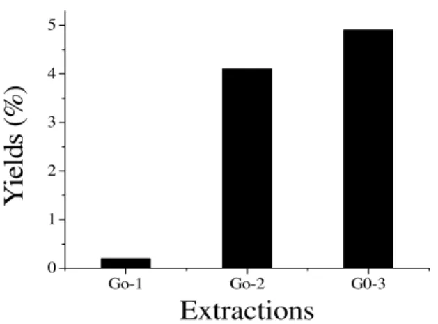
The total yield of SPs extracted from the red alga
G. ornata at room temperature (25°C) and
sequentially at 80°C (twice) was 9.2% (w/w%). Lower yields were obtained in Go-2 and Go-3 (4.1 and 4.9%, respectively), while the lowest was 0.2% (Go-1) (Fig. 1). This species presented a total SPs yield higher to that from the red seaweeds Gelidium crinale (2.6%) (Pereira et al.
2005), Botryocladia occidentalis (4%) (Farias et
al. 2000) and G. birdiae (6.5%) (Maciel et al.
2008), but lower than those of G. cornea (21.4%)
(Melo et al. 2002), G. bursa-pastoris (38.8%), G. dura (33.5%), G. gracilis (30.0%)
(Marinho-Soriano 2001), Halymenia pseudofloresia
(47.14%) (Rodrigues et al. 2009) and Halymenia
sp (53.96%) (Rodrigues et al. 2010), respectively. In a recent study, Amorim et al. (2011) performed three differential successive aqueous extractions from H. floresia and obtained that at high
temperatures (80°C), 34.6% SPs were extracted against 4% at 25°C.
Figure 1 - Yields of crude SPs extracts (Go-1, Go-2 and Go-3) isolated by sequential extractions in
water (25°C and 80°C) from the red seaweed Gracilaria ornata. Yields are express as
percentage weight of alga desihydrated weight.
The lowest yield in Go-1 (25°C) could be due to the presence of precursors or other non-gel promoting structural elements (Murano et al. 1997; S.F-Tischer et al. 2006), whereas in 2 and Go-3 (80°C) suggested the presence of floridean starch granules in G. ornata (Mazumder et al. 2002;
S.F-Tischer et al. 2006). According to Rodrigues et al. (2009, 2010), the employment of successive extractions could be a valuable strategy for the
identification of new SPs. However, Chotigeat et al. (2004) reported that accentuated variations in the yield of seaweeds SPs could also occur among different species. SPs yield from Gracilaria
species has been correlated with the environmental factors (Marinho-Soriano and Bourret 2003). From this point of view, G. ornata SPs deserved further
economical fields (Farias et al. 2000; Mazumder et al. 2002; Duarte et al. 2004; Pereira et al. 2005; S.F-Tischer et al. 2006; Araújo et al. 2008; Siqueira et al. 2011). The high cost of these polymers in the international market encourages to discover new natural sources of phycocolloids for biotechnology (Melo et al. 2002; Maciel et al. 2008; Campo et al. 2009; Silva et al. 2010).
Chemical analysis
The chemical composition varied among the crude SPs extracts obtained (Table 1). Extract Go-1 had the lowest FS content (5.88%) and highest CPs content (3.70%) compared to Go-2 and Go-3 extracts. These results reported were in accordance with others SPs from Gracilaria species
(Mazumder et al. 2002; Melo et al. 2002; Maciel et al. 2008), but the sulfate content of algae SPs could be variable among the different species (Pereira et al. 2005; S.F-Tischer et al. 2006; Zhang et al. 2010; Amorim et al. 2011). The CPs content of the extracts was very small, suggesting the presence of amino acids (Mazumder et al. 2002; Ghosh et al. 2004; Amorim et al. 2011).
Specific rotation
The SPs solutions of G. ornata crude extracts
(Go-1, Go-2 and Go-3) prepared at concentration of 0.2% in deionized water (25°C) showed SR raging from –19.0 to –56.5 (Tab. 1), suggesting that the SPs from G. ornata belonged to the L-series,
similar to reported for other natural SPs found in
Gracilaria species (Pomin and Mourão 2008) and
in the brown seaweed Padina tetrastomatica
(Karmakar et al. 2009).
Infrared spectroscopy
The IR spectrums of crude SPs extracts are shown in Fig. 2. This technique is considered as an useful information for partial characterization of seaweeds SPs (Melo et al. 2002; Maciel et al. 2008; Campo et al. 2009; Silva et al. 2010), such as in the determination of sulfate and 3,6-anhydrogalactose contents in SPs (Mazumder et al. 2002). The original bands at 845-850 cm-1 (C-O-S,
secondary axial sulfate) were extended and showed the presence of galactose-4-sulfate in the IR spectra of all the crude SPs extracts. Furthermore, the intensity of this signal was corroborated by the FS content (Table 1). This meant that the sulfate occurred at the position C-4 of D-galactose. The IR spectrums of SPs also showed absorption bands at 1250 cm-1, denoting the presence of sulfate ester (Mazumder et al. 2002; Melo et al. 2002; Ghosh et al. 2004; Chattopadhyay et al. 2007a; Karmakar et al. 2009; Silva et al. 2010).
Band absorbance provides information on the occurrence in the crude extracts of 3,6 anhydrogalactose at 930 cm-1 (Melo et al. 2002; Silva el al. 2010) and characteristics of agarocolloids at 1375, 1153, 1070, 890 and 761 cm-1 based on Melo et al. (2002) and Maciel et al. (2008). 1030 cm-1 corresponded to the glycosidic linkage stretch vibration of C-O-H (Ananthi et al. 2010). 3400-3423 (OH stretching) and 2922-2926 (CH stretching) cm-1 were also detected (data not shown). However, there were no absorption bands or shoulders detected at 820 and 805 cm-1, showing that 2-sulfate galactose, galactose-6-sulfate and galactose-6-sulfate on C-2 of 3,6-anhydrogalactose were not present. Pomin and Mourão (2008) reported that the chemical structure of SPs from red seaweeds could occur as agaran (L-series), carrageenan (D-series) or both (hybrids D-/L-series).
Therefore, the little differences found in this experiment could be determined by the technique employment. The SPs yield and chemical and IR analyses varied among the differential extractions, with low values recorded at 25°C (Go-1) (Maciel et al. 2008) when compared to those found at 80°C (Go-2 and Go-3) (Figs. 1 and 2, Table 1) (Amorim et al. 2011), suggesting the occurrence, at least, of two “populations” of cell-wall SPs (Ray and Lahaye 1995; Rodrigues et al. 2009; Graça et al. 2011; Siqueira et al. 2011), and that high temperature was an important parameter for G.
ornata SPs extraction (Amorim et al. 2011).
Table 1 - Chemical composition of crude SPs extracts from red seaweed Gracilaria ornata
Chemical composition (%)
SPs °C * [∝]D20° TSs a FS b CPs c
Go-1 25 -19.00 33.14 5.88 3.70
Go-2 80 -51.00 57.71 10.30 1.30
Go-3 80 -56.50 62.20 10.30 0.10
a – Dosage by Dubois et al.’ method using D-galactose as standard; b – Dosage by Dodgson and Price’ method using NaSO3 as standard; c –
Figure 2 – FT-IR spectrums of Go-1 (A), Go-2 (B) and Go-3 (C) crude SPs extracts from red
seaweed Gracilaria ornata.
These data were not in accordance with other studied Gracilaria species SPs. Mazumder et al.
(2002) isolated and investigated chemically several SPs present in G. corticata. The authors
observed that the SPs extracted with water presented the sulfated groups at positions C-4 of the 1,3-linked D-galactose units and C-6 of the 1,4-linked L-galactose residues. In contrast, when the SPs was extracted by alkaline treatment, there was absence of the band at 850 cm-1 (sulfate-4-galactose). The obtained SPs alkaline presented a signal more intense at 930 cm-1 (3,6-anhydrogalactose). In fact, it has been suggested that 6-sulfate-α-L-galactose may be chemically
converted to 3,6-anhydrogalactose after alkaline treatment.
In another study, the FT-IR spectra of crude SP and its obtained fractions from papain digestion from G. cornea was reported by Melo et al.
(2002). The results revealed the possible presence of sulfate-4-galactose and a shoulder close to 820 cm-1 suggestive of sulfate-6-galactose. In contrast, 2-sulfate galactose and sulfate on C-2 of 3,6,-anhydrogalactose were not identified. The results showed that the ratio between galactose and anhydrogalactose was quite far from the ideal agarose ratio, and this chemical aspect was attributed to the absence of gelation in aqueous solutions. According to Percival and McDowell (1967), possibly the use of crude enzyme
preparations could modify the structure of the residual polysaccharide.
Diverse others marine organisms are rich sources in SPs possessing highly complex and heterogeneous structures. Pereira et al. (2005) reported the occurrence of -4-α-Galp-(1-3)-β
-Galp1, but with a variable sulfation pattern, having
2,3-di-sulfated and 2-sulfated, for the red alga G. crinale. The presence of 2-sulfated, 3-linked α
-L-galactan, was also characteristics of some marine invertebrates (Pereira et al. 2002). More recently, Aquino et al. (2005) identified sulfated galactans from the marine angiosperms (Ruppia maritima, Halodule wrightii, and Halophila decipiens). The
authors concluded that those from the sea grass R.
maritima were constituted by a regular
tetrasaccharide repeating unit that had an intermediate structure when compared to those presents in marine invertebrates and red marine algae.
Sinha et al. (2010) extracted the polysacharides from Sargassum tenerrimum (Phaeophyta). The
FT-IR spectrum of a fraction also contained a band at 1420 (COO-) cm-1 characteristic of alginate, being the first report for the presence of guluronate in Sargassaceae.
The absence of 2-sulfate galactose, sulfate-6-galactose and sulfate on C-2 of 3,6-anhydrogalactose in the IR spectra (Fig. 2) in the present study suggested the hypothesis of relating 9 7 0
8 9 0
7 0 5 7 6 1
8 4 5 9 3 0 1 0 3 0
1 0 7 0
1 1 5 3
1 2 5 0 1 3 7 0
1 4 0 0 1 2 0 0 1 0 0 0 8 0 0 6 0 0
C o m p r i m e n t o d e O n d a ( c m- 1
) Wave number (cm-1)
B B A
the structural studies of SPs as supplement in morphology, anatomy and life history studies, as auxiliary tools in the elucidation of taxonomic position of these organisms (Usov 1998). It has been reported that different extraction methods could influence the extraction of these polymers (Mazumder et al. 2002; Melo et al. 2002), as observed in this study (Fig. 1). In this context, a more detailed investigation is needed.
The absence of an absorption band in the IR spectra for galactose-6-sulfate was also noted (Fig. 2). The presence of this signal in the algae IR spectra indicated that the sulfate was linked at position C-6 of galactose (Mazumder et al. 2002). In general, the Caulerpa SPs occurred with this
structural feature (Ghosh et al. 2004; Chattopadhyay et al. 2007b).
Microbiological assays
In recent years, the risks of the ingestion of contaminated food by human have been significantly increased. Among the major agents are bacteria (Gram-positive and -negative bacteria). Bacteria cause huge damage in several economic fields, including food industry, fish and shrimp farms, etc (Chotigeat et al. 2004; Carvalho et al. 2009). Although the antibiotics are widely used for bacterial control, their indiscriminate administration is considered a problem of public health (Harakeh et al. 2006; Carvalho et al. 2009). The hypothesis of a possible biological effect of Go-3, which presented the highest yield (Fig. 1) and low CPs content (Table 1), from G. ornata,
was similar to that reported by Graça et al. (2011), studying the effects of a crude SP extract (named Hf2s) from red seaweed Halymenia floresia on
gastrointestinal smooth muscle contractility (in vitro and in vivo). These authors, based on a
previous study performed by Amorim et al. (2011), noticed that this crude polysaccharide contained a highest yield and sulfate content, and low CPs content. In addition, low CPs content in
G. ornata (Go-3) could be important in the
solubility behavior of this crude polysaccharide (Ray and Lahaye 1995).
Based on these considerations, the effects of a crude SPs extract (Go-3) on the growth of seven bacteria (Bacillus subtilis, Staphylococcus aureus
(Gram-positive), Enterobacter aerogens,
Escherichia coli, Pseudomonas aeruginosa,
Salmonela choleraesuis and Salmonela typhi
(Gram-negative) was studied.
In this study, no antimicrobial effect was obtained by Go-3 through plate diffusion method, suggesting that the high viscosity of the G. ornata
SPs in the cultivate medium interfered (Cappuccino et al. 1986). Also, no growth of bacteria was observed in the presence of Go-3 in the mineral medium. This indicated that all the tested bacteria were not capable of utilizing the G. ornata SPs as carbon source.
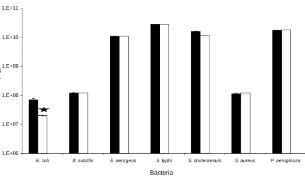
In order to still verify some bactericidal action of Go-3, the method using chemical agents in liquid medium was tested. However, as in the plate diffusion and mineral medium methods, Go-3 had also no effect. Surprisingly, when Go-3 was tested in another inhibition assay, G. ornata SPs
presented inhibitory effect against E. coli bacteria
(Gran-negative) (p<0.05) (Fig 3). No morphological alteration in the cultured cell in the presence of Go-3 was also observed.
There are a few reports about the antimicrobial effects of seaweeds SPs. According to some literature data, the extracts from the marine algae
Padina gymnospora, Dictyota dichotoma (Rao and
Parekh 1981), Dunaliella bardawill, Isochrysis
galbana (Fabregas et al. 1999), Sargassum
muticum (Hellio et al. 2001) and S. polycystum
(Chotigeat et al. 2004) possessed antimicrobial effects.
Chotigeat et al. (2004) evaluated S. polycystum
against three bacteria (E. coli, Staphylococcus aureus and Vibrio harveyi) and found that the S. polycystum extract had ability to inhibit the growth
of all these bacteria, which caused diseases in shrimp Litopenaeus vannamei.
The present study demonstrated that Go-3 only inhibited the growth of E. coli (Fig. 3). It showed
1,E+06 1,E+07 1,E+08 1,E+09 1,E+10 1,E+11
E. coli B. sub tilis E. aerogens S. typhi S. choleraesuis S. aureus P. aeruginosa
Bactérias
U
F
C
(
lo
g
)
Bacteria
C
F
U
(
lo
g
)
Figure 3 – Effect of the crude SPs extract (Go-3) from the red alga Gracilaria ornata on the
growth Gran-positive and -negative bacteria. Control group ; Go-3 . *P<0.05 statistically significant.
SPs occur in several marine organisms and comprise a group of structurally heterogeneous macromolecules with a diversity of biological activities. SPs from different algal species have demonstrated that each polysaccharide possesses a particular biological action, and as a consequence of the presence of sulfate radicals, these polymers deserve to be evaluated using different biological assays (Mourão and Pereira 1999).
The occurrence of -4-α-Galp-(1-3)-β-Galp1, but
with a variable sulfation pattern, having 2,3-di-sulfated and 2-2,3-di-sulfated, has been attributed as a structural requirement for the anticoagulant action of galactans from red algae (Pereira et al. 2005), and as a structure–anticoagulant relationship that also indicate 2-sulfated, 3-linked α-L-galactan,
being a potent thrombin inhibitor mediated by antithrombin or heparin cofactor II, for marine invertebrates (Pereira et al. 2002).
More recently, Fonseca et al. (2008) compared a SP isolated from B. occidentalis having
anticoagulant activity (90 IU mg-1) with that of G. crinale (65 IU mg-1) (Pereira et al. 2005)
(Rhodophyta) on specific coagulation assays and experimental models of thrombosis. The results indicated that slight differences in the proportion
and/or distribution of sulfated residues in these polysaccharides chain was critical for interaction between proteases, inhibitors and activators of the coagulation system, resulting therefore in a distinct pattern in anti- and procoagulant activities and in the antithrombotic action.
It is believed that the absence of antimicrobial activity of seaweeds SPs was related to charge the repulsion between the sulfated groups and cell-wall of bacteria. The inhibition of growth of the bacteria by the addition of G. ornata SPs (Go-3)
suggested the hypothesis of the presence of glycoprotein-receptors in the cell-surface capable of recognizing and binding to the charged compounds presents in the cell-surface of bacteria (Rostand and Esko 1997). Another hypothesis is the production of enzyme by the bacteria (heparin lyases, chondroitin lyases and chondroitinases) capable of removing SPs on the host cell-surface (Rostand and Esko 1997; Huang et al. 1999). Therefore, current data indicated that Go-3 from the red alga G. ornata exerted selectivity on the
growth of E. coli. The action mechanism of G. ornata SPs on the antimicrobial effect (E. coli),
ACKNOWLEDGMENTS
This work was funded by the grants from the National Scientific and Technological Development Council (CNPq), Northeast Biotechnology Network (RENORBIO), Coordination for the Improvement of Higher Education Personnel (CAPES), and Ceará State Scientific and Technological Development Foundation (FUNCAP). Paula, R.C.M.; Melo, V.M.M. and Benevides, N.M.B. are senior investigators of CNPq/Brazil.
REFERENCES
Amorim RCN, Rodrigues JAG, Holanda ML, Mourão PAS, Benevides NMB. Anticoagulant properties of a crude sulfated polysaccharide from the red marine alga Halymenia floresia (Clemente) C. Agardh. Acta
Scien Biol Scien. 2011; 33(3): 255-261.
Ananthi S, Raghavendran HRB, Sunil AG, Gayathri V, Ramakrishnan G, Vasanthi HR. In vitro antioxidant
and in vivo anti-inflammatory potential of crude
polysaccharide from Turbinaria ornata (Marine
Brown Alga). Food Chem Toxicol. 2010; 48(1):
187-192.
Aquino RS, Landeira-Fernandez AM, Valente AP, Andrade IR, Mourão PAS. Occurrence of sulfated galactans in marine angiosperms: evolutionary implications. Glycobiol. 2005; 15(1): 11-20.
Araújo GS, Farias WRL, Rodrigues JAG, Torres VM, Pontes GC. Administração oral dos polissacarídeos sulfatados da rodofícea Gracilaria caudata na
sobrevivência de pós-larvas de tilápia. Rev Ciên
Agron. 2008; 39(4): 548-554
Bezerra AF, Marinho-Soriano E. Cultivation of the red seaweed Gracilaria birdiae (Gracilariales,
Rhodophyta) in tropical waters of northeast Brazil.
Biom Bioen. 2010; 34(12): 1813-1817.
Bradford MM. A rapid and sensitive method for the quantification of microgram quantities of protein utilizing the principle of protein-dye binding. Anal
Biochem. 1976; 22(1-2): 248-254.
Campo VL, Kawano DF, Silva DB, Carvalho I. Carrageenans: Biological properties, chemical modifications and structural analysis – a review.
Carbohydr Polym. 2009; 77(2): 167–180.
Cappuccino JP. Agar plate sensitivity method. Microbial A Laboratory Manual. The Benjamin Cummings Publishing: California; 1986.
Carvalho FCT, Barreto NSSE, Reis CMF, Hofer E, Vieira RHSF. Susceptibilidade antimicrobiana de
Salmonella spp. isoladas de fazendas de
carciniculturas no Estado do Ceará. Rev Ciênc Agron.
2009; 40(4): 549-556.
Chattopadhyay K, Mandal P, Lerouge P, Driouich A, Ghosal P, Ray B. Sulphated polysaccharides from Indian samples of Enteromorpha compressa (Ulvales,
Chlorophyta): Isolation and structural features. Food
Chem. 2007a; 104(3): 928-935.
Chattopadhyay K, Adhikari U, Lerouge P, Ray B. Polysaccharides from Caulerpa racemosa:
Purification and structural features. Carbohydr
Polym. 2007b; 68(3): 407-415.
Chotigeat W, Tongsupa S, Supamataya K, Phongdara A. Effect of fucoidan on disease resistance of black shrimp. Aquacult. 2004; 233(1-4): 23-30.
Duarte MER, Cauduro JP, Noseda DG, Noseda MD, Gonçalves AG, Pujol CA, et al. The structure of the agaran sulfate from Acanthophora spicifera
(Rhodomelaceae, Ceramiales) and its antiviral activity. Relation between structure and antiviral activity in agarans. Carbohydr Res. 2004; 339(2):
335-347.
Dodgson KS, Price RG. A note on the determination of the ester sulfate content of sulfated polysaccharides.
Biochem J. 1962; 84(1): 106-110.
Dubois M, Gilles KA, Hamilton JK, Rebers PA, Smith F. Colorimetric method for determination of sugars and related substances. Anal Chem. 1956; 28(3):
350-356.
Farias WRL, Valente AP, Pereira MS, Mourão PAS. Structure and anticoagulant activity of galactans: isolation of a unique sulfated galactan from the red algae Botryocladia occidentalis and comparison of its
anticoagulant action with that of sulfated galactans from invertebrates. J Biol Chem. 2000; 275(38):
29299-29307.
Fernández LE, Valiente OG, Mainardi V, Bello JL. Isolation and characterization of an antitumor active agar-type polysaccharide of Gracilaria dominguensis.
Carbohydr Res. 1989; 190(1): 77-83.
Fonseca RJC, Oliveira SNMCG, Melo FR, Pereira MG, Benevides NMB, Mourão PAS. Slight differences in sulfatation of algal galactans account for differences in their anticoagulant and venous antithrombotic activities. Thromb Haemost. 2008; 99(3): 539-545.
Ghosh P, Adhikari U, Ghossal PK, Pujol CA, Carlucci MJ, Damonte EB, et al. In vitro anti-herpetic activity
of sulfated polysaccharide fractions from Caulerpa
racemosa. Phytochem. 2004; 65(23): 3151-3157.
Graça JRV, Bezerra MM, Lima V, Rodrigues JAG, Monteiro DLS, Quinderé ALG, et al. Effect of a crude sulfated polysaccharide from Halymenia
floresia (Rhodophyta) on gastrointestinal smooth
muscle contractility. Braz Arch Biol Technol. 2011;
54(5): 907-916.
Harakeh S, Yassine H, El-Fadel M. Antimicrobial resistant patters of Escherichia coli and Salmonella
strains in the aquatic Lebanese environments.
Hellio C, De La Broise D, Dufosse L, Le Gal Y, Bourgougnon N. Inhibition of marine bacteria by extracts of macroalgae: potential use for environmentally friendly antifouling paints. Mar
Environ Res. 2001; 52(3): 231-247.
Huang W, Matte A, Li Y, Kim YS, Linhardt JJ, Su H, et al. Crystal structure of chondroitinase B from
Flavobacterium heparinum and its complex with a
disaccharide product at 1.7 Å resolution. J Molec
Biol. 1999; 294(5): 1257-1269.
Karmakar P, Ghosh T, Sinha S, Saha S, Mandal P, Ghosal PK, et al. Polysaccharides from the brown seaweed Padina tetrastromatica: Characterization of
a sulfated fucan. Carbohydr Polym. 2009; 78(3):
416-421.
Leite E.L, Medeiros MGL, Rocha HAO, Farias GGM, Silva LF, Chanvante SF, et al. Structure and pharmacological activities of a sulfated xylofucoglucuronan from the alga Spatoglossum
schröederi. Plant Scien. 1998; 132(2): 215-228.
Leite YFM, Silva LMCM, Amorim RCN, Freire EA, Jorge DMM, Grangeiro TB, et al. Purification of a lectin from the marine red alga Gracilaria ornata and
its effect on the development of the cowpea weevil
Callosobruchus maculates (Coleoptera: Bruchidae).
Biochim Biophys Acta. 2005; 1724(1-2): 137-145.
Maciel JS, Chaves LS, Souza BWS, Teixeira DIA, Freitas ALP, Feitosa JAP, et al. Structural characterization of cold extracted fraction of soluble sulfated polysaccharides from red seaweed
Gracilaria birdiae. Carbohydr Res. 2008; 71(4):
559-565.
Marinho-Soriano B. Agar polysaccharides from
Gracilaria species (Rhodophyta), Gracilariaceae). J
Biotech. 2001; 89(1): 81-84.
Marinho-Soriano E, Bourret E. Effects of season on the yield and quality of agar from Gracilaria species
(Gracilariaceae, Rhodophyta). Biores Technol. 2003;
90(3): 329-333.
Marinho-Soriano, E, Bourret, E. Polysacchrides from the red seaweed Gracilaria dura (Gracilariales,
Rhodophyta). Biores Technol. 2005; 96(3): 379-382.
Mazumder S, Ghosal PK, Pujol CA, Carlucci M, Damonte EB, Ray B. Isolation, chemical investigation and antiviral activity of polysaccharides from Gracilaria corticata (Gracilariaceae,
Rhodophyta). Inter J Biol Macromol. 2002; 31(1-3):
87-95.
Mourão PAS. Use of sulfated fucans as anticoagulant and antithrombotic agents: future perspectives. Curr
Pharmac Des. 2004; 10(9): 967-981.
Mourão PAS, Pereira MS. Searching for alternatives to heparin: sulfated fucans from marine invertebrates.
Tren Cardiov Med. 1999; 9(8): 225-232.
Melo MRS, Feitosa JPA, Freitas ALP, Paula RCM. Isolation and characterization of soluble sulfated polysaccharide from the red seaweed Gracilaria
cornea. Carbohydr Polym. 2002; 49(4): 491-498.
Murano E, Toffanin R, Cecere E, Rizzo R. Investigation of the carrageenans extracted from
Solieria filiformis and Agardhiell subulata from Mar
Piccolo, Taranto. Mar Chem. 1997; 58(3-4): 319-325.
Nishino T, Aizu Y, Nagumo T. The influence of sulfated content and molecular weight of a fucan sulfate from the brown seaweed Ecklonia kurome.
Thromb Res. 1991; 64(6): 723-731.
Percival E, McDowell RH. Chemistry and enzymology of marine algal polysaccharides. Academic Press: New York; 1967.
Pereira MS, Vilela-Silva ACES, Valente AP, Mourão PAS. A 2-sulfated, 3-linked α-L-galactan is an anticoagulant polysaccharide. Carbohydr Res. 2002;
337(21-23): 2231-2238.
Pereira MG, Benevides NMB, Melo MRS, Valente AP, Melo FR, Mourão PAS. Structure and anticoagulant activity of a sulfated galactan from the red alga,
Gelidium crinale. Is there a specific structural
requirement for the anticoagulant action? Carbohydr Res. 2005; 340(12):2015-2023.
Pomin VH, Mourão PAS. Structure, biology, evolution, and medical importance of sulfated fucans and galactans. Glycobiol. 2008; 18(12): 1016-1027.
Qi H, Zhang Q, Zhao T, Chen R, Zhang H, Niu X, et al. Antioxidant activity of different sulfate content derivates of polysaccharide extracted from Ulva
pertusa (Chlorophyta) in vitro. Inter J Biol Macrom.
2005; 37(4):195-199.
Rao PS, Parekh KS. Antibacterial activity of Indian seaweed extracts. Bot Mar. 1981; 24(11): 577-582.
Ray B, Lahaye M. Cell-wall polysaccharides from the marine green alga Ulva “rigida” (Ulvales,
Chlorophyta). Extraction and chemical composition.
Carbohydr Res. (1995); 274:251-261.
Rodrigues JAG, Torres VM, Alencar DB, Sampaio AH, Farias WRL. Extração e atividade anticoagulante dos polissacarídeos sulfatados da alga marinha vermelha
Halymenia pseudofloresia. Rev Ciên Agron. 2009;
40(2):224-231.
Rodrigues JAG, Torres VM, Alencar DB, Sampaio AH, Farias WRL. Heparinoides naturais isolados de rodofíceas (Halymenia sp.) arribadas na costa
cearense. Acta Scien Biol Scien. 2010; 32(3):
235-242.
Rodrigues JAG, Araújo IWF, Paula GA, Lima TB, Bessa EF, Benevides NMB. Carragenana da epífita
Hypnea musciformis obtida do cultivo experimental
de Solieria filiformis em Flecheiras, Estado do Ceará,
Brasil. Acta Scien Technol. 2011; 33(2):137-144.
Rodrigues JAG, Vanderlei ESO, Quinderé ALG, Queiroz, INL, Bessa EF, Benevides NMB. Analysis of two drying methods on the yield and activity of sulfated polysaccharides extracted from Halymenia
sp. (Rhodophyta). Acta Scien Biol Scien. 2012; 34(1):
Rostand K, Esko JD. Microbial adherence to and invasion through proteoglycans. Infect Immun. 1997;
65(1):1-8.
Salles JP, Scherner F, Yoshimura Y, Fanganiello M, Bouzon ZL, Horta PA. Cultivation of native seaweed
Gracilaria domingensis (Rhodophyta) in Southern
Brazil. Braz Arch Biol Technol. 2010; 53(3):
633-640.
Silva FRF, Dore CMPG, Marques CT, Nascimento MS, Benevides NMB, Rocha HAO, et al. Anticoagulant activity, paw edema and pleurisy induced carrageenan: Action of major types of commercial carrageenans. Carboh Polym. 2010; 79(1):26-33.
Sinha S, Astani A, Ghosh T, Schnitzler P, Ray B. Polysaccharides from Sargassum tenerrimum:
Structural features, chemical modification and anti-viral activity. Phytochem. 2010; 71(2-3):235-242.
Siqueira RCL, Silva MSJ, Alencar DB, Pires AF, Alencar NMN, Pereira MG, et al. In vivo
anti-inflammatory effect of a sulfated polysaccharide isolated from the marine algae Lobophora variegata.
Pharm Biol. 2011; 49(2): 167-174.
Soares JB, Casimiro AR, Aguiar LMBA. Microbiologia. Edições UFC: Fortaleza; 1987.
S.F-Tischer PC, Talarico LB, Noseda MD, Guimarães SMPB, Damonte EB, Duarte MER. Chemical structure and antiviral activity of carrageenans from
Meristiella gelidium against herpes simplex and
dengue virus. Carbohydr Polym. 2006; 63(4):
459-465.
Usov AI. Structural analysis of red seaweed galactans of agar and carragenan group. Food Hydrocoll. 1998;
12(3): 301-308.
Zhang HJ, Mao WJ, Fang F, Li HY, Sun HH, Chen Y, et al. Chemical characteristics and anticoagulant activities of a sulfated polysaccharide and its fragments from Monostroma latissimum, Carbohydr
Polym. 2008; 71(3):428-434.
Zhou G, Sun Y, Xin H, Zhang Y, Li Z, Xu Z. In vivo
antitumor and immunomodulation activities of different molecular weight lambda-carrageenans from
Chondrus ocellatus. Pharm Res. 2004; 50(1): 47–53.
Zhou Z, Wang F, Wang X, Liu X, Hou Y, Zhang Q. Extraction of the polysaccharides from five algae and their potential antioxidant activity in vitro. Carbohydr
Polym. 2010; 82(1):118-121.