Printed version ISSN 0001-3765 / Online version ISSN 1678-2690 http://dx.doi.org/10.1590/0001-3765201720160407
www.scielo.br/aabc
Bone turnover markers in sheep and goat: A review of the scientific literature
JOSÉ A. CAMASSA1, CAMILA C. DIOGO1, CRISTINA P. SOUSA2, JORGE T. AZEVEDO3,4, CARLOS A. VIEGAS1,5
, RUI L. REIS6,7
, NUNO DOURADO8
and ISABEL R. DIAS1,5
1
Department of Veterinary Sciences, Agricultural and Veterinary Sciences School/ ECAV, University of Trás-os-Montes and Alto Douro/ UTAD, Quinta de Prados, 5000-801 Vila Real, Portugal
2
Center Hospitalar of Porto, Largo Prof. Abel Salazar, 4099-001 Porto, Portugal 3
Department of Animal Sciences/ ECAV, UTAD, Quinta de Prados, 5000-801 Vila Real, Portugal 4
Centre for Animal Sciences and Veterinary Studies/ CECAV, UTAD, Quinta de Prados, 5000-801 Vila Real, Portugal 5
Centre for the Research and Technology of Agro-Environmental and Biological Sciences/ CITAB, UTAD, Quinta de Prados, 5000-801 Vila Real, Portugal
6
3B’s Research Group - Biomaterials, Biodegradables and Biomimetics, Department of Polymer Engineering, University of Minho, Avepark - Parque de Ciência e Tecnologia, Zona Industrial da Gandra, 4805-017 Barco GMR, Portugal
7
Life and Health Sciences Research Institute/ ICVS, School of Health Sciences, University of Minho Campus de Gualtar 4710-057 Braga, Portugal 8
Department of Mechanical Engineering, University of Minho, Azurém Campus, 4804-533 Guimarães, Portugal
Manuscript received on June 25, 2016; accepted for publication on October 11, 2016
ABSTRACT
Bone turnover markers (BTMs) are product of bone cell activity and are generally divided in bone formation
and bone resorption markers. The purpose of this review was to structure the available information on the use
of BTMs in studies on small ruminants, especially for monitoring their variations related to diet, exercise,
gestation and metabolic lactation state, circadian and seasonal variations, and also during skeletal growth.
Pre-clinical and translational studies using BTMs with sheep and goats as animal models in orthopaedic
research studies to help in the evaluation of the fracture healing process and osteoporosis research are
also described in this review. The available information from the reviewed studies was systematically
organized in order to highlight the most promising BTMs in small ruminant research, as well as provide
a wide view of the use of sheep and goat as animal models in orthopaedic research, type of markers and
commercial assay kits with cross-reactivity in sheep and goat, method of sample and storage of serum and
urine for bone turnover markers determination and the usefulness and limitations of bone turnover markers
in the different studies, therefore an effective tool for researchers that seek answers to different questions
while using BTMs in small ruminants.
Key words: Bone formation markers, bone resorption markers, bone metabolism, small ruminants.
Correspondence to: Isabel R. Dias E-mail: [email protected]
INTRODUCTION
In the last decades, small ruminants - sheep and
goats - have been widely accepted as animal
The suitability of small ruminants as animal
models for orthopaedic research results mainly
from having the most similar body weight and
long bones with dimensions compatible with
application of implants and prostheses developed
for humans (Newman et al. 1995, Anderson et al.
1999, van der Donk et al. 2001). In this manner,
compared with other species used in orthopaedic
research, sheep and goats have an adequate body
weight and long bones, with a macrostructure more
similar to humans (Newman et al. 1995), despite
the bone microstructure of small ruminants being
less similar to humans than other animal models
such as dogs (Pearce et al. 2007). Sheep have a
predominance of plexiform bone until 3 to 4 years
of age (Newman et al. 1995) due to fast growth in
weight and size (Reinwald and Burr 2011)
and just
a predominance of secondary Haversian systems
after 7 to 9 years of age with the presence of bone
remodelling (Newman et al. 1995). Sheep also
presents a trabecular bone density, mineralization
and subsequently elevated strength relative to
humans, that are variable according to skeletal
location (Nafei et al. 2000, Liebschner 2004),
nevertheless the bone mineral composition being
apparently similar between small ruminants and
humans (Ravaglioli et al. 1996).
Despite these macro- and micro-structural
differences in bone tissue, studies with small
ruminants used as animal models in orthopaedic
research have increased considerably (Pearce et
al. 2007), and more recently they have also been
used for studying bone turnover markers (BTMs)
(Sousa et al. 2014a). The BTMs are proteins which
indicate bone metabolism (Sousa et al. 2014b),
and are generally divided into collagenous bone
formation markers, bone resorption markers and
osteoclast regulatory protein markers (Leeming
et al. 2006). Analysis of BTMs might supply
information in a fast, effective, sensitive, specific,
and low cost manner (Allen 2003). Nowadays, it is
used in human medicine to help evaluate fracture
risk, delayed fracture healing and consolidation
process, and development of metabolic bone
diseases (Vasikaran et al. 2011).
These similarities in biochemistry,
biomechan-ics, and bone histology make BTMs a resource in
sheep and goats for pre-clinical and/or translational
orthopaedic research studies and veterinary and
animal science studies (Turner 2007b).
Neverthe-less, the reported biological variability of BTMs
among age, gender, disease, recent fractures,
ex-ercise, time (Seibel 2005), diet (Nicodemo et al.
1999, Liesegang and Risteli 2005, Liesegang et al.
2013), seasonal changes (Arens et al. 2007) and
circadian variation (Liesegang et al. 2003), which
can contribute substantially to the variability of
these parameters (Smith et al. 2011), are their main
limitation (Cremers et al. 2008).
Therefore, the aim of this review was to collect
the studies published in scientific literature until
the present date concerning the use of BTMs in
small ruminant research or to investigate the
clini-cal effectiveness of BTMs in pre-cliniclini-cal or transla
-tional experimental orthopaedic research related to
human medicine when sheep and goat are used as
experimental animal models for this latter purpose.
BONE TURNOVER MARKERS
Bone tissue undergoes turnover along the animal
lifespan (Seibel 2006) and that process is divided
into two parts: modelling and remodelling (Clarke
2008).
time and location (Raggatt and Partridge 2010).
Remodelling is a process of bone replacement
where bone formation outpaces bone resorption
(Altman et al. 2015), to maintain bone strength and
mineral homeostasis, regulated by osteoclasts and
osteoblasts that sequentially carry out resorption of
old bone and formation of new bone, keeping the
new bone healthy (Clarke 2008). Bone remodelling
predominates when bone is reaching maturity
(Iglesias et al. 2011), but it does not influence the
size and shape, although the internal architecture
may have slight changes caused by external
forces (Hadjidakis and Androulakis 2006). Bone
formation and resorption are present in same site,
but not at the same time in order to maintain bone
mass (Raggatt and Partridge 2010).
The proteins produced during bone turnover
are detectable mainly in serum in bone formation
markers, whereas many of the bone resorption
markers are detectable in both serum and urine
(Allen 2003), and there are a significant number of
commercial kits developed for use in humans that
have cross-reactivity with other species, including
sheep and goats (Tables I to III).
TABLE I
Bone formation markers, method of analysis and available commercial assay kits.
Marker Tissue of origin Sample Method of analysis Available commercial Assay kit Cross-Reactivity Sheep / Goat
BALP Bone Serum Colorimetric No commercial kit available NO / NO
Electrophoretic No commercial kit available NO / NO Precipitation No commercial kit available NO / NO CLA LIAISON BAP Ostase, Stillwater, MN, USA ? / ?
ELISA MicroVue BAP, Quidel Corporation, San
Diego, CA, USA YES / YES
RIA Tandem-R-Ostase, Beckman Coulter, Brea,
CA, USA YES / ?
OC Bone Serum CLA LIAISON Osteocalcin, Stillwater, MN, USA ? / ?
RIA BTI Human Osteocalcin RIA, Biomedical
Technologies Inc, Stoughton, MA, USA ? / ? ELISA MicroVue Osteocalcin, Quidel Corporation,
San Diego, CA, USA YES / YES
BTI Intact Osteocalcin, Biomedical
Technologies Inc, Stoughton, MA, USA ? / ? Osteocalcin, SIGMA, Saint Louis, Missouri,
USA ? / ?
Osteocalcin, GenWay Biotech, San Diego,
CA, USA NO / NO
PINP Bone, soft tissue Serum CLA PINP Roche Diagnostics, Penzberg, Germany ? / ?
RIA UniQ Intact PINP, Orion Corporation, Espoo,
Finland ? / ?
Serum or urine ELISA PINP, Neobiolab Inc, Cambridge MA, UK YES / YES PICP Bone, soft tissue Serum RIA PICP, Orion Corporation, Espoo, Finland ? / ?
PICP DiaSorin, Stillwater, MN, USA YES / ?
ELISA MicroVue CICP, Quidel Corporation, San
Diego, CA, USA YES / ?
Serum or urine PICP, Neobiolab Inc, Cambridge MA, UK YES / YES
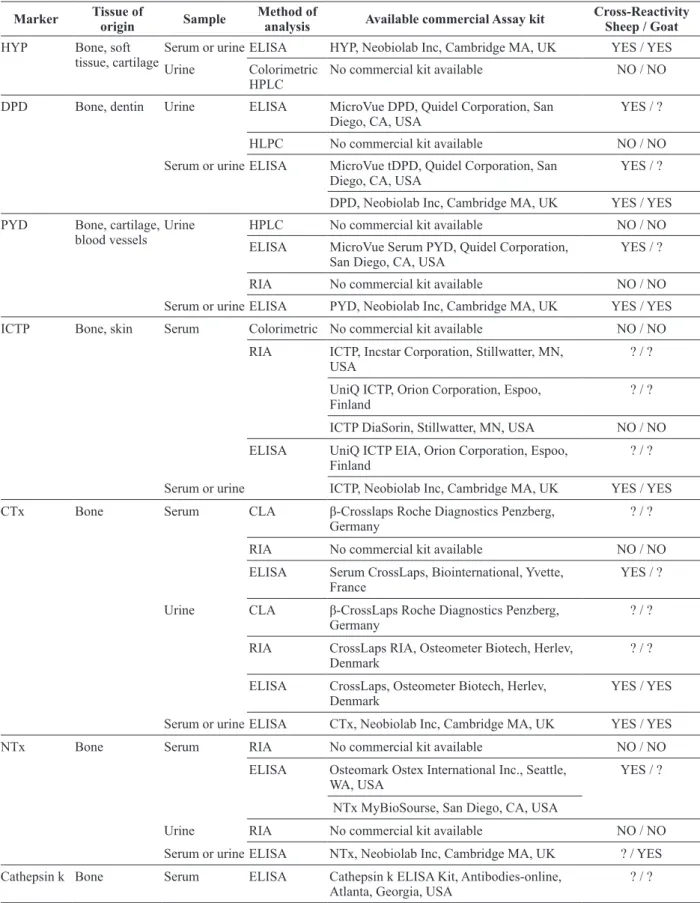
TABLE II
Bone resorption markers, method of analysis and available commercial assay kits.
Marker Tissue of
origin Sample
Method of
analysis Available commercial Assay kit Cross-Reactivity Sheep / Goat HYP Bone, soft
tissue, cartilage
Serum or urine ELISA HYP, Neobiolab Inc, Cambridge MA, UK YES / YES
Urine Colorimetric HPLC
No commercial kit available NO / NO
DPD Bone, dentin Urine ELISA MicroVue DPD, Quidel Corporation, San Diego, CA, USA
YES / ?
HLPC No commercial kit available NO / NO
Serum or urine ELISA MicroVue tDPD, Quidel Corporation, San Diego, CA, USA
YES / ?
DPD, Neobiolab Inc, Cambridge MA, UK YES / YES
PYD Bone, cartilage, blood vessels
Urine HPLC No commercial kit available NO / NO
ELISA MicroVue Serum PYD, Quidel Corporation, San Diego, CA, USA
YES / ?
RIA No commercial kit available NO / NO
Serum or urine ELISA PYD, Neobiolab Inc, Cambridge MA, UK YES / YES
ICTP Bone, skin Serum Colorimetric No commercial kit available NO / NO
RIA ICTP, Incstar Corporation, Stillwatter, MN, USA
? / ?
UniQ ICTP, Orion Corporation, Espoo, Finland
? / ?
ICTP DiaSorin, Stillwatter, MN, USA NO / NO
ELISA UniQ ICTP EIA, Orion Corporation, Espoo, Finland
? / ?
Serum or urine ICTP, Neobiolab Inc, Cambridge MA, UK YES / YES
CTx Bone Serum CLA β-Crosslaps Roche Diagnostics Penzberg,
Germany
? / ?
RIA No commercial kit available NO / NO
ELISA Serum CrossLaps, Biointernational, Yvette, France
YES / ?
Urine CLA β-CrossLaps Roche Diagnostics Penzberg, Germany
? / ?
RIA CrossLaps RIA, Osteometer Biotech, Herlev, Denmark
? / ?
ELISA CrossLaps, Osteometer Biotech, Herlev, Denmark
YES / YES
Serum or urine ELISA CTx, Neobiolab Inc, Cambridge MA, UK YES / YES
NTx Bone Serum RIA No commercial kit available NO / NO
ELISA Osteomark Ostex International Inc., Seattle, WA, USA
YES / ?
NTx MyBioSourse, San Diego, CA, USA
Urine RIA No commercial kit available NO / NO
Serum or urine ELISA NTx, Neobiolab Inc, Cambridge MA, UK ? / YES
Cathepsin k Bone Serum ELISA Cathepsin k ELISA Kit, Antibodies-online, Atlanta, Georgia, USA
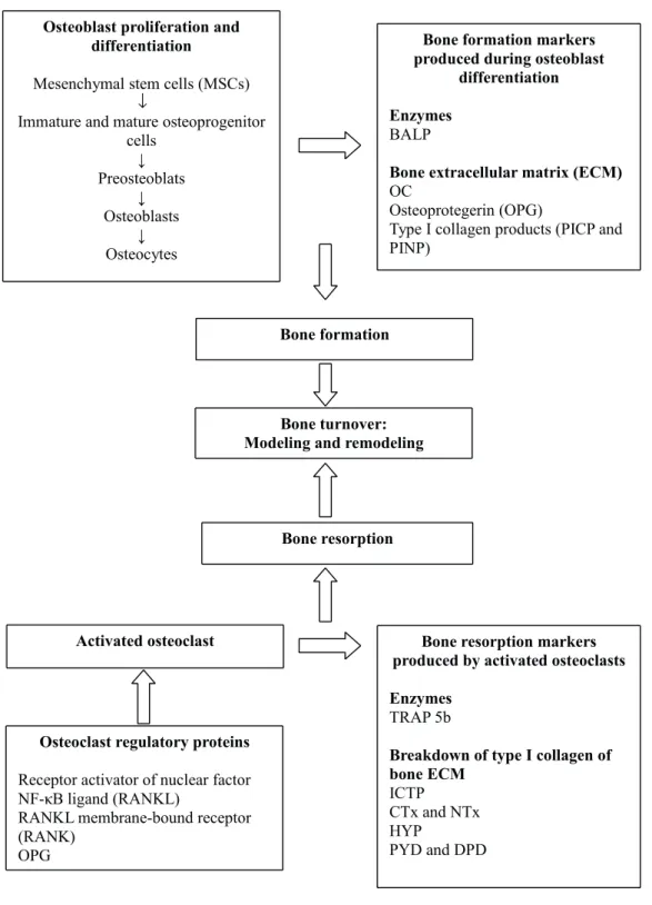
During the process of bone formation by
osteo-blasts, formation markers are represented by serum
total (ALP) and the bone-specific isoform of alka
-line phosphatase (BALP), serum osteocalcin (OC)
and two molecules which are
released during the
type I collagen molecule synthesis
–
serum
procol-lagen type I carboxy- and amino-terminal
propep-tides (PICP and PINP, respectively) (Seibel 2002).
In the bone resorption process there is a breakdown
of type I collagen, so resorption markers are
rep-resented by serum C-terminal telopeptide of type
I collagen (serum ICTP), urinary collagen type I
cross-linked C- and N-telopeptide (CTx and NTx),
urinary hydroxyproline (HYP), total and free
uri-nary pyridinoline and deoxypyridinoline (PYD
and DPD) and also by serum tartrate-resistant acid
phosphatase (TRAP) as an enzyme produced by
osteoclasts during their bone resorption activity
(Seibel 2002) (Figure 1).
BONE FORMATION MARKERS
ALKALINE PHOSPHATASE
Alkaline phosphatase (ALP) is a glycoprotein that
is connected to the extracellular surface of cells
and is synthesized in a variety of tissues, such as
intestines, placenta, and germ cells (Millan 2006).
Animals have four isoforms of ALP –
bone-specific
Marker Tissue oforigin Sample
Method of
analysis Available commercial Assay kit Cross-Reactivity Sheep / Goat
TRAP Bone Serum RIA No commercial kit available NO / NO
ELISA MicroVue TRAP 5b, Quidel Corporation, San Diego, CA, USA
YES / YES
Osteolink-TRAP b, Nitto Boseki Corporation, Tokio, Japan
? / ?
Bone TRAP SBA, Science, Boldon, UK ? / ? Serum or urine TRAP, Neobiolab Inc, Cambridge MA, UK YES / YES
HYP: Hydroxyproline; DPD: Deoxypyridinoline; PYD: Pyridinoline; ICTP: Carboxy-terminal telopeptide of type I collagen; CTx: cross-linked C-terminal telopeptides of type I collagen; NTx: cross- linked N-terminal telopeptides of type I collagen; TRAP 5b: Tartrate-resistant acid phosphatase isoenzyme 5b; RIA: Radioimmunoassay; ELISA: Enzyme-linked immunosorbant assay; CLA: Chemiluminescence immunoassay; HPLC: High-performance liquid chromatography.
TABLE II (continuation)
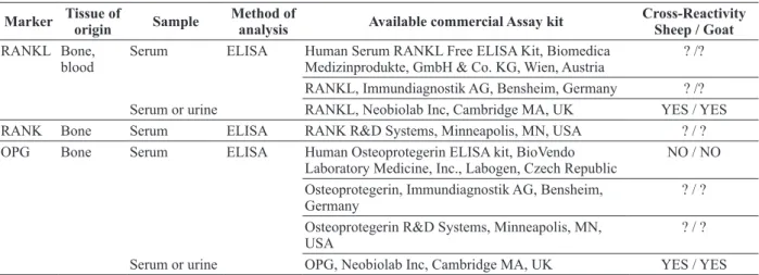
TABLE III
Osteoclast regulatory proteins, method of analysis and available commercial assay kits.
Marker Tissue of
origin Sample
Method of
analysis Available commercial Assay kit Cross-Reactivity Sheep / Goat RANKL Bone,
blood
Serum ELISA Human Serum RANKL Free ELISA Kit, Biomedica Medizinprodukte, GmbH & Co. KG, Wien, Austria
? /?
RANKL, Immundiagnostik AG, Bensheim, Germany ? /? Serum or urine RANKL, Neobiolab Inc, Cambridge MA, UK YES / YES RANK Bone Serum ELISA RANK R&D Systems, Minneapolis, MN, USA ? / ? OPG Bone Serum ELISA Human Osteoprotegerin ELISA kit, BioVendo
Laboratory Medicine, Inc., Labogen, Czech Republic
NO / NO
Osteoprotegerin, Immundiagnostik AG, Bensheim, Germany
? / ?
Osteoprotegerin R&D Systems, Minneapolis, MN, USA
? / ?
Serum or urine OPG, Neobiolab Inc, Cambridge MA, UK YES / YES
ALP (BALP), intestinal ALP, liver ALP, and in
dogs also the corticosteroid-induced ALP. This
variation would render difficult the interpretation
of possible variations of the ALP isoenzymes
(Allen 2003). Bone ALP has been used due to its
high sensitivity as bone formation marker (Seibel
2006). It is produced by osteoblasts (Millan
2006) and is involved in the calcification of bone
↓↓
↓
↓
κ
matrix (Masrour and Mahjoub 2012) through the
hydrolysis of phosphate esters on the osteoblast cell
surface, resulting in a high extracellular inorganic
phosphate concentration (Whyte 1994).
OSTEOCALCIN
Osteocalcin (OC) is synthesized by mature
osteo-blasts, odontoosteo-blasts, and hypertrophic
chondro-cytes and it is vitamin K dependent protein. It has
three residues of the calcium-binding amino acid, γ
- carboxyglutamic acid (Gla). Its function is poorly
understood, although it is primarily deposited in
the bone extracellular matrix (ECM), with a small
amount present in the blood stream (Cremers et
al. 2008). Serum OC is a marker of osteoblastic
activity and its serum level thus reflects the rate of
bone formation (Seebeck et al. 2005), influences
bone mineralization by binding calcium and
conse-quently hydroxyapatite (Neve et al. 2013).
PRO-COLLAGEN TYPE I PROPEPTIDES
Collagen type I is produced by osteoblasts in the
last stage of new bone formation (Allen 2003).
The procollagen undergoes enzymatic cleavage
producing the C- and N-terminal procollagen type
I extension peptides (PICP and PINP, respectively),
both extension are cleared by the liver and may be
added to the bone ECM (Watts 1999). Nevertheless,
type I collagen does not depend exclusively on the
bone tissue turnover because it is also a component
of other soft tissues as fibro-cartilage, tendon,
skin, gum, intestine, heart valve, large vessels,
and muscle. However, as the metabolism of type
I collagen is faster in the bone tissue than in other
tissues, changes in type I collagen are considered
representative of bone collagen synthesis (Cremers
et al. 2008). It is suggested that PINP is useful
in early detection of non union processes with
potential for study of the fracture healing process
(Coulibaly et al. 2010), although in humans it is
unknown whether there exists a correspondence
between PINP and the progression of fracture
healing (Moghaddam et al. 2011).
BONE RESORPTION MARKERS
DEOXYPYRIDINOLINE AND PYRIDINOLINE
The collagen fibrils recently deposited in bone
ECM are stabilized by intra- and intermolecular
cross links helping to build the mature collagen
molecule (Cepelak and Cvorišcec 2009).
The pyridinium cross links – deoxypyridinoline
(DPD) and pyridinoline (PYD) are formed during
extracellular maturation of fibrillar collagens
(Gerrits et al. 1995). The PYD is found in bone
and cartilage tissues and ligaments (Watts 1999)
while DPD is found in bone and dentin (Delmas et
al. 2000), so in the bloodstream PYD is generally
more abundant (Cremers et al. 2008), although DPD
is more specific as a resorption marker for bone
tissue (Seibel et al. 1992). In a study with sheep
after ovariectomy, this animal model demonstrated
relevance as a model for osteoporosis due to the
values of PYD and OC found (Newton et al. 2004).
CARBOXY-TERMINAL TELOPEPTIDE OF COLLAGEN TYPE I AND AMINO-TERMINAL TELOPEPTIDE OF COLLAGEN TYPE I
The N-terminal (NTx) and C-terminal telopeptide
of collagen type I (CTx) are fragments of the type
I collagen molecule composed by a short peptide
sequence from the non-helical domain of this
molecule (Chubb 2012), attached by a pyridinium
crosslink (Allen et al. 2000). Both markers are
sensitive and reliable indicators of the bone
resorption process (Cremers et al. 2008) and final
products of the metabolism of bone ECM, amino
acids, and free or peptide-bound PYD or DPD
(Allen et al. 2000).
The CTx is not specific as a resorption marker
other types of collagen (Chubb 2012). However,
CTx could be used for monitoring the bone healing
process because it was detected that variations in
its levels corresponded to bone resorption in an
experimental fracture healing study performed in
dogs where two different osteosynthesis techniques
were used (Paskalev and Krastev 2010).
CARBOXY-TERMINAL TELOPEPTIDE OF TYPE I COLLAGEN - MATRIX METALLOPROTEINASE
Cleavage of the type I collagen molecule by the
matrix metalloproteinases (MMP) results in the
formation of cross-linked C-terminal telopeptide of
type I collagen (CTX-MMP or ICTP) (Cremers et
al. 2008), suitable to represent osteoclastic activity
(Allen et al. 2000).
The ICTP is an indicator for mobilization of
bone tissue around parturition and at the beginning
of lactation in sheep and goats (Liesegang et al.
2007). In dogs with osteosarcoma (Hintermeister
et al. 2008)
and horses during physical training,
this marker has not revealed itself suitable for
determining bone resorption since it did not show
correlation with other resorption markers, however
it was an indicator of the rate of bone turnover
(Price et al. 1995).
TARTRATE-RESISTANT ACID PHOSPHATASE
Tartrate-resistant acid phosphatase (TRAP) is a
bone resorption marker, but not originated from
the degradation of type I collagen (Hannon et al.
2004). It is a glycoprotein produced by osteoclasts,
activated macrophages, and dendritic cells
(Leeming et al. 2006). There is an isoenzyme 5,
from a total of 6 isoenzymes of the acid phosphate
identified by electrophoresis, which through
protease cleavage presents two isoforms (a, b) –
the TRAP 5a is sialylated and TRAP 5b is produced
by osteoclasts, and the latter proposed to reflect
osteoclast activity (Delmas et al. 2000, Leeming et
al. 2006). The TRAP could be a suitable resorption
marker for detection of normal or delayed fracture
healing process in sheep (Seebeck et al. 2005)
or
dogs (Sousa et al. 2011).
CATHEPSIN K
Cathepsin K is part of the cysteine protease family
and has the ability to cleave both helical and
telopeptide regions of collagen type I (Leeming et al.
2006). This enzymatic cleavage is able to degrade,
at low pH, several proteins of the bone ECM,
namely the telopeptide and helical regions of the
collagen type I molecule, the OC and osteopontin
(Cremers et al. 2008). This marker could be used as
a tool to measure bone resorption, such as in canine
osteosarcoma clinical cases (Schmit et al. 2012).
VARIABILITY OF BONE TURNOVER MARKERS
The BTMs could suffer constant variation through
-out the lifetime of an individual (Sousa et al.
2014b). However, variation between individuals is
also a great cause of oscillation in markers, specifi
-cally due to biological variability, together with the
analytical variability introduced by the different as
-say techniques (Vasikaran et al. 2011).
Biological variability can be influenced by
many uncontrollable factors (Cremers et al. 2008),
such as growth (Sousa et al. 2014a), geographical
location (Liesegang et al. 2013), pregnancy and
lactation (Liesegang et al. 2006, Liesegang et
al. 2007), and controllable factors, such as diet
(MacLeay et al. 2004a, b, Liesegang et al. 2013),
and season of the year (Arens et al. 2007), which
can be mitigated in clinical studies (Liesegang
2008). In short, biological variability is affected
by any factor that influences the bone remodelling
(Watts 1999).
The high inter-individual variability of BTMs
is their main limitation for clinical use due to the
difficulty to establish reference ranges for serum
and urinary BTM levels (Souberbielle et al. 1999),
although bone markers are an effective tool in
clinical studies due to reliable, fast, non-invasive,
and cost effective assays with improved sensitivity
and specificity (Wheater et al. 2013).
SAMPLE AND STORAGE
Blood collection for measuring BTMs must be done
at a specific time (morning) to avoid the influence
of circadian variations (Klein et al. 2004, Seebeck
et al. 2005, Dias et al. 2008, Sousa et al. 2014a, b).
Blood samples can be collected from the cephalic
vein (Klein et al. 2004) or jugular vein (Dias et al.
2008, Sousa et al. 2014a, b) into serological tubes
containing no anticoagulant (Vernon et al. 2010),
and centrifuged (3000 rpm for 10 min) within 30
min of collection (Liesegang et al. 2007). Urine
can be obtained using a special external urine
collector (Windhagen et al. 2002) or collected by
cystocentesis (Allen et al. 2000). Urine and serum
samples should be stored at -20°C for mineral
analyses (Chanetsa et al. 2000, Taylor et al. 2009,
Sousa et al. 2014a) and at -80°C until determination
of BTMs (Seebeck et al. 2005, Tatara 2008, Sousa
et al. 2014b), which provides molecular stability
for several months (Lomeo and Bolner 2000).
ANIMAL AND VETERINARY SCIENCE STUDIES
Characteristics of the animal and veterinary science
studies regarding population, type of studies,
time, and conclusion (Table SIV – Supplementary
Material).
DIET
According to Liesegang and Risteli (2005)
Liesegang et al. (2013), MacLeay et al. (2004a, b)
and Nicodemo et al. (1999) nutritional studies using
BTMs were influenced by different diets, though
this influence was not statistically significant.
MacLeay et al. (2004a) concluded that during the
administration of a diet that induced metabolic
acidosis in mature ewes, there were no significant
changes in serum BALP and DPD levels. In another
study by Liesegang et al. (2013) with sheep grazing
at different altitudes, it was not possible to confirm
the interference of diet in the serum variation
of ICTP or BALP, but high bone turnover was
confirmed. Also, in a study by Liesegang and Risteli
(2005) where a diet with varying calcium content
was used, it was not possible to demonstrate the
influence of the diet on bone mineral metabolism
in growing goats and sheep, possibly due to the
short duration of this study, where only the sheep
showed a variation in BMD due to an increase in
calcium intake. However, Wilkens et al. (2010)
demonstrated that sheep were a suitable model for
studies with varying diets, calcium deficiency, and
calcitrol.
EXERCISE
Liesegang and Risteli (2005) demonstrated that
sheep in pasture at high altitudes had an increase
in bone turnover and bone mineral content without
clear cause, one possible factor being the increase
in exercise. In another study in lambs, Vernon et al.
(2010) concluded that the markers used were not
adequate to indicate the effects of forced exercise.
GESTATION AND LACTATION
Liesegang et al. (2006) noticed that the interval
between parturition and early lactation in sheep and
goats required a high nutritional value of calcium
due to losses to the fetus and lactation, occurring
inefficiency of calcium absorption, leading to
increases in bone remodelling to help replace
maternal bone loss classified as a physiological
mechanism. During a second pregnancy, bone
loss was less significant compared with the first
to the adaptation of the organism (Liesegang et al.
2007). Finally, it was concluded that sheep were
more adapted to the loss of calcium in comparison
to goats, that had a lower bone mineral density and
bone mineral content before parturition (Liesegang
and Risteli 2005) increased bone turnover, resulting
in a higher activity of bone metabolism and
sensitivity to changes in calcium during pregnancy
and lactation (Liesegang et al. 2003).
CIRCADIAN AND SEASONAL VARIATION
Chavassieux et al. (1991) reported that bone
remodelling was influenced by the photoperiod,
with decrease in bone remodelling occurring
between spring and summer. Arens et al. (2007)
confirmed that bone mass increases in summer and
decreases in winter, so taking seasonal variation
into account is fundamental in studies using BTMs.
Liesegang et al. (2003) reported an increase in the
rate of bone formation during the evening and night,
indicating the influence of the circadian rhythm in
bone turnover. Sousa et al. (2014b) concluded that
the short-term variability should be considered
during interpretation of data, such as circadian and
seasonal variations, nevertheless the short-term
biological variability do not represent a limitation
for the use of BTMs.
SKELETAL GROWING
Pastoureau et al. (1991) mention that sheep are a
good model to study the bone growth in growing
lamb. It was reported that goats showed a more
accelerated bone remodelling that sheep, which
was demonstrated by ICTP, CTx (Liesegang et al.
2003), BALP (Liesegang et al. 2003, Sousa et al.
2014a), and OC determinations in various studies
(Pastoureau et al. 1991, Liesegang et al. 2003).
Collignon et al. (1996) demonstrated that bone
growth since the fetal stage produces alterations in
serum OC and BALP, confirming the usefulness of
these markers in bone formation and growth. Scott
et al. (1997) reported that OC, BALP, DPD, and
PYD may be useful for detection of changes in bone
growth caused by deficient diets, and Wan Zahari
et al. (1994) reported that high phosphorus diets
resulted in increased bone resorption (increased
TRAP) in lambs. However, Chanetsa et al. (2000)
exposed castrated lambs to an oestrogen agonist. In
this study, bone growth was observed, but no effect
on markers of bone remodelling was noticed.
PRE-CLINICAL AND TRANSLATIONAL ORTHOPAEDIC RESEARCH STUDIES
The characteristics of the pre-clinical and
transla-tional orthopaedic research studies, such as
popula-tion, type of studies, time, and conclusion
are listed
in Table SIV.
FRACTURE HEALING PROCESS
Tralman et al. (2013) and Windhagen et al. (2002)
reported that the serum markers of bone formation
are useful for reflecting the bone healing process,
and Goebel et al. (2009) suggested that FGF23
is a good marker to indicate the healing process.
Seebeck et al. (2005) stated that degradation of soft
callus can be determined by serum PIIINP during
the bone fracture healing process and Schmidt et
al. (2008) concluded that it is possible to monitor
the maturation of bone callus with the total ALP
and NTx. However, without individual reference
values, BTMs become a weak tool to determine the
prognosis of bone consolidation (Klein et al. 2004).
OSTEOPOROSIS
Newton et al. (2004) reported that ovariectomized
(OVX) ewe were a useful model due to alterations
in trabecular bone architecture along with the
decrease in oestrogen levels, which resemble
women in early menopause and Turner (2001)
suggested that old OVX ewes could be a valid
model for bone loss due to oestrogen deficiency.
OVX in sheep there was a decrease in alveolar
bone BMD which became serious during the next
6 months. However, Sigrist et al. (2007) reported
that in sheep, 6 months after the OVX, the markers
for formation and resorption returned to baseline,
indicating that the model was not appropriate for
human postmenopausal osteoporosis. Kreipke et
al. (2014) reported that OVX induces the necessary
changes in bone microarchitecture for studying
osteoporosis, but after a year, the changes in
architecture stabilize in ovine. Chavassieux et al.
(2001) reported that in goats, remodelling occurred
only in the cortical bone tissue regions, which was
also demonstrated by increased levels of CTx one
month after OVX and OC three months after OVX.
Ding et al. (2010) and Andreasen et al. (2015)
stated that the induction by glucocorticoids in
sheep is similar to the change in the microstructure
of human bone also induced by long-term
glucocorticoid treatment, therefore being a useful
model. MacLeay et al. (2004b) though not knowing
what the true mechanism is involved in diets that
induce metabolic acidosis in bone loss, concluded
that the sheep model is useful for studies of
osteoporosis induced by diet.
Therefore, small ruminant models are important
for the study of human osteoporosis (Chavassieux
et al. 1997, Lill et al. 2002a, b, Andreasen et al.
2015, Kielbowicz et al. 2015, 2016) induced by
OVX and with attention to continuous treatment
with glucocorticoids to maintain the osteoporotic
bone condition (Ding et al. 2010).
CONCLUSIONS
The suitability of the determination of BTMs in
small ruminants is already confirmed in numerous
animal and veterinary sciences studies and also
in preclinical and/or translational studies in
orthopaedic research, in addition to imaging,
mechanical, histological and histomorphometric
analyses. Their advantage relies on a fast and
non-invasive assessment via biochemical analysis
of serum or urine samples, although the referred
negative aspect of using BTMs in the clinical setting
is related with their high biological variability.
Particularly in sheep, BTMs have been used to
estimate the extent of the osteogenic response at a
local level at the fracture healing site, as precocious
indicators of possible bone healing disturbances.
BTMs could provide important information
concerning bone metabolism at a systemic level,
namely about bone remodelling process during
induction of osteoporosis and its treatment in
experimental orthopaedic studies. Recently it was
developed a study by Baharuddin et al. (2014)
in sheep with osteoclast regulatory protein –
receptor activator of nuclear factor NF-κB ligand
(RANKL) produced by osteocytes, osteoblasts
and immune system cells, its membrane-bound
receptor (RANK) in the osteoclast precursor cells
and osteoprotegerin (OPG) as new potential bone
markers in future (Sousa et al. 2015), nevertheless
more studies would be necessary to assess the
usefulness of BTMs in this scientific field.
ACKNOWLEDGMENTS
José Arthur de A. Camassa acknowledges to the
Conselho Nacional de Desenvolvimento Científico
e Tecnológico (CNPq), Brazil, for his PhD
scholarship 202248/2015-1.
REFERENCES
ALLEN MJ. 2003. Biochemical markers of bone metabolism in animals: uses and limitations. Vet Clin Pathol 32: 101-113.
ALLEN MJ, ALLEN LC, HOFFMANN WE, RICHARDSON DC AND BREUR GJ. 2000. Urinary markers of type I collagen degradation in the dog. Res Vet Sci 69: 123-127. ALTMAN AR, TSENG WJ, DE BAKKER CM, CHANDRA
A, LAN S, HUH BK, LUO S, LEONARD MB, QIN L AND LIU XS. 2015. Quantification of skeletal growth, modelling, and remodelling by in vivo micro computed tomography. Bone 81: 370-379.
VERBOUT AJ. 1999. Critical size defect in the goat’s os ilium. A model to evaluate bone grafts and substitutes. Clin Orthop Relat Res 364: 231-239.
ANDREASEN CM, DING M, OVERGAARD S, BOLLEN P AND ANDERSEN TL. 2015. A reversal phase arrest uncoupling the bone formation and resorption contributes to the bone loss in glucocorticoid treated ovariectomised aged sheep. Bone 75: 32-39.
ARENS D, SIGRIST I, ALINI M, SCHAWALDER P, SCHNEIDER E AND EGERMANN M. 2007. Seasonal changes in bone metabolism in sheep. Vet J 174: 585-591. BAHARUDDIN NA, COATES DE, CULLINAN M,
SEYMOUR G AND DUNCAN W. 2014. Localization of rank, rankl and osteoprotegerin during healing of surgically created periodontal defects in sheep. J Periodont Res 50: 211-219.
CEPELAK I AND CVORIŠCEC D. 2009. Biochemical markers of bone remodeling - review. Biochem Medica 19: 17-35.
CHANETSA F, HILLMAN LS, THOMAS MG AND KEISLER DH. 2000. Estrogen agonist (zeranol) treatment in a castrated male lamb model: effects on growth and bone mineral accretion. J Bone Miner Res 15: 1361-1367. CHAVASSIEUX P, BUFFET A, VERGNAUD P, GARNERO
P AND MEUNIER PJ. 1997. Short-term effects of corticosteroids on trabecular bone remodeling in old ewes. Bone 20: 451- 455.
CHAVASSIEUX P, GARNERO P, DUBOEUF F, VERGNAUD P, BRUNNER-FERBER F, DELMAS PD AND MEUNIER PJ. 2001. Effects of a new selective estrogen receptor modulator (MDL 103,323) on cancellous and cortical bone in ovariectomized ewes: a biochemical, histomorphometric, and densitometric study. J Bone Miner Res 16: 89-96.
CHAVASSIEUX P, PASTOUREAU P, BOIVIN G, DELMAS PD, CHAPUY MC AND MEUNIER PJ. 1991. Effects of ossein-hydroxyapatite compound on ewe bone remodelling: Biochemical and histomorphometric study. Clin Rheumatol 10: 269-273.
CHUBB SA. 2012. Measurement of C-terminal telopeptide of type I collagen (CTX) in serum. Clin Biochem 45: 928-935.
CLARKE B. 2008. Normal bone anatomy and physiology. Clin J Am Soc Nephrol 3: 131-139.
COLLIGNON H, DAVICCO MJ AND BARLET JP. 1996. Metacarpal growth and systemic markers of bone metabolism in the ovine fetus. Reprod Fertil Dev 8: 287-295.
COULIBALY MO, SIETSEMA DL, BURGERS TA, MASON J, WILLIAMS BO AND JONES CB. 2010. Recent advances in the use of serological bone formation markers to monitor callus development and fracture healing. Crit Rev Eukaryot Gene Expr 20: 105-127.
CREMERS S, GARNERO P AND SEIBEL MJ. 2008. Biochemical markers of bone metabolism. In: Bilezikian JP, Raisz LG and Martin TJ (Eds), Bone Biology, San Diego: Elsevier, San Diego, USA, p. 1857-1882.
DELMAS PD, EASTELL R, GARNERO P, SEIBEL MJ AND STEPAN J. 2000. The use of biochemical markers of bone turnover in osteoporosis. Committee of Scientific Advisors of the International Osteoporosis Foundation. Osteoporos Int 11: 2-17.
DIAS IR, VIEGAS CA, DE AZEVEDO JT, COSTA EM, LOURENCO P, RODRIGUES A AND CABRITA AS. 2008. Assessment of markers of bone formation under controlled environmental factors and their correlation with serum minerals in adult sheep as a model for orthopaedic research. Lab Anim 42: 465-472.
DING M, CHENG L, BOLLEN P, SCHWARZ P AND OVERGAARD S. 2010. Glucocorticoid induced osteopenia in cancellous bone of sheep: validation of large animal model for spine fusion and biomaterial research. Spine 35: 363-370.
GERRITS MI, THIJSSEN JH AND VAN RIJN HJ. 1995. Determination of pyridinoline and deoxypyridinoline in urine, with special attention to retaining their stability. Clin Chem 41: 571-574.
GOEBEL S, LIENAU J, RAMMOSER U, SEEFRIED L, WINTGENS KF, SEUFERT J, DUDA G, JAKOB F AND EBERT R. 2009. FGF23 is a putative marker for bone healing and regeneration. J Orthop Res 27: 1141-1146. HADJIDAKIS DJ AND ANDROULAKIS II. 2006. Bone
remodelling. Ann NY Acad Sci 1092: 385-396.
HANNON RA, CLOWES JA, EAGLETON AC, AL HA, EASTELL R AND BLUMSOHN A. 2004. Clinical performance of immunoreactive tartrate-resistant acid phosphatase isoform 5b as a marker of bone resorption. Bone 34: 187-194.
HINTERMEISTER JG, JONES PD, HOFFMANN WE, SIEGEL AM, DERVISIS NG AND KITCHELL BE. 2008. Measurement of serum carboxyterminal cross-linked telopeptide of type I collagen concentration in dogs with osteosarcoma. Am J Vet Res 69: 1481-1486.
IGLESIAS L, YEH JK, CASTRO-MAGANA M AND ALOIA JF. 2011. Effects of growth hormone on bone modeling and remodeling in hypophysectomized young female rats: a bone histomorphometric study. J Bone Miner Metab 29: 159-167.
JOHNSON RB, GILBERT JA, COOPER RC, PARSELL DE, STEWART BA, DAI X, NICK TG, STRECKFUS CF, BUTLER RA AND BORING JG. 2002. Effect of estrogen deficiency on skeletal and alveolar bone density in sheep. J Periodontol 73: 383-391.
KIEłBOWICZ Z, PIąTEK A, BIEżYńSKI J,
2015. The experimental osteoporosis in sheep - Clinical approach. Pol J Vet Sci 3: 645-654.
KIEłBOWICZ Z, PIąTEK A, KuROPKA P, MYTNIK E, NIKODEM A, BIEżYńSKI J, SKRZYPCZAK P,
PEZOWICZ C, KURYSZKO J AND REICHERT P. 2016. Experimental osteoporosis in sheep - Mechanical and histological approach. Pol J Vet Sci 19: 109-118. KLEIN P, BAIL HJ, SCHELL H, MICHEL R, AMTHAUER
H, BRAGULLA H AND DUDA GN. 2004. Are bone turnover markers capable of predicting callus consolidation during bone healing? Calcif Tissue Int 75: 40-49.
KREIPKE TC, RIVERA NC, GARRISON JG, EASLEY JT, TURNER AS AND NIEBUR GL. 2014. Alterations in trabecular bone microarchitecture in the ovine spine and distal femur following ovariectomy. J Biomech 47: 1918-1921.
LEEMING DJ, ALEXANDERSEN P, KARSDAL MA, QVIST P, SCHALLER S AND TANKO LB. 2006. An update on biomarkers of bone turnover and their utility in biomedical research and clinical practice. Eur J Clin Pharmacol 62: 781-792.
LIEBSCHNER MA. 2004. Biomechanical considerations of animal models used in tissue engineering of bone. Biomaterials 25: 1697-1714.
LIESEGANG A. 2008. Influence of anionic salts on bone metabolism in periparturient dairy goats and sheep. J Dairy Sci 91: 2449-2460.
LIESEGANG A, HUTTENMOSER D, RISTELI J, LEIBER F, KREUZER M AND WANNER M. 2013. Influence of high-altitude grazing on bone metabolism of growing sheep. J Anim Physiol Anim Nutr (Berl) 97: 58-66. LIESEGANG A AND RISTELI J. 2005. Influence of different
calcium concentrations in the diet on bone metabolism in growing dairy goats and sheep. J Anim Physiol Anim Nutr (Berl) 89: 113-119.
LIESEGANG A, RISTELI J AND WANNER M. 2006. The effects of first gestation and lactation on bone metabolism in dairy goats and milk sheep. Bone 38: 794-802.
LIESEGANG A, RISTELI J AND WANNER M. 2007. Bone metabolism of milk goats and sheep during second pregnancy and lactation in comparison to first lactation. J Anim Physiol Anim Nutr (Berl) 91: 217-225.
LIESEGANG A, SASSI ML AND RISTELI J. 2003. Diurnal variation in concentrations of various markers of bone metabolism in growing female goats and sheep. Anim Scien 77: 197-203.
LILL CA, FLUEGEL AK AND SCHNEIDER E. 2002a. Effect of ovariectomy, malnutrition and glucocorticoid application on bone properties in sheep: a pilot study. Osteoporosis Int 13: 480-486.
LILL CA, GERLACH UV, ECKHARDT C, GOLDHAHN J AND SCHNEIDER E. 2002b. Bone changes due to glucocorticoid application in an ovariectomized animal
model for fracture treatment in osteoporosis. Osteoporosis Int 13: 407-414.
LOMEO A AND BOLNER A. 2000. Stability of several biochemical markers of bone metabolism. Clin Chem 46: 1200-1202.
MACLEAY JM, OLSON JD, ENNS RM, LES CM, TOTH CA, WHEELER DL AND TURNER AS. 2004a. Dietary-induced metabolic acidosis decreases bone mineral density in mature ovariectomized ewes. Calcif Tissue Int 75: 431-437.
MACLEAY JM, OLSON JD AND TURNER AS. 2004b. Effect of dietary-induced metabolic acidosis and ovariectomy on bone mineral density and markers of bone turnover. J Bone Miner Metab 22: 561-568.
MASROUR RJ AND MAHJOUB S. 2012. Quantification and comparison of bone-specific alkaline phosphatase with two methods in normal and paget’s specimens. Caspian J Intern Med 3: 478-483.
MILLAN JL. 2006. Alkaline Phosphatases: Structure, substrate specificity and functional relatedness to other members of a large superfamily of enzymes. Purinergic Signal 2: 335-341.
MOGHADDAM A, MULLER U, ROTH HJ, WENTZENSEN A, GRUTZNER PA AND ZIMMERMANN G. 2011. TRACP 5b and CTX as osteological markers of delayed fracture healing. Injury 42: 758-764.
NAFEI A, KABEL J, ODGAARD A, LINDE F AND HVID I. 2000. Properties of growing trabecular ovine bone. Part II: architectural and mechanical properties. J Bone Joint Surg (Br) 82: 921-927.
NEVE A, CORRADO A AND CANTATORE FP. 2013. Osteocalcin: Skeletal and extra-skeletal effects. J Cell Physiol 228: 1149-1153.
NEWMAN E, TURNER AS AND WARK JD. 1995. The potential of sheep for the study of osteopenia: current status and comparison with other animal models. Bone 16: 277-284.
NEWTON BI, COOPER RC, GILBERT JA, JOHNSON RB AND ZARDIACKAS LD. 2004. The ovariectomized sheep as a model for human bone loss. J Comp Pathol 130: 323-326.
NICODEMO ML, SCOTT D, BUCHAN W, DUNCAN A AND ROBINS SP. 1999. Effects of variations in live weight gain on bone growth and composition and on markers of bone turnover in lambs. Exp Physiol 84: 579-587.
O’LOUGHLIN PF, MORR S, BOGUNOVIC L, KIM AD, PARK B AND LANE JM. 2008. Selection and development of preclinical models in fracture-healing research. J Bone Joint Surg (Am) 90(1): 79-84.
PASTOUREAU P, MEUNIER PJ AND DELMAS PD. 1991. Serum osteocalcin (bone Gla-protein), an index of bone growth in lambs. Comparison with age-related histomorphometric changes. Bone 12: 143-149.
PEARCE AI, RICHARDS RG, MILZ S, SCHNEIDER E AND PEARCE SG. 2007. Animal models for implant biomate-rial research in bone: a review. Eur Cell Mater 13: 1-10. PRICE JS, JACKSON B, EASTELL R, WILSON AM,
RUSSELL RG, LANYON LE AND GOODSHIP AE. 1995. The response of the skeleton to physical training: a biochemical study in horses. Bone 17: 221-227.
RAGGATT LJ AND PARTRIDGE NC. 2010. Cellular and molecular mechanisms of bone remodelling. J Biol Chem 285: 25103-25108.
RAVAGLIOLI A, KRAJEWSKI A, CELOTTI GC, PIANCASTELLI A, BACCHINI B, MONTANARI L, ZAMA G AND PIOMBI L. 1996. Mineral evolution of bone. Biomaterials 17: 617-622.
REICHERT JC, SAIFZADEH S, WULLSCHLEGER ME, EPARI DR, SCHUTZ MA, DUDA GN, SCHELL H, VAN GM, REDL H AND HUTMACHER DW. 2009. The challenge of establishing preclinical models for segmental bone defect research. Biomaterials 30: 2149-2163. REINWALD S AND BURR DB. 2011. Other large animal
models. In: Duque G and Watanabe K (Eds), Osteoporosis Research, London: Springer-Verlag, London, UK, p. 159-174.
SCHMIDT J, WINKER H, SCHMIDT I, EKKERNKAMP A AND MERK H. 2008. Biochemical control of callus maturation: An experimental animal study. Z Orthop Unfall 146: 490-497.
SCHMIT JM, PONDENIS HC, BARGER AM, BORST LB, GARRETT LD, WYPIJ JM, NEUMANN ZL AND FAN TM. 2012. Cathepsin K expression and activity in canine osteosarcoma. J Vet Intern Med 26: 126-134.
SCOTT D, LOVERIDGE N, NICODEMO L, BUCHAN W, MILNE J, DUNCAN A, NICOL P AND ROBINS P. 1997. Effect of diets varying in nitrogen or phosphorus content on indicators of bone growth in lambs. Exp Physiol 82: 193-202.
SEEBECK P, BAIL HJ, EXNER C, SCHELL H, MICHEL R, AMTHAUER H, BRAGULLA H AND DUDA GN. 2005. Do serological tissue turnover markers represent callus formation during fracture healing? Bone 37: 669-677. SEEMAN E. 2009. Bone modelling and remodelling. Crit Rev
Eukaryot Gene Expr 19: 219-233.
SEIBEL MJ. 2002. Nutrition and molecular markers of bone remodelling. Curr Opin Clin Nutr Metab Care 5: 525-531. SEIBEL MJ. 2005. Biochemical markers of bone turnover:
Part I: Biochemistry and variability. Clin Biochem Rev 26: 97-122.
SEIBEL MJ. 2006. Clinical application of biochemical markers of bone turnover. Arq Bras Endocrinol Metabol 50: 603-620.
SEIBEL MJ, LANG M AND GEILENKEUSER WJ. 2001. Inter-laboratory variation of biochemical markers of bone turnover. Clin Chem 47: 1443-1450.
SEIBEL MJ, ROBINS SP AND BILEZIKIAN JP. 1992. Urinary pyridinium crosslinks of collagen: specific markers of bone resorption in metabolic bone disease. Trends Endocrinol Metab 3: 263-270.
SIGRIST IM, GERHARDT C, ALINI M, SCHNEIDER E, EGERMANN M. 2007. The long-term effects of ovariectomy on bone metabolism in sheep. J Bone Miner Metab 25: 28-35.
SMITH SY, VARELA A AND JOLETTE J. 2011. Nonhuman Primate Models of Osteoporosis. In: Duque G, Watanabe K (Eds), Osteoporosis Research, London: Springer-Verlag, London, UK, p. 135-158.
SOUBERBIELLE JC, CORMIER C AND KINDERMANS C. 1999. Bone markers in clinical practice. Curr Opin Rheumatol 11: 312-319.
SOUSA C, ABREU H, VIEGAS C, AZEVEDO J, REIS R, GOMES ME AND DIAS I. 2011. Serum total and bone alkaline phosphatase and tartrate-resistant acid phosphatase activities for the assessment of bone fracture healing in dogs. Arq Bras Med Vet Zootec 63: 1007-1011. SOUSA CP, AZEVEDO JT, SILVA AM, VIEGAS CA, REIS
RL, GOMES ME AND DIAS IR. 2014a. Serum total and bone alkaline phosphatase levels and their correlation with serum minerals over the lifespan of sheep. Acta Vet Hung 62: 205-214.
SOUSA CP, DE AZEVEDO JT, REIS RL, GOMES ME AND DIAS IR. 2014b. Short-term variability in biomarkers of bone metabolism in sheep. Lab Anim (NY) 43: 21-26. SOUSA CP, DIAS IR, LOPEZ-PEÑA M, CAMASSA JA,
LOURENÇO PJ, JUDAS FM, GOMES ME AND REIS RL. 2015. Bone turnover markers for early detection of fracture healing disturbances: A review of the scientific literature. An Acad Bras Cienc87: 1049-1061.
TATARA MR. 2008. Neonatal programming of skeletal development in sheep is mediated by somatotrophic axis function. Exp Physiol 93: 763-772.
TAYLOR MS, KNOWLTON KF, MCGILLIARD ML, SWECKER WS, FERGUSON JD, WU Z AND HANIGAN MD. 2009. Dietary calcium has little effect on mineral balance and bone mineral metabolism through twenty weeks of lactation in Holstein cows. J Dairy Sci 92: 223-237.
TRALMAN G, ANDRIANOV V, AREND A, MANNIK P, KIBUR RT, NOUPUU K, UKSOV D AND AUNAPUU M. 2013. A novel combined method of osteosynthesis in treatment of tibial fractures: a comparative study on sheep with application of rod-through-plate fixator and bone plating. Anat Histol Embryol 42: 80-89.
TURNER AS. 2007a. Experiences with sheep as an animal model for shoulder surgery: strengths and shortcomings. J Shoulder Elbow Surg 16: S158-S163.
TURNER AS. 2007b. Seasonal changes in bone metabolism in sheep: Further characterization of an animal model for human osteoporosis. Vet J 174: 460-461.
TURNER AS, ALVIS M, MYERS W, STEVENS ML AND LUNDY MW. 1995. Changes in bone mineral density and bone-specific alkaline phosphatase in ovariectomized ewes. Bone. 17: 395S-402S.
VAN DER DONK S, BUMA P, ASPENBERG P AND SCHREURS BW. 2001. Similarity of bone ingrowth in rats and goats: a bone chamber study. Comp Med 51: 336-340.
VASIKARAN S, COOPER C, EASTELL R, GRIESMACHER A, MORRIS HA, TRENTI T AND KANIS JA. 2011. International osteoporosis foundation and international federation of clinical chemistry and laboratory medicine position on bone marker standards in osteoporosis. Clin Chem Lab Med 49: 1271-1274.
VERNON KL, RIGGS L, COVERDALE J, BODINE AB AND GIBBONS J. 2010. The effects of forced exercise on collagen type II fragments, lysyl oxidase concentrations, and total protein concentrations in sera and synovial fluid of lambs. J Equine Vet Sci 30: 266-274.
WAN ZAHARI M, SCOTT D, LOVERIDGE N, BUCHAN W AND MILNE J. 1994. The effect of high phosphorus
intake on calcium and phosphorus retention and bone turnover in growing lambs. Exp Physiol 79: 175-181. WATTS NB. 1999. Clinical utility of biochemical markers of
bone remodelling. Clin Chem 45: 1359-1368.
WHEATER G, ELSHAHALY M, TUCK SP, DATTA HK AND VAN LAAR JM. 2013. The clinical utility of bone marker measurements in osteoporosis. J Transl Med 11: 201.
WHYTE MP. 1994. Hypophosphatasia and the role of alkaline phosphatase in skeletal mineralization. Endocr Rev 15: 439-461.
WILKENS MR, MROCHEN N, BREVES G AND SCHRODER B. 2010. Effects of 1,25-dihydroxyvitamin D3 on calcium and phosphorus homeostasis in sheep fed diets either adequate or restricted in calcium content. Domest Anim Endocrinol 38: 190-199.
W I N D H A G E N H , W I T T E F, H U R S C H L E R C , MACIEJEWSKI O, LINNENBERG D AND THOREY F. 2002. Bone turnover during distraction osteogenesis in an experimental sheep model. Arch Orthop Trauma Surg 122: 279-282.
SUPPLEMENTARY MATERIAL