Vol.48, n. 1 : pp. 1-6, January 2005
ISSN 1516-8913 Printed in Brazil
BRAZILIAN ARCHIVES OF
BIOLOGY AND TECHNOLOGY
A N I N T E R N A T I O N A L J O U R N A L
Pulsed Field Gel Electrophoresis Reveals Chromosome
Length and Number Differences in Brazilian Strains of
Metarhizium Anisopliae
Vanessa Kava-Cordeiro
1*, Marisa Vieira de Queiroz
2, Aline Aparecida Pizzirani-Kleiner
3and João Lúcio Azevedo
31
Laboratório de Genética de Microrganismos; Departamento de Genética; Universidade Federal do Paraná; C. P. 19071; 81.531-990; Curitiba - PR - Brazil. 2 Departamento de Microbiologia; Universidade Federal de Viçosa; Viçosa - MG - Brazil. 3 Departamento de Genética; Escola Superior de Agricultura “Luiz de Queiroz”; Universidade de São Paulo; Piracicaba - SP - Brazil
ABSTRACT
Electrophoretic karyotypes of eight wild-type strains of Metarhizium anisopliae var. anisopliae were obtained by pulsed-field gel electrophoresis. These strains were isolated from insects of six different Brazilian states. The chromosomal DNA molecules of three strains were separated into seven bands and of five strains into eight bands. Chromosome length polymorphisms were also observed. The size of the chromosomal DNA of all strains varied between 7.7 and 0.9 Mb using the Aspergillus nidulans chromosomes as size standards. The total genome size of these strains was estimated in at least 29.7 Mb. Some correlations between differences in karyotype and occurrence of parasexual cycle likewise the host specificity were discussed.
Key words: Metarhizium anisopliae, pulsed-field gel electrophoresis, chromosome polymorphisms, parasexual cycle, entomopathogenic fungus
*
Author for correspondence
INTRODUCTION
Metarhizium anisopliae is an entomopathogenic hyphomycete fungus, which has been recognized as a powerful tool in the control of the pests in agriculture. In Brazil, M. anisopliae var.
anisopliae has been mainly employed to control
Cercopidae: Homoptera, such as Mahanarva
posticata in sugarcane and Deois flavopicta and
Zulia entreriana in pasture grasses. The nature and extent of genetic variation in this species have been assessed in several ways: isoenzyme analysis (De Conti, et al., 1980; St. Leger et al., 1992);
pyrolisis-gas chromatography (Messias et al.,
among soil isolates, suggesting that this fungus was developed a certain degree of host specificity. An effective technique designated pulsed-field gel electrophoresis (PFGE), was used to separate
chromosome-sized DNA molecules of M.
anisopliae (Shimizu et al., 1992 and Valadares-Inglis and Peberdy, 1998). However, so far only a few isolates have been tested by this technique and most of them derived from a single species host. The present work was carried out in an attempt to improve the conditions used in the chromosome separation for this fungus; to study the possible chromosome-sized DNA molecules differences in number and size of eight isolates of M. anisopliae
var. anisopliae from different insect-hosts and regions and find some correlation between the occurrence of parasexual cycle described by other authors and the karyotype found in the present work using the same strains.
MATERIALS AND METHODS
Strains
Strains of M. anisopliae used in this work were obtained from the “Laboratório de Genética de Microrganismos” (ESALQ/University of São Paulo, Brazil). The eight wild-type strains were isolated from insects of six different states of Brazil and were designated as follows: E6 and E9 (from the state of Espirito Santo), M5 (Pernambuco), MT (Mato Grosso), RJ (Rio de
Janeiro) and AL (Alagoas), all from Deois
flavopicta. Strains A4 and A19 were isolated from Bahia state, from Mahanarva posticata and Deois schach, respectively. The MSE strain from
Aspergillus nidulans was used as source of chromosomal DNA size standard.
Preparation of intact chromosomal DNA
Agarose plugs containing A. nidulans intact chromosome DNAs were prepared as described by Brody and Carbon (1989). The protoplasts from the M. anisopliae strains were obtained as described by Silveira and Azevedo (1987) in a 0.7M KCl phosphate buffer as osmotic stabilizer. Chromosome-sized DNA was prepared based on procedures currently used for A. nidulans, with modifications. Protoplast suspensions were centrifuged for 10 minutes at 4000 rpm and
protoplasts were resuspended in GMB buffer (0.125M EDTA pH 7.5, 0.9M sorbitol) and centrifuged again for five minutes under the same conditions. This procedure was repeated twice to remove lytic enzymes used in protoplast production. The protoplasts were then resuspended in GMB buffer, to obtain a final concentration of 109 cells per mL. The suspension was then placed at 42o C and an equal volume of 1.4% agarose (LGT-Low Gelling Temperature) at the same temperature was added. The mixture was gently homogenized, placed into a plug mould (Bio-Rad) and kept on an ice-bath for ten minutes. The plugs were then removed and incubated in NDS buffer (0.5M EDTA, pH 8.0; 10mM Tris-HCl, pH 9.5; 1% (w/v) sodium N-lauroylsarcosinate)
containing proteinase K (1mg/mL) at 50oC
overnight. The NDS plus proteinase K was replaced by 50mM EDTA (pH 8.0) plus 1% (w/v) sodium N-lauroylsarcosinate and incubated at 50o C for 30 minutes. Finally, the plugs were washed three times in 50mM EDTA (pH 8.0) at 50o C with one hour interval between each wash with occasional swirling. The plugs were stored in 50mM EDTA (pH 8.0) at 4oC.
Pulsed Field Gel Electrophoresis conditions Electrophoresis was performed in a CHEF-DRII (Bio-Rad) system. Gels of 0.6% and 0.8% (w/v) chromosomal grade agarose (Sigma) were prepared and run in 0.5x TBE buffer (44.5mM Tris, 44.5mM boric acid, 1mM EDTA). The plugs were inserted into the gel wells and sealed with the same agarose used to prepare the gel. The electrophoresis conditions (pulse intervals and durations) were: A) 50 min, 45min and 37 min, during 73h, 18h and 73h, respectively, with a voltage of 46V; B) 90 and 60 min during 72h each one with a voltage of 40V (Shimizu et al., 1992). During the run, the temperature was kept at 12o C. Gels were stained in ethidium bromide (0.5 mg/mL) for 20 min, destained in distilled water for 20 min and photographed under ultraviolet transillumination using Ilford 50 film.
RESULTS AND DISCUSSION
of M. anisopliae var. anisopliae proved to be efficient considering that good resolution bands were obtained for all the analyzed strains. Using parameters described by Shimuzi et al.(1992) we were not able to resolve the chromosomal DNAs in seven bands as described by these authors. Three strains of M. anisopliae isolated in Japan were from soil and two from insects (Bombyx mori and
Anomala cuprea). Each of these strains showed a unique banding profile. Valadares-Inglis and Peberdy (1998) using four strains of M. anisopliae, isolated from Deois sp. in Brazil, were able to resolve chromosomal DNA into eight bands for one strain and seven bands for others. They used linearly ramped initial and final pulse lengths of 3000 s and 1300 s, respectively, for 167 hours. Among three groups of M. anisopliae chromosomal DNA, Group I showed two to three bands corresponding to molecules between 0.9 and
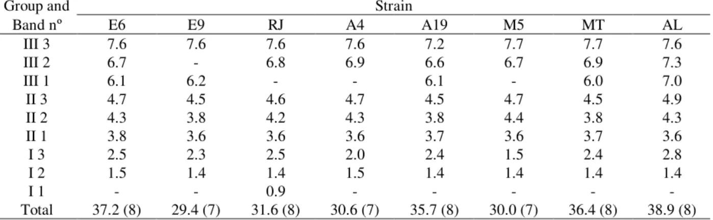
2.8Mb; Group II had three bands corresponding to molecules between 3.6 and 4.9 Mb and, Group III had two to three bands corresponding to molecules between 6.0 and 7.7 Mb (Table 1 and Fig. 1). Three bands in Group III were only resolved using B electrophoresis conditions (Shimizu et al., 1992) (data not shown). These parameters were useful to show that some single bands showed in Group III with condition A were doublets. In Group I and Group III polymorphisms were observed in the length and in the number of chromosomal DNA while in Group II only length polymorphisms were detected. The total genome size of these strains was estimated in at least 29.4 Mb. Molecular sizes of the chromosomal DNAs were estimated with reference to the size standards, using Kodak 1D Imaging Analysis Software.
Table 1 - Size estimated (Mb) and number of M. anisopliae chromosomal DNAs as determined by CHEF analysis.
Group and Strain
Band nº E6 E9 RJ A4 A19 M5 MT AL
III 3 7.6 7.6 7.6 7.6 7.2 7.7 7.7 7.6
III 2 6.7 - 6.8 6.9 6.6 6.7 6.9 7.3
III 1 6.1 6.2 - - 6.1 - 6.0 7.0
II 3 4.7 4.5 4.6 4.7 4.5 4.7 4.5 4.9
II 2 4.3 3.8 4.2 4.3 3.8 4.4 3.8 4.3
II 1 3.8 3.6 3.6 3.6 3.7 3.6 3.7 3.6
I 3 2.5 2.3 2.5 2.0 2.4 1.5 2.4 2.8
I 2 1.5 1.4 1.4 1.5 1.4 1.4 1.4 1.4
I 1 - - 0.9 - - - -
-Total 37.2 (8) 29.4 (7) 31.6 (8) 30.6 (7) 35.7 (8) 30.0 (7) 36.4 (8) 38.9 (8)
Differences in chromosome number and length are
common not only in Metarhizium anisopliae
(Shimizu et al., 1992, Valadares-Inglis and Peberdy, 1998) but also in other fungi such as
Cladosporium fulvum (Talbot et al. 1991),
Neurospora crassa (Orbach et al., 1988),
Colletotrichum gloeosporioides (Masel et al., 1990), Ustilago hordei (McCluskey and Mills, 1990), Fusarium solani (Bruschi and Nazareth, 1994; Suga et al. 2002) and Fusarium oxysporum
(Davière et al., 2001). These differences were attributed mainly to translocations (Orbach et al., 1988 and Talbot et al. 1991) and other chromosomal rearrangements (Masel et al., 1990). Davière et al. (2001) observed a correlation between the high level of chromosomal
polymorphism in F. oxysporum and concentrations of transposables elements (TEs) but these elements were not detected in M. anisopliae.
Apart from translocations, other factors may account for degree of incompatibility between RJ and E6 strains. Pamphile (1992) was not able to produce recombinants between different strains like E6 and MT, which showed a very similar karyotype.
The similarities and differences found in the molecular karyotypes of the strains studied in the
present work, partialy agreed with results from other authors. De Conti et al. (1980), using esterase profile analysis, distinguished three groups of strains, the first one including E6 and E9 strains, a second one comprising A19 strain and a third one including A4 strain. In the present work, it was not possible to compare E9 with others, but E6, A4 and A19 showed different karyotypes.
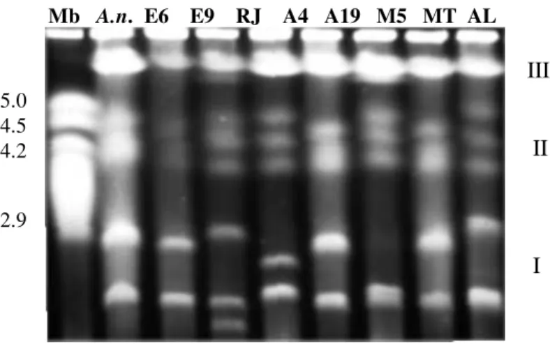
Mb A.n. E6 E9 RJ A4 A19 M5 MT AL
Figure 1 - Separation of Metarhizium anisopliae intact chromosomal DNAs on a CHEF gel. Gel (0.6% w/v) was electrophoresed in A condition. Group I) two to three bands corresponding to molecules between 0.9 and 2.8Mb; Group II) three bands corresponding to molecules between 3.6 and 4.9 Mb and, Group III) with these parameters presents two bands corresponding to molecules between 6.0 and 7.7 Mb.
A.n. shows the Aspergillus nidulans chromosome size standards.
The RAPD analysis between E6, E9 M5, AL and RJ strains showed distinct profile for each strain, except for E6 and E9 that showed the same profile. UPGMA dendrogram data showed that E6/E9 and M5 were the most similar to AL in the same group and RJ strain was the least similar (Fungaro et al., 1996). In the present work, distinct electrophoretic karyotypes were found from these strains, derived from the same host, D. flavopicta, and RJ was the only strain that showed a small chromosomal band (0.9 Mb).
These results were in part in accordance wit h the work of Fungaro et al. (1996). The strain A19 isolated from another species of Deois (D. schach) showed almost the same electrophoretic karyotype from MT and E6. These strains were isolated from different regions far apart from each other. Based on the present data, it is not possible to do some correlation between the electrophoretic karyotype
of each strain and their hosts or the geographic localization. Using data derived from AFLP, amplified fragment length polymorphism, Muro et al. (2003) were not able to do some correlation between 50 isolates of the entomopathogenic
fungus Beauveria bassiana and hosts or
geographical origins.
The data shown here indicated that this species presented a wide genetic diversity. The genus
Metarhizium could be basically divided in three species: M. anisopliae, M. flavoviride and M. albus, with some described varieties inside these species. A more detailed study including electrophoretic karyotype should be carried out, and may prove to be valuable for a better definition of the Metarhizium genus taxonomy.
III
II
I
5.0 4.5 4.2
ACKNOWLEDGMENTS
The present work was supported by Capes (scholarship to V.K.C.) and CNPq.
RESUMO
Cariótipos de oito linhagens selvagens do fungo entomopatogênico Metarhizium anisopliae var.
anisopliae foram obtidos em gel, por eletroforese em campo pulsado. As linhagens foram isoladas de insetos provenientes de seis estados brasileiros. As moléculas de DNA cromossômico de três linhagens foram separadas em sete bandas e, de cinco linhagens, em oito bandas. Polimorfismo de tamanho cromossômico também foi observado. O tamanho do DNA cromossômico de todas as linhagens variou de 7,7 a 0,9 Mb, utilizando-se DNA cromossômico de Aspergillus nidulans como padrão. O tamanho do genoma total foi estimado em pelo menos 29,7 Mb. Algumas correlações entre semelhanças e diferenças no cariótipo eletroforético e a ocorrência do ciclo parassexual como também a especificidade com insetos hospedeiros foram discutidas.
REFERENCES
Bagagli, E.; Valadares, M. C. C.and Azevedo, J. L. (1991), Parameiosis in the entomopathogenic fungus
Metarhizium anisopliae (Metsch.) Sorokin. Rev. Brasil. Genet., 14, 261-271.
Bidochka, M. J.; McDonald, M. A.; St Leger, R. J. and Roberts, D. W. (1994), Differentiation of species and strains of entomopathogenic fungi by random amplification by polymorphic DNA. Curr. Genet.,
25, 107-113.
Brody, H. and Carbon, J. (1989), Electrophoretic karyotype of Aspergillus nidulans. Proc. Natl. Acad. Sci. USA, 86,6260-6263.
Bruschi, C. V. and Nazareth, S. W. (1994), Electrophoretic karyotype of Fusarium solani.
Microbiology, 140, 1373-1375.
Daoust, R. A. and Roberts, D. W. (1982), Virulence of natural and insect passaged strains of Metarhizium anisopliae to mosquito larvae. J. Invertebr. Pathol.,
40, 107-117.
Davière, J. M.; Langin, T. and Daboussi, M. J. (2001), Potential role of transposable elements in the rapid reorganization of the Fusarium oxysporum genome.
Fung. Genet. Biol., 34, 177-192.
De Conti, E.; Messias, C. L.; De Souza, H. M. L. and Azevedo, J. L. (1980), Electrophoretic variation in esterases and phosphatases in eleven wild-type strains of Metarhizium anisopliae. Experientia,
36, 293-294.
Fegan, M.; Manners, J. M.; MacLean, D. J.; Irwin, J. A. G.; Samuels, K. D. Z., Holdom, D. G. and Li, D. P. (1993), Random amplified polymorphic DNA markers reveal a high degree of genetic diversity in the entomopathogenic fungus
Metarhizium anisopliae var. anisopliae. J. Gen. Microbiol., 139, 2075-2081.
Fungaro, M. H. P.; Vieira, M. L. C.; Pizzirani-Kleiner, A. A. and Azevedo, J. L. (1996), Diversity among soil and insect isolates of Metarhizium anisopliae
var. anisopliae detected by RAPD. Lett. Appl. Microbiol., 22, 389-392.
Huxam, I. M.; Samuels, K. D. Z.; Heale, J. B. and McCorkindale, N. J. (1989), In vivo and in vitro
assays for pathogenicity of wild-type and mutant strains of Metarhizium anisopliae for three insect species. J. Invertebr. Pathol., 53, 143-151.
Masel, A.; Braithwaite, K.; Irwin, J. and Manners, J. (1990), Highly variable molecular karyotypes in the plant pathogen Colletotrichum gloeosporioides.
Curr. Genet., 18, 81-86.
McCluskey, K. and Mills, D. (1990), Identification and characterization of chromosome length polymorphisms among strains representing fourteen races of Ustilago hordei. Mol. Plant-Microbe Interact., 3, 366-373.
Messias, C. L. and Azevedo, J. L. (1980), Parasexuality in the deuteromycete Metarhizium anisopliae. Trans. Br. Mycol. Soc., 75, 473-477.
Messias, C. L.; Roberts, D. W. and Grefig, T. (1983), Pyrolisis-gas chromatography of the fungus
Metarhizium anisopliae: an aid to strain
identification. J. Invertebr. Pathol., 42, 393-396. Muro, M. A.; Mehta, S. and Moore, D. (2003), The use
of amplified fragment polymorphism for molecular analysis of Beauveria bassiana isolates from Kenya and other countries, and their correlation with host and geographical origin. FEMS Microbiol. Lett.,229, 249-257.
Orbach, M. J.; Vollrath, D.; Davis, R. W. and Yanofsky, C. (1988), An electrophoretic karyotype of
Neurospora crassa. Mol. Cell Biol., 8, 1469-1473. Pamphile, J. A. (1992), Estudos genéticos no fungo
entomopatogênico Metarhizium anisopliae var.
Pipe, N. D.; Chandler, D.; Bainbridge, B. W. and Heale, J. B. (1995), Restriction fragment length polymorphisms in the ribosomal RNA gene complex of isolates of the entomopathogenic fungus
Metarhizium anisopliae. Mycol. Res. 99, 485-491. Rosato, Y. B.; Messias, C. L. and Azevedo, J. L.
(1981), Production of extracellular enzymes by isolates of Metarhizium anisopliae. J. Invertebr. Pathol., 38, 1-3.
Shimizu, S.; Aray, Y. and Matsumoto, T. (1992), Electrophoretic karyotype of Metarhizium anisopliae.
J. Invertebr. Pathol., 60, 185-187.
Silveira, W. D. and Azevedo, J. L. (1987), Protoplast fusion and genetic recombination in Metarhizium anisopliae. Enzyme Microb. Technol., 9, 149-152. St Leger, R. J.; May, B.; Allee, L. L.; Frank, D. C.;
Staples, R. C. and Roberts, D. W. (1992), Genetic differences in allozymes and information of infection structures among isolates of the entomopathogenic fungus Metarhizium anisopliae.
J. Invertebr. Pathol., 60, 89-101.
Suga, H.; Ikeda, S.; Taga, M.; Kageyama, K. and Hyakumachi, M. (2002), Electrophoretic karyotyping and gene mapping of seven formae speciales in
Fusarium solani. Curr. Genet., 41, 254-260.
Talbot, N. J.; Oliver, R. P. and Coddington, A. (1991), Pulsed field gel electrophoresis reveals chromosome length differences between strains of Cladosporium fulvum (syn. Fulvia fulva) Mol. Gen. Genet.,
229, 267-272.
Valadares-Inglis, M. C. and Azevedo, J. L. (1997), Amylase and protease secretion in recombinant strains of Metarhizium anisopliae var. anisopliae
following parasexual crosses. Rev. Braz. J. Genet.,
20, 171-175.
Valadares-Inglis, M. C. and Peberdy, J. F. (1998), Variation in the electrophoretic karyotype of Brazilian strains of Metarhizium anisopliae.
Genet. Mol. Biol., 21, 11-14.