Time domain heart rate variability in Boxer dogs with arrhythmogenic
right ventricular cardiomyopathy
Variabilidade da frequência cardíaca no domínio do tempo em cães da raça Boxer com cardiomiopatia arritmogênica do ventrículo direito
Evandro Zacché1* Thais Cristine Alves de Assumpção2 Talita Beani Corsini1 Aparecido Antonio Camacho1
ISSNe 1678-4596
INTRODUCTION
Boxer arrhythmogenic right ventricular cardiomyopathy (ARVC) is inherited as an autosomal dominant disease with incomplete penetrance that can lead to syncope or sudden death as a consequence
of ventricular arrhythmias, caused by a series of ultrastructural changes in cardiomyocytes (MEURS, 2010; OXFORD et al., 2011). In 2004 ARVC was described as a spontaneous animal model for the disease observed in human patients, with very similar clinical and pathological characteristics (BASSO et al., 2004).
1Departamento de Clínica e Cirurgia Veterinária, Faculdade de Ciências Agrárias e Veterinárias, Universidade Estadual Paulista (UNESP), Campus de Jaboticabal, s/n, 14884 900, Jaboticabal, SP, Brasil. E-mail: [email protected]. *Corresponding author.
2Vet Support, Terapia Intensiva Veterinária, São Paulo, SP, Brasil.
ABSTRACT: The aim of the present study was to assess heart rate variability (HRV) in Boxer dogs affected by arrhythmogenic right
ventricular cardiomyopathy (ARVC). Fourteen Boxer dogs classified as affected and 28 classified as unaffected were included in a prospective case-control study. Dogs underwent 24-hour ambulatory ECG and were classified as affected (>1,000 VPCs/24 hours) or unaffected (<20 VPCs/24 hours) by ARVC based on the number of ventricular arrhythmias. HRV was assessed using 24-h Holter ECG monitoring; the studied parameters were SDNN, SDANN, SDNNIDX, rMSSD and pNN50. Data were submitted to logarithmic transformation and HRV parameters were compared between groups and correlated according to the disease status, number and severity of ventricular arrhythmias using Student’s t test, linear regression and Spearman’s test. There was no interaction between the HRV parameters and the number and severity of ventricular
arrhythmias. SDNNlog (2.35±0.14 vs. 2.46±0.12, P=0.01), SDNNIDXlog (2.18±0.14 vs. 2.24±0.10, P=0.002) and pNN50log (1.47±0.19 vs.
1.64±0.13, P=0.002) were significantly lower in the affected group compared with the unaffected. According to this study, HRV are different in a population of Boxers dogs affected by ARVC compared to a population of unaffected dogs, and these differences are not consequences of
low cardiac output caused by ventricular arrhythmias since animals that had higher number and complexity of arrhythmias were not those who had lower values of HRV.
Key words: autonomic nervous system, ventricular arrhythmia, Ambulatory ECG.
RESUMO: O objetivo do presente estudo foi avaliar a variabilidade da frequência cardíaca (VFC) em cães da raça Boxer acometidos pela cardiomiopatia arritmogênica do ventrículo direito (CAVD). Para isso, foram incluídos, em estudo prospectivo caso-controle, 14 cães classificados como acometidos pela CAVD e 28 classificados como não acometidos. Os cães foram submetidos à eletrocardiografia ambulatorial de 24 horas e então classificados como acometidos (quando apresentaram mais de 1.000 complexos ventriculares prematuros em 24 horas) ou não acometidos (quando apresentaram menos de 20 complexos ventriculares prematuros em 24 horas) pela CAVD, com base no número de arritmias ventriculares. A VFC foi avaliada mediante monitoramento Holter de 24 horas. Os parâmetros estudados foram SDNN, SDANN, SDNNIDX, rMSSD e pNN50. Os dados foram submetidos à transformação logarítmica e os parâmetros da VFC foram comparados e correlacionados de acordo com a presença ou não da doença, número e severidade das arritmias pelo teste t de Student, regressão linear e teste de Spearman. Não houve interação entre as variáveis da VFC e o número e a severidade das arritmias ventriculares. Porém, as variáveis
SDNNlog (2,35±0,14 vs. 2,46±0,12, P=0,01), SDNNIDXlog (2,18±0,14 vs. 2,24±0,10, P=0.002) e pNN50log (1,47±0,19 vs. 1,64±0,13, P=0,002)
foram significativamente menores no grupo de animais acometidos pela doença. De acordo com os resultados do presente estudo, a VFC é menor nos Boxers acometidos pela CAVD em relação aos cães não acometidos, e essa diferença não pode ser atribuída ao baixo débito cardíaco, supostamente causado pelas arritmias ventriculares, uma vez que os animais que apresentaram maior número e complexidade das arritmias não foram os que apresentaram os menores valores da VFC.
Palavras-chave: sistema nervoso autônomo, arritmia ventricular, ECG ambulatorial.
Since the observation that catecholamines favor the development of ventricular arrhythmias and these arrhythmias could be suppressed by beta-adrenergic blockers (WICHTER et al., 1992), the sympathetic nervous system has been reported to play an important role in the genesis of ventricular arrhythmias in patients with ARVC (WICHTER et al., 2000).
A well-recognized method for evaluating the imbalance in the sympathetic and parasympathetic nervous system is heart rate variability (HRV), defined as the temporal variation between sequences of consecutive heartbeats (TASK FORCE OF THE EUROPEAN SOCIETY OF CARDIOLOGY AND THE NORTH AMERICAN SOCIETY OF PACING AND ELECTROPHYSIOLOGY, 1996). A reduction in HRV has been reported in several cardiological and noncardiological diseases, both in humans and animals, (MALIK et al., 2000; KUDAT et al., 2006; OLIVEIRA et al., 2012). Studies in humans with ARVC identified that affected patients with lower HRV parameters are more prone to arrhythmic events (BATTIPAGLIA et al., 2012).
ARVC has been widely investigated in Boxer dogs (BASSO et al., 2004; SPIER & MEURS, 2004; MEURS et al., 2010, KAYE et al., 2015), however, little information is available regarding the autonomic status in affected animals (SPIER & MEURS, 2004). In view of this fact, we hypothesized that dogs with ARVC suffer from autonomic dysfunction that is reflected as a reduction in HRV parameters. Thus, the aim of the present study was to evaluate whether Boxer dogs affected by ARVC exhibit changes in HRV reflecting abnormalities in autonomic modulation compared with unaffected Boxers.
MATERIALS AND METHODS
Patient selection criteria
Client-owned purebred Boxer dogs aged 4 years and older were recruited from local pet owners, university students and veterinary teaching hospital employees for a prospective study. Besides 24-hours Holter monitoring, dogs were evaluated by physical examination, 10-lead electrocardiography and 2-dimensional echocardiography. Exclusion criteria included any alteration in the physical exam other than syncope, such as congenital heart disease, valve
insufficiency, systolic dysfunction (considered when
fractional shortening <20%) or increased cardiac dimensions during the echocardiographic evaluation and bradyarrhythmias at the electrocardiographic
evaluation. Dogs receiving antiarrhythmic drugs were also excluded.
Holter monitoring
Twenty-four-hour ECGs were obtained with a 4-lead, 3-channel electrode system and
acquired with a digital recordera. The area of
electrode placement was prepared by shaving and cleaning the skin with alcohol. Leads were arranged to approximate the frontal leads I, II, and III (SPIER & MEURS, 2004). A bandage and a specially designed vest were used to secure the Holter recorder and leads to the dog.
During the period of monitoring, dogs were maintained individually in a kennel in the veterinary
hospital with water ad libitum and food according
to owners’ recommendation. Recordings were initially processed and analyzed automatically by a
commercially available softwareb and then manually
reviewed for arrhythmias and artefacts and checked
for proper classification of every QRS complexes by
a veterinary cardiologist (E.Z.). Were included only those recordings with >23 hours and <3% of artifacts.
Study groups
Patients were distributed into 2 groups according to their frequency of ventricular premature complexes (VPCs) as observed during 24-hour ECG. Animals that exhibited less than 20 VPCs/24 hours were included in the unaffected group. Those animals that exhibited over 1,000 VPCs/24 hours were
classified as affected by ARVC and included in the
affected group. Dogs with more than 20 and less than 1,000 VPCs were not included in the study.
According to the complexity of the ventricular arrhythmia, dogs were also distributed into three different subgroups. Animals with only single VPCs were allocated in subgroup A. Dogs with couplets as the most complex ventricular pattern were allocated in subgroup B. Dogs with non-sustained ventricular tachycardia as the most complex ventricular pattern were allocated in subgroup C.
Time domain heart rate variability
After careful revision of the recording, time domain HRV was analyzed using a commercially available softwareb designed for people. The automatic
HRV analysis excluded complexes of ventricular and supraventricular (supraventricular premature complexes and supraventricular tachycardia) origin and included only intervals between adjacent normal
QRS complexes (NN-intervals) of sinus origin.
of all RR intervals (SDNN), standard deviation of the average RR intervals calculated over a period of 5 minutes (SDANN), the mean of the 5 minutes standard deviation of the RR intervals calculated over 24 hours (SDNNIDX), the square root of the mean squared differences of successive RR intervals (rMSSD) and the percentage of RR intervals differing >50 ms from the preceding interval (pNN50).
Statistical analyses
Initially, data were submitted to Cramer-von Mises normality test and checked for outliers. Variables were summarized as the mean ± SD when Gaussian distribution was assumed or median and range when not normally distributed. Student’s t test was used to compare the mean value of each time domain variable among the affected and unaffected groups as well as age between groups. Time domain heart rate variability variables were submitted to logarithmic transformation, to ensure normal distribution and equal variances for Student’s t test analysis. Linear regression and Spearman’s correlation were used to evaluate the relationship between HRV parameters and complexity and the number of ventricular arrhythmias within the affected
group. Significance was defined as P<0.05.
RESULTS
Initially, 57 animals were recruited. Of these, 5 were excluded due to chronic mitral valve
insufficiency, 3 due to less than 23 hours of Holter
recording, 2 for 2nd degree atrioventricular block and 5 due to total count of VPCs between 20 and 1000. Thus, forty-two dogs were included in the study. The
group of animals classified as affected consisted of
14 dogs, 6 males and 8 females. The group of animals
classified as unaffected comprised 28 dogs, 7 males
and 21 females. The group of affected animals aged 6 to 12 years (mean age, 9 years) and the group of unaffected dogs aged between 4 and 11 years (mean
age, 7 years) (P=0.007).
At 24-hour Holter evaluation, animals from the affected group exhibited between 1,004 and 36,445 VPCs/24-hours. One animal (7.14%) exhibited only isolated VPCs (subgroup A), 6 animals (42.85%) exhibited couplets (subgroup B) and 7 animals (50%) exhibited non-sustained ventricular tachycardia (subgroup C) as the most complex pattern of arrhythmia. Animals in the affected group exhibited between 0 and 20 isolated VPCs/24-hour.
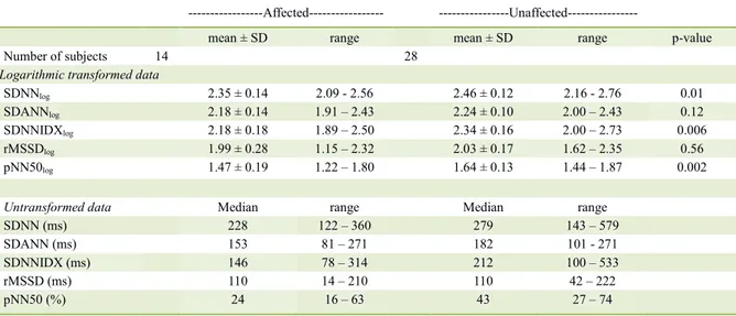
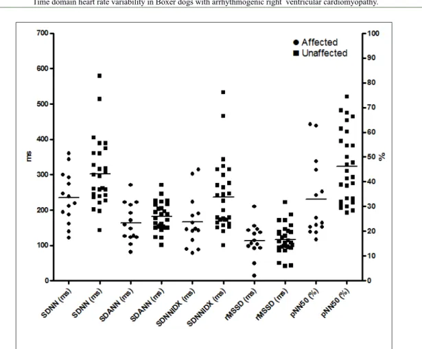
Descriptive data for HRV parameters are
presented in table 1 and depicted in figure 1. Results
of Student’s t test indicated a significant decrease
in SDNNlog, SDNNIDXlog and pNN50log in the
affected group. There was no significant correlation
between the subgroups according to complexity of ventricular arrhythmia and each parameter of HRV.
Correlation coefficients for the linear regression
between subgroups of ventricular arrhythmia complexity and SDNN, SDANN, SDNNIDX,
rMSSD and pNN50 were r2<0.01 and P=0.99;
r2<0.01 and P=0.86; r2<0.01 and P=0.85; r2=0.03
and P=0.53; and r2<0.01 and P=0.92, respectively.
Spearman’s correlation coefficient between the
number of ventricular arrhythmias and the HRV parameters were as follows: SDNN, Spearman r=0.29 and P=0.33; SDANN, Spearman r=0.008 and P=0.97; SDNNIDX, Spearman r=0.23 and P=0.40; rMSSD, Spearman r= -0.09 and P=0.73; and pNN50, Spearman r=0.07 and P=0.80.
DISCUSSION
According to the results obtained in the present study, animals affected by ARVC
can exhibit autonomic imbalance, reflected
by decreased SDNN, SDNNIDX and pNN50 compared with unaffected Boxers.
Among the methods available to assess the status of the autonomic nervous system, HRV has emerged as a simple and noninvasive method to assess the sympathovagal activity at sinoatrial level (TASK FORCE OF THE EUROPEAN SOCIETY OF CARDIOLOGY AND THE NORTH AMERICAN SOCIETY OF PACING AND ELECTROPHYSIOLOGY, 1996). HRV parameters derived from the difference between consecutives RR intervals (pNN50 and rMSSD) predominantly reflect vagal action. Variables derived from the direct measurement of RR intervals (SDNN and SDANN SDNNIDX) are global indices of HRV and reflect long-term components, circadian rhythm, physical activity and postural changes (SZTAJZEL, 2004). Thus, it can be assumed that the decrease in HRV exhibited by the ARVC dogs may reflect a reduction in vagal tone and/or a predominance of sympathetic influences on the heart, conditions known to be arrhythmogenic (ZIPES & WELLENS 1998).
cleft confirmed the presence of abnormalities in cardiac sympathetic innervation in this disease (WICHTER et al., 1994; WICHTER et al., 2000). More recently, in studies assessing HRV in humans with ARVC, the authors observed a decrease in HRV parameters in patients who experienced arrhythmic events (BATTIPAGLIA et al., 2012).
The precise reason why patients with ARVC exhibit sympathetic abnormalities and consequently exhibit reduced HRV remains speculative. Among the potential mechanisms responsible for abnormalities in cardiac sympathetic innervation and reduced HRV, such changes may be due to the following: myocardial degeneration and fibrofatty infiltration beginning in the subepicardium and damaging sympathetic nervous fibers located on the same site (WICHTER et al., 1994); increased sympathetic activity secondary to the increased release of catecholamines in the synaptic cleft and/or increased myocardial sensitivity to catecholamines (WICHTER et al., 2000); or ventricular dysfunction with hemodynamic impairment (FAUCHIERAND et al., 1997). Although it was not the aim of this study to identify the reason for the decrease in HRV in patients affected by ARVC, the theory that the sympathetic nervous system may have been activated to compensate for a low cardiac output, resulted from the high number of arrhythmias
or systolic dysfunction, does not seem appropriate because no animal with contractile dysfunction was introduced in the study and no correlation between HRV parameters and the number and severity of ventricular arrhythmias were observed.
Although the present study suggests autonomic dysfunction in animals with ARVC, similar that which occurs in human patients, our study is inconsistent with the only other study to assess HRV in Boxers dogs with ARVC. In 2004, SPIER & MEURS measured HRV in Boxers with ARVC (>1,000 VPCs/24-hour and normal ventricular size and function) and in Boxers with congestive heart failure (CHF) and only observed a reduction in HRV parameters in animals with CHF. However, the authors did not rule out the possibility of involvement of the autonomic nervous system in the genesis of ventricular arrhythmias and attributed the non-reduction of HRV parameters simply to the lack of persistently high sympathetic tone (SPIER & MEURS, 2004). Unlike the study cited above, in our study, the animals were kept in the hospital environment, away from their owners and in contact with other dogs. This environment may have functioned as a “stress test”, leading to a persistent state of sympathetic activation in dogs with some degree of autonomic dysfunction, i.e., the ARVC-affected animals.
Table 1 - Assessment of heart rate variability (mean ± SD) in ARVC-affected (>1,000 VPCs/24 hour; n=14) and unaffected (<20 VPCs/24-hour; n=28) Boxer dogs.
---Affected--- ---Unaffected---
mean ± SD range mean ± SD range p-value
Number of subjects 14 28
Logarithmic transformed data
SDNNlog 2.35 ± 0.14 2.09 - 2.56 2.46 ± 0.12 2.16 - 2.76 0.01
SDANNlog 2.18 ± 0.14 1.91 – 2.43 2.24 ± 0.10 2.00 – 2.43 0.12
SDNNIDXlog 2.18 ± 0.18 1.89 – 2.50 2.34 ± 0.16 2.00 – 2.73 0.006
rMSSDlog 1.99 ± 0.28 1.15 – 2.32 2.03 ± 0.17 1.62 – 2.35 0.56
pNN50log 1.47 ± 0.19 1.22 – 1.80 1.64 ± 0.13 1.44 – 1.87 0.002
Untransformed data Median range Median range
SDNN (ms) 228 122 – 360 279 143 – 579
SDANN (ms) 153 81 – 271 182 101 - 271
SDNNIDX (ms) 146 78 – 314 212 100 – 533
rMSSD (ms) 110 14 – 210 110 42 – 222
pNN50 (%) 24 16 – 63 43 27 – 74
According to previous study that assessed HRV in dogs with myxomatous mitral valve disease, the parameters of HRV SDNN and pNN50 of healthy dogs had average values of 287 ms and 62.14%, respectively (OLIVEIRA et al., 2012). Thus, one can note observing figure 1 that a significant number of animals, of both groups, showed relatively low values of HRV parameters compared to healthy dogs of that study, which indicates that the environment where they were subjected to examination was indeed stressful. Despite the relatively low levels of HRV in both groups, only animals classified as affected showed extremely low values on certain parameters.
A major limitation of the study described here was the method of diagnosis of ARVC. Despite the observation of cardiac chamber enlargement and systolic dysfunction, which could suggest the
presence of dilated cardiomyopathy, the dogs did not undergo genetic evaluation or any identification of myocardial fibrofatty infiltration for confirmation of the disease; thus, the number of ventricular arrhythmias in 24 hours was the only diagnostic criterion used. Careful observation of figure 1 and the results of pNN50 demonstrates that among the affected animals, a portion exhibited very low values not displayed by animals in the other group, and another portion exhibited higher values, suggesting that animals with higher values have ventricular arrhythmias caused by abnormalities other than ARVC or simply that the involvement of the sympathetic nervous system is less severe due to different genetic backgrounds. Despite have been identified reduction of HRV in dogs affected by CAVD, such analysis does not seem to be promising
as a diagnostic tool due to the large overlap of data between affected and unaffected Boxer dogs.
CONCLUSION
In this study, our results demonstrated that Boxer dogs affected by CAVD have autonomic changes reflected by reduction of HRV parameters, and this reduction is not a consequence of the number or severity of ventricular arrhythmias.
SOURCES AND MANUFACTURES
a - Cardioflash® digital – Cardios Sistemas – São Paulo, Brazil.
b - CardioSmart 540 ® – Cardios Sistemas – São Paulo, Brazil.
BIOETHICS AND BIOSSECURITY COMMITTEE APPROVAL
The study was approved by the Ethical Committee on the Use of Animals of the Universidade Estadual Paulista and is in accordance with the Ethical Principles of Animal Experimentation adopted by the Brazilian College Experimentation under the protocol number 002965/10.
REFERENCES
BASSO, C. et al. Arrhythmogenic right ventricular cardiomyopathy causing sudden cardiac death in boxer dogs: a new animal model of human disease. Circulation, v.109, p.1180-1185, 2004. Available from:<http://circ.ahajournals.org/content/109/9/1180.full.pdf+html>. Accessed: Sept. 02, 2014. doi: 10.1161/01.CIR.0000118494.07530.65.
BATTIPAGLIA, I. et al. Association of heart rate variability with arrhythmic events in patients with arrhythmogenic right ventricular cardiomyopathy/dysplasia. Circulation Journal, v.76, p.618-623, 2012. Available from: <https://www.jstage.jst.go.jp/article/ circj/76/3/76_CJ-11-1052/_pdf>. Accessed: Sept. 02, 2014. doi: 10.1253/circj.CJ-11-1052.
KUDAT, H. et al. Heart rate variability in diabetes patients. Journal of International Medical Research, v.34, p.291-296, 2006. Available from: <http://imr.sagepub.com/content/34/3/291.long>. Accessed: Sept. 02, 2014. doi: 10.1177/147323000603400308.
MALIK, M. et al. Depressed heart rate variability identifies post infarction patients who might benefit from prophylactic
treatment with amiodarone. Journal of American College of Cardiology, v.35, p.1263-1275, 2000. Available from: <http:// www.sciencedirect.com/science/article/pii/S0735109700005714>. Accessed: Sept. 02, 2014. doi: 10.1016/S0735-1097(00)00571-4.
MEURS, K.M. et al. Genome-wide association identifies a
deletion in the 3’ untranslated region of Striatin in a canine model
of arrhythmogenic right ventricular cardiomyopathy: identification
of Striatin deletion in canine ARVC. Human Genetics, v.128, p.315-324, 2010. Available from: <http://link.springer.com/ article/10.1007%2Fs00439-010-0855-y>. Accessed: Sept. 02, 2014.
MEURS, K. Genetics of cardiac disease in the small animal patient. Veterinary Clinics of North America: Small Animal Practice, v.43, p.701-715, 2010. Available from: <http://www.sciencedirect.
com/science/article/pii/S0195561610000343>. Accessed: Sept. 02, 2014. doi: 10.1016/j.cvsm.2010.03.006.
OLIVEIRA, M.S. et al. Heart rate variability parameters of myxomatous mitral valve disease in dogs with and without heart failure obtained using 24-hour Holter electrocardiography. Veterinary Record, v.170, p.622-625, 2012. Available from: <http://www.ncbi.nlm.nih.gov/pubmed/22645158>. Accessed: Sept. 02, 2014. doi: 10.1136/vr.100202.
OXFORD, E.M. et al. Ultrastructural chances in cardiac myocytes from Boxer dogs with arrhythmogenic right ventricular cardiomyopathy. Journal of Veterinary Cardiology, v.13, p.101-113, 2011. Available from: <http://www.sciencedirect.com/science/article/pii/S1760273411000300>. Accessed: Sept. 02, 2014. doi: 10.1016/j.jvc.2011.03.002.
SPIER, A.W.; MEURS, K.M. Assessment of heart rate variability in Boxers with arrhythmogenic right ventricular cardiomyopathy. Journal of the American Veterinary Medical Association, v.224, p.534-535, 2004. Available from: <http://www.ncbi.nlm.nih.gov/ pubmed/14989545>. Accessed: Sept. 02, 2014.
SZTAJZEL, J. Heart rate variability: a noninvasive electrocardiographic method to measure the autonomic nervous system. Swiss Medical Weekly, v.134, p.514-522, 2004. Available from: <http://www.ncbi. nlm.nih.gov/pubmed/15517504>. Accessed: Sept. 02, 2014. doi: 2004/35/smw-10321.
TASK FORCE OF THE EUROPEAN SOCIETY OF CARDIOLOGY AND THE NORTH AMERICAN SOCIETY OF PACING AND ELECTROPHYSIOLOGY. Heart rate variability: standards of measurement, physiological interpretation and clinical use. Circulation, v.93, p.1043-1065, 1996. Available from: <http:// www.ncbi.nlm.nih.gov/pubmed/8598068>. Accessed: Sept. 02, 2014. doi: 10.1161/01.CIR.93.5.1043.
WICHTER, T. et al. Efficacy of antiarrythmic drugs in patients with
arrhythmogenic right ventricular disease. Circulation, v.86, p.29-37, 1992. Available from: <http://circ.ahajournals.org/content/86/1/29. long>. Accessed: Sept. 02, 2014. doi: 10.1161/01.CIR.86.1.29.
WICHTER, T. et al. Regional myocardial sympathetic dysinnervation in arrhythmogenic right ventricular cardiomyopathy. An analysis using 123 I-meta-iodobenzylguanidine scintigraphy. Circulation, v.89, p.667-683, 1994. Available from: <http:// circ.ahajournals.org/content/89/2/667.long>. Accessed: Sept. 02, 2014. doi: 10.1161/01.CIR.89.2.667.
WICHTER, T. et al. Abnormalities of cardiac sympathetic innervation in arrhythmogenic right ventricular cardiomyopathy: quantitative assessment of presynaptic norepinephrine reuptake and postsynaptic B-adrenergic receptor density with positron emission tomography. Circulation, v.101, p.1552-1558, 2000. Available from: <http:// circ.ahajournals.org/content/101/13/1552.long>. Accessed: Sept. 02, 2014. doi: 10.1161/01.CIR.101.13.1552.
ZIPES, D.P.; WELLENS, H.J . Sudden cardiac death. Circulation, v.98, p.2334-2351, 1998. Available from: <http://circ.ahajournals. org/content/98/21/2334>. Accessed: Sept. 02, 2014. doi: 10.1161/01.CIR.98.21.2334.