ARTIGO DE REVISÃO
ABSTRACT
Introduction: COVID-19 is caused by the coronavirus SARS-CoV-2. Ocular manifestations have been reported including conjunctivitis and retinal changes. Therefore, it is of the utmost importance to clarify eye involvement in COVID-19 in order to help with its diagnosis and to further prevent its transmission. The purpose of this review is to describe the structure and transmission of SARS-CoV-2, re-ported ocular findings and protection strategies for ophthalmologists.
Material and Methods: Literature search on PubMed for relevant articles using the keywords ‘COVID-19’, ‘coronavirus’, and ‘SARS-CoV-2’ in conjunction with ‘ophthalmology’ and ‘eye’. Moreover, official recommendations of ophthalmological societies were reviewed.
Results: Although the conjunctiva is directly exposed to extraocular pathogens, and the mucosa of the ocular surface and upper respi-ratory tract are connected by the nasolacrimal duct, the eye is rarely involved in human SARS-CoV-2 infection and the SARS-CoV-2 RNA positive rate by RT-PCR test in tears and conjunctival secretions from patients with COVID-19 is also extremely low.
Discussion: The eye can be affected by SARS-CoV-2, which is supported by some reports of conjunctivitis and retinal changes, but its role in the spread of the disease is still unknown.
Conclusion: Given the current scarce evidence, more research is needed to clarify the relationship between SARS-CoV-2 and the eye. Keywords: Conjunctivitis; Coronavirus Infections; COVID-19; Eye Diseases; Ophthalmology; SARS-COV-2
RESUMO
Introdução: COVID-19 é o nome atribuído à doença causada pelo novo coronavírus - SARS-CoV-2. Esta infeção rapidamente atingiu uma disseminação mundial, face ao aumento da globalização e adaptação do vírus a ambientes distintos. Foram descritas manifesta-ções oftalmológicas em doentes com COVID-19, nomeadamente, conjuntivite e alteramanifesta-ções retinianas. Assim, é fundamental esclarecer o envolvimento ocular na COVID-19, contribuindo para o seu diagnóstico precoce e limitando a sua transmissão. O objetivo desta re-visão é descrever a estrutura e o modo de transmissão do SARS-CoV-2, assim como manifestações oculares reportadas e estratégias de proteção para oftalmologistas.
Material e Métodos: Revisão dos artigos relevantes publicados na PubMed usando as palavras-chave ‘COVID-19’, ‘coronavirus’ e ‘SARS-CoV-2’ em associação com as palavras ‘ophthalmology’ e ‘eye’. Além disso, foi feita uma revisão das recomendações oficiais de várias sociedades oftalmológicas a nível mundial.
Resultados: Apesar da conjuntiva estar diretamente exposta a patógenos exógenos, e da mucosa da superfície ocular e do trato respiratório superior estarem conectados pelo canal nasolacrimal, o olho raramente parece ser afetado pelo SARS-CoV-2. A infeção por SARS-CoV-2 e a taxa de positividade para a pesquisa do RNA do SARS-CoV-2 pelo teste de RT-PCR em lágrimas e secreções conjuntivais de pacientes com COVID-19 também são extremamente baixas.
Discussão: O olho pode ser afetado pelo SARS-CoV-2, dada a descrição de casos de conjuntivite e alterações retinianas, mas o seu papel na disseminação da doença ainda é desconhecido.
Conclusão: Dada a escassa evidência atual, são necessários mais estudos para esclarecer a relação entre o SARS-CoV-2 e o globo ocular.
Palavras-chave: Conjuntivite; COVID-19; Infecções por Coronavírus; Oftalmologia; Oftalmopatias; SARS-COV-2
SARS-COV-2 in Ophthalmology: Current Evidence and
Standards for Clinical Practice
SARS-COV-2 em Oftalmologia: Evidência Atual e
Recomendações para a Prática Clínica
1. Department of Ophthalmology. Centro Hospitalar Universitário de São João. Porto. Portugal. 2. Department of Surgery and Physiology. Faculty of Medicine. University of Porto. Porto. Portugal. Autor correspondente: Manuel Falcão. [email protected]
Recebido: 13 de maio de 2020 - Aceite: 016 de junho de 2020 | Copyright © Ordem dos Médicos 2020
Sónia TORRES-COSTA1, Mário LIMA-FONTES1, Fernando FALCÃO-REIS1,2, Manuel FALCÃO1,2
Acta Med Port 2020 Sep;33(9):593-600 ▪ https://doi.org/10.20344/amp.14118
INTRODUCTION
On the 31st December 2019, the World Health
Organi-zation (WHO) was notified of a cluster of 41 cases of unex-plained pneumonia happening in Wuhan, Hubei Province, China. Most of the infected people were confirmed to be related to the South China Seafood Market. On the 7th
Janu-ary 2020, the Chinese Centre for Disease Control and Pre-vention (CDC) isolated through a throat swab the pathogen causative of this disease as a novel type of coronavirus, later called severe acute respiratory syndrome coronavi-rus 2 (SARS-CoV-2).1,2 Based on phylogenomic analysis,
it was suggested that this new pathogen was closely
re-lated to two coronavirus that were isore-lated in bats, prob-ably sharing a common ancestor.3 The disease associated
with SARS-CoV-2 was named coronavirus disease 2019 (COVID-19) and continued to spread in China and progres-sively all over the world. On the 11th March 2020, WHO
rec-ognized COVID-19 as a pandemic. On the 21st May 2020,
COVID-19 was identified in 4 893 186 people globally, re-sulting in 323 256 deaths.4 Currently, there are some
re-ports in the medical literature that identify a direct relation-ship between SARS-CoV-2 and the eye.2,5-9 Therefore, the
ARTIGO DE REVISÃO COVID-19 by ophthalmologists and others may help in the
diagnosis and prevention of disease transmission. The pur-pose of this review is to describe the SARS-CoV-2 structure, transmission, reported ocular manifestations and protection strategies for ophthalmologists.
MATERIAL AND METHODS
We performed a literature search on PubMed for rele-vant articles using the keywords ‘COVID-19’, ‘coronavirus’, and ‘SARS-CoV-2’ in conjunction with ‘ophthalmology’ and ‘eye’. Moreover, official recommendations of ophthalmologi-cal societies were systematiophthalmologi-cally reviewed.
Coronavirus family and SARS-CoV-2 structure
Coronaviruses get their name from the Latin word co-rona, which means crown and its characteristic structure is due to surface projections on the viral envelope. These pos-itive-sense RNA viruses belong to the subfamily Coronaviri-nae, in the family Coronaviridae of the order Nidovirales. They have four main subgroups - alpha, beta, gamma, and delta - based on their genomic structure and affect birds and mammals. Coronaviruses may obtain mutations which facilitate the transmission from animal species to humans and this has made them zoonotic pathogens of concern.10,11
Up to December 2019, only six different coronaviruses were known to infect humans. Four of these (HCoV-NL63, HCoV-229E, HCoV-OC43 and HKU1) usually caused mild common cold-type symptoms in immunocompetent people whilst the other two have caused notorious epidemics in the past two decades.12 In 2002 – 2003, the severe acute
res-piratory syndrome coronavirus (now termed SARS-CoV-1) was responsible for 774 deaths in 26 countries, affecting more than 8000 people around the world.13 Similarly, the
Middle East respiratory syndrome coronavirus (MERS-CoV) caused a devastating epidemic in 2012, with over 2400 cases reported and a 37% mortality rate.14
SARS-CoV-2 transmission
While animals are considered the original source, SARS-CoV-2 is believed to spread primarily via person-to-person through larger respiratory droplets, generally above 5 µm diameter, which are subject to gravitational forces.15
These droplets can be directly inhaled by other people or land on surfaces that others may contact with, where the virus may remain viable from hours to days.13 Respiratory
particles may be spread while breathing, speaking, cough-ing or sneezcough-ing. Aerosol and fomite transmission of SARS-CoV-2 is also plausible and should not be neglected.16
Airborne transmission occurs when smaller respiratory par-ticles (generally < 5 µm) circulate in the air for prolonged periods. Viral particles could be absorbed via the respira-tory mucosa and potentially across the conjunctivae.15 The
coronavirus is not currently considered to be an airborne virus unless aerosols are created. In those circumstances, the virus could spread more than 2 meters and additional protective measures should be taken.15
Viral RNA has also been found in stool samples from
in-fected patients, raising the possibility of a faecal/oral trans-mission route.17 SARS-COV-2 transmission could happen
in asymptomatic carrier patients, making the prevention of COVID-19 infection challenging.18
The biochemical interactions and the pathogenesis of CoV-2 are believed to be similar to those of SARS-CoV-1.19 The spike (S) protein of coronaviruses facilitates
viral entry into target cells. Entry depends on binding of the surface unit, S1, of the S protein to a cellular receptor, which facilitates viral attachment to the surface of target cells. In addition, entry requires S protein priming by cellular pro-teases, which entails spike protein cleavage at the S1/S2 and the S2’ site and allows fusion of viral and cellular mem-branes, a process driven by the S2 subunit. It was known that SARS-CoV-1 engages angiotensin-converting enzyme 2 (ACE2) as the entry receptor and employs the cellular serine protease TMPRSS2 for S protein priming.19-22 A
re-cent study with human cell lines reported that SARS-CoV-2 uses the same mechanism to infect cells and that inhibition of TMPRSS2 blocks viral entry. Moreover, sera from con-valescent CoV-1 patients cross-neutralized SARS-CoV-2-driven entry.23
The most common symptoms of infection are fever, cough and shortness of breath.24 Other symptoms include
nasal congestion, headache, sore throat, sputum produc-tion, fatigue, myalgias, arthralgias, nausea, vomiting, diar-rhoea, anosmia and ageusia.25,26 Despite this vast array of
symptoms, a significant number of infected individuals may be asymptomatic.18
Coronavirus family and ocular disease
It is known that infection by different coronaviruses may lead to ocular involvement.
In 2004, HCoV-NL63 was isolated for the first time from a 7-month-old child with bronchiolitis and conjunctivitis.27
The association between this coronavirus and conjunctivi-tis was corroborated in 2015, in France, by a retrospective study that found an incidence of 17% for conjunctivitis in a population of children infected with HCoV-NL63.28
Ocular involvement has not been reported in MERS-CoV and SARS-CoV-1 infection. Nevertheless, in 2004, a study was conducted to assess the presence of SARS-CoV-1 RNA in tears of infected patients by RT-PCR. Three out of 36 patients tested positive which led to the hypothesis that the conjunctiva could be a direct inoculation site of the vi-rus from infected droplets; other options included migration from the upper respiratory tract through the nasolacrimal duct; or hematogenous infection via the lacrimal gland.29,30
A similar assessment was performed in a study published in the same year. However, it failed to demonstrate positive results for SARS-CoV-1 RNA in the tears of the tested pa-tients.31
In the current pandemic of SARS-CoV-2, there are al-ready some reports of ocular infection. A member of the Chinese national expert panel on pneumonia developed conjunctivitis during an inspection of Wuhan and later test-ed positive for the SARS-CoV-2, despite being fully gowntest-ed
ARTIGO DE REVISÃO with a protective suit and N95 respirator.8 This report
alert-ed healthcare professionals in China for the importance of eye protection and resulted in a call for research into ocu-lar infection as a possible alternative route of SARS-CoV-2 transmission.
The WHO-China Joint Mission on COVID-19 estimated the incidence of conjunctival congestion at 0.8%, based on a study in 55 924 laboratory confirmed cases.9
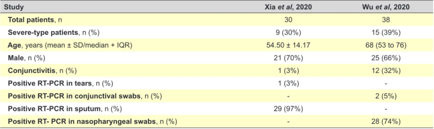
Two papers studied the presence of SARS-CoV-2 RNA in the ocular surface of infected patients. The first study evaluated 30 infected patients and reported a positive RT-PCR test in tears in only one of them, which happened to be the only one who showed signs of conjunctivitis.2 The
second study analysed a sample of 38 confirmed Covid-19 cases (based on the 5th edition of the National Guideline on
Prevention and Control of the Novel Coronavirus Pneumo-nia published by the National Health Commission of China on February 8, 2020), with 28 patients yielding positive find-ings for Covid-19 on RT-PCR from nasopharyngeal swabs but only two testing positive for SARS-CoV-2 in their con-junctival swabs. Twelve (31.6%) patients showed ocular manifestations consistent with conjunctivitis, including con-junctival hyperaemia, chemosis, epiphora, or increased se-cretions. Conjunctivitis was the first symptom of COVID-19 in one of them. Patients with ocular symptoms were more likely to have higher white blood cell and neutrophil counts and higher levels of procalcitonin, C-reactive protein, and lactate dehydrogenase than patients without ocular symp-toms, which could predict a worse prognosis. From the 12 patients with ocular abnormalities, 11 had positive results for SARS-CoV-2 on RT-PCR from nasopharyngeal swabs and only two had positive results for SARS-CoV-2 on RT-PCR from conjunctival swabs (and also from nasopharyn-geal swabs).7 Both studies are compared in Table 1.
Since the human eye has its own renin-angiotensin system, it has been theorized that this new coronavirus could use ACE2 receptors to infect ocular tissues in simi-larity with the SARS-CoV-1 mechanism of infection of the respiratory tract. Although it was thought that ACE2 was mainly expressed in the posterior tissues of the eye, such as the retina and the retinal pigment epithelium, recently, published evidence demonstrated consistent expression of
ACE2 and TMPRSS2 genes in the conjunctival cells and pterygium cells from patients with pterygium and in corneas of mouses, reintroducing the possibility that the ocular sur-face could work as an entry point for SARS-CoV-2.32,33 This
evidence could support the theory that the ocular surface may be a direct infection route to SARS-CoV-2.
One different theory defends that indirect transmission from droplets could occur when viral particles in tears are drained through the nasolacrimal duct into the respiratory tract.34
Despite all the described evidence, little is known about the characteristics of COVID-19 conjunctivitis. One paper described the ocular manifestations of a hospitalized patient with confirmed COVID-19 in more detail. Thirteen days af-ter disease onset (with sore throat and diarrhoea being the first symptoms), the patient reported redness, foreign body sensation and tearing in both eyes without blurred vision. At slit lamp examination, bilateral moderate conjunctival injection, watery discharge, inferior palpebral conjunctival follicles and tender palpable preauricular lymph nodes were noted. No subconjunctival haemorrhage or pseudomem-branes were observed. No lesions on the corneal or ante-rior chamber inflammation were detected. Fundoscopy was normal. All ocular symptoms and signs resolved by day 19 of illness. Conjunctival swabs for detection of SARS-CoV-2 RNA were collected on days 13, 14, 17 and 19. All the pa-tient’s samples tested positive except the last one which correlated with the resolution of the ocular clinical picture.6
This is the first report of the duration of COVID-19 associ-ated conjunctivitis. Since there is no knowledge of specific treatments, general supportive measures should be rec-ommended. No evidence was found about ocular surface sequela or corneal involvement or about uveitis linked to COVID-19. In May 2020, the first paper on ocular manifes-tations other than conjunctivitis in patients with COVID-19 was published. The authors reported retinal findings in 12 patients infected with SARS-CoV-2. In all patients, optical coherence tomography (OCT) showed hyper-reflective le-sions at the level of ganglion cell and inner plexiform layers, which were more prominent in the papillomacular bundle in both eyes. Furthermore, four patients presented subtle cotton wool spots and microhaemorrhages along the retinal
Table 1 – Data comparison between Xia et al (2020) and Wu et al (2020) studies about ocular findings in COVID-19
Study Xia et al, 2020 Wu et al, 2020
Total patients, n 30 38
Severe-type patients, n (%) 9 (30%) 15 (39%)
Age, years (mean ± SD/median + IQR) 54.50 ± 14.17 68 (53 to 76)
Male, n (%) 21 (70%) 25 (66%)
Conjunctivitis, n (%) 1 (3%) 12 (32%)
Positive RT-PCR in tears, n (%) 1 (3%)
Positive RT-PCR in conjunctival swabs, n (%) - 2 (5%)
Positive RT-PCR in sputum, n (%) 29 (97%)
Positive RT- PCR in nasopharyngeal swabs, n (%) - 28 (74%)
ARTIGO DE REVISÃO arcades. Visual acuity and pupillary reflexes were normal in
all eyes, and no symptoms or signs of intraocular inflamma-tion were detected.5
Currently, there is increasing evidence that SARS-Cov-2 can involve the nervous system. Recent studies suggested that neurotropic potential is one common feature of the coronavirus family.35 Therefore, the infection mechanisms
and neurotropism previously found for other coronaviruses like MERS and SARS-CoV could be seen in SARS-CoV-2.36
Regarding the clinical features, a previous study in Wuhan, demonstrated that 77 of 214 patients (36%) hospitalised for COVID-19 developed neurological symptoms or secondary cerebral events.36 Other studies reported several categories
of central and peripheral neurological disorders in COVID-19 patients.36 Reported non-specific and systemic neurological
symptoms included headache, myalgia, dizziness, fatigue and hyposmia, hypogeusia and visual dysfunction.24,26,37,38
These symptoms range from 30% to 45.5%, and are more common in severe stages of disease.36 Other severe
neu-rologic manifestations, recently, reported in COVID-19 patients are encephalopathy, epilepsy, paralysis and con-sciousness disorders, several categories of cerebrovascu-lar events, such as, intra-cerebral haemorrhage, ischemic stroke, cerebral venous thrombosis and also acute necrotiz-ing encephalopathy, mennecrotiz-ingitis, encephalitis and Guillain-Barré syndrome.36,38,39 Hypothesized mechanisms of the
neuro-invasion and manifestations on the nervous system include dissemination across the cribriform plate of the ethmoid bone36; movement of the virus to the brain via the
olfactory bulb40 supported by symptoms of hyposmia and
hypogeusia reported in COVID-19 patients41,42; other
possi-ble mechanisms are the hematological pathway and ACE2 receptors. In fact, SARS-CoV-2 has been shown to use the ACE2 receptor as a cellular entry way.36 This receptor has
been detected in neurons and glial cells, which make it a potential target for SARS-CoV-2. Moreover, SARS-CoV-2 S proteins may interact with ACE2 expressed in the capillary endothelium; this could lead to blood-brain barrier damage and central nervous system (CNS) involvement through the circulatory system.43
Due to neuroinvasive and neurotrophic properties, we may hypothesize that SARS-CoV-2 may affect other neu-ronal structures in the eye including the optic nerve, sub-ba-sal corneal nerve plexus, nerves to the extraocular muscles and to the autonomic system. However, as far as we know, there are no reports of involvement of ocular neuronal struc-tures.
The risks of COVID-19 to ophthalmologists and patients
By the end of March 2020, up to 10% of the reported cases in China and up to 9% of all cases in Italy have been identified among healthcare workers.44 Curiously, one of the
first alarms about COVID-19 was made by Li Wenliang, a Chinese ophthalmologist caring for patients in Wuhan. He died at age 34 years allegedly from the disease.45 In fact,
ophthalmologists are a risk group due to their proximity to the patient during observation in the slit lamp.
Between the 10th and the 12th March 2020, a survey of
ophthalmology practitioners on current COVID-19 guidance at three major UK eye hospitals was performed. It showed a lack of confidence and understanding of COVID-19 Pub-lic Health guidance amongst practicing ophthalmologists, along with significant anxiety regarding exposure risk in the ophthalmic setting.46 One Italian ophthalmologist reported
the dramatic conditions experienced, where adequate per-sonal protective equipment (PPE) was missing.47 Adequate
protection in healthcare workers, including ophthalmolo-gists, is essential.
The strategies adopted during the pandemic could be divided in three different types of measures:
1. To protect healthcare workers with PPE;
2. To prevent virus spread between patients and health-care workers;
3. Environmental cleaning and disinfection in healthcare facilities.
(1) Personal protective equipment
Although, there was some controversy initially regard-ing the appropriate PPE for ophthalmologists durregard-ing their clinical practice, several organizations currently agree and recommend the use of masks, long sleeved gowns, gog-gles and breath shields, and gloves.44 Respiratory
protec-tion could be subdivided into surgical masks (which pro-tect against infectious agents transmitted by droplets) and facial filtering piece (FFP) respirators (which prevent the wearer from inhaling aerosols). In Europe, FFP respira-tors are classified in three classes based on filter efficacy and face adhesion: FFP 1, 2 and 3 with filter efficacy of 80%, 94%, and 99%, respectively.48 In the United States
of America, the respirator classification is subdivided in N (not resistant to aerosols), R (somewhat resistant to oil-aerosols) and S (resistant to oil-oil-aerosols) series, and each one of these should have a minimum filter efficiency of 95%, 99% and 99.97%, respectively.49 The American equivalent
of the FFP2 is the N95. This respirator blocks at least 95 percent of very small (0.3 micron) test particles. Regarding the potential viral spread by asymptomatic COVID-19 car-riers, surgical masks are recommended for all patients and healthcare workers.48 If in contact with a suspected or
con-firmed COVID-19 case, healthcare workers should wear, if available, an FFP2 or N95 respirator tested for fitting or, a surgical mask, if no respirator is available. With the excep-tion of aerosol generating procedures, it is unclear whether FFP respirators (class 2 or 3) provide better protection than surgical masks against other coronaviruses and respira-tory viruses such as influenza.44 Therefore, in the event of
widespread community transmission leading to shortages of PPE, a rational approach would necessarily prioritize the use of FFP2/3 or N95 respirators for care activities involv-ing a higher perceived risk of transmission, such as durinvolv-ing aerosol generating procedures or in intensive care.44
Healthcare workers should strictly follow the procedures for putting on (donning) and safely removing (doffing) PPE in the correct sequence. Active assistance during donning
ARTIGO DE REVISÃO and doffing will help minimize the risk of accidental
con-tamination. Hands should be washed immediately after the removal of PPE.44
(2) Preventing spread of the virus between patients and healthcare professionals
Deferred all non-urgent appointments and surgeries: Many institutions worldwide have recommended delay-ing all elective and nonurgent appointments and delaydelay-ing all elective surgical and procedural cases. These appoint-ments and procedures should only be rescheduled upon recommendation of public health authorities. Only emer-gent and uremer-gent appointments or surgeries should be per-formed. Regarding the definition of ‘elective’, although it can vary between ophthalmologists, the American Academy of Ophthalmology (AAO) defines it as an appointment or pro-cedure that can be postponed for two months without sub-stantive risk to the patient’s vision, material functioning, or general health.50
Staff reorganization: To split the staff into two or three segregated teams, keeping them separated in order to min-imize infection risk, as recommended by the Portuguese Society of Ophthalmology.51 In particular, colleagues with
increased risk (including pregnant women, immunosup-pressed people and those with underlying health condi-tions) should be allocated to a lower risk area or provided with measures to avoid contact with patients.48
Teleophthalmology: In the case of routine appointments, the use of telemedicine during the COVID-19 public health crisis could be an alternative. This system allows an effec-tive screening, decreasing patient travel and limiting pro-vider exposure. In some cases, telehealth information may be enough to make treatment recommendations. In other cases, telemedicine may allow ophthalmologists to suspect or even identify a specific ocular disease, such as corneal disease, retinal or neuro-ophthalmological symptoms, and guide the patient for observation by subspeciality trained ophthalmologists.50
Patient triage: Patients who come to an appointment should answer a survey, at the main hospital entrance, about the presence of risks factors or symptoms suggestive of COVID-19: (1) fever, acute onset or persistent or continu-ous cough, hoarseness, sore throat, shortness of breath, wheezing, anosmia, headache, myalgia, gastrointestinal symptoms; (2) symptoms of conjunctivitis including red eye, pruritus, foreign body sensation, watering; (3) close con-tact with suspected or confirmed COVID-19. Patients that fulfil any criteria, should be sent home, and recommended to stay in isolation 14 days until symptoms resolve. At the hospital main entrance, temperature should be measured, hands disinfected, and a surgical face mask should be given to the patient. Only the patient can enter the hospital. Fam-ily members or friends must wait at the main entrance.48
In special situations in which a caregiver is necessary, the caregiver should also disinfect his hands and wear a mask. Ophthalmology waiting room: Advise seated patients to remain at least two meters apart from one another. The
healthcare workers should regularly disinfect surfaces. During ophthalmological examination: Ophthalmolo-gists should wear PPE, as described previously. The use of slit-lamp barriers is recommended. If possible, all patients should wear masks during examination and ophthalmolo-gists should inform their patients not to speak during the slit-lamp observation. The time required to observe the patient should be minimized. Only essential procedures should be performed. Special tests such as visual field test, optical coherence tomography, corneal topography, ultra-sound, should only be requested when critical to making a clinical decision.
Hand hygiene: The WHO recommends washing hands regularly with alcoholic gel solutions for both healthcare workers and patients.
(3) Environmental cleaning and equipment disinfec-tion recommendadisinfec-tions
Rooms and instruments should be thoroughly disin-fected after each patient observation. Healthcare workers should wear disposable gloves when cleaning and disinfect-ing surfaces and discard the gloves after use. Slit lamps, including controls and accompanying breath shields, should be disinfected, immediately after each patient evaluation. According to WHO recommendations and current prac-tice of several centres, all shared equipment having contact with ocular tissues, such as slit lamps and b-scan probes, should be disinfected with 70% ethyl alcohol after the clini-cal examination.3,52 The current CDC recommendations for
disinfectants specific to COVID-19 include diluted house-hold bleach 5 tablespoons bleach per gallon/per 3.8 litres of water and alcohol solutions with at least 70% alcohol.50
Oth-er common United Stated Environment Protection Agency (EPA) registered disinfectants currently recommended for use against SARS-CoV-2 can be seen in the EPA recom-mendations.53
SARS-CoV-2 is an enveloped virus, unlike adenovi-ruses that are much more resistant to alcohol. Therefore, 70% alcohol solutions should be effective for disinfecting to-nometer tips.50 However, considering the risk of conjunctival
transmission, tonometry should be limited to cases strictly necessary. In that case, AAO recommends single-use, dis-posable tonometer tips or preferably using the retropulsion tonometer with disposable nozzles.50 Non-contact
tonom-etry is a potential source of microaerosols, and therefore, it is prudent to suspend its use.50
Therapy adjustment in ocular inflammatory disease
According to the recommendations of the Portuguese Group of Ocular Inflammation and in line with the Interna-tional Uveitis Study Group (IUSG), the InternaInterna-tional Ocular Inflammation Society (IOIS) and the Ocular Foster Inflam-mation Society (FOIS), the following measures are recom-mended in patients with known ophthalmological inflam-matory disease: to individualize the therapeutic strategy on a case-by-case basis, depending on the intraocular in-flammatory activity, involvement of the posterior segment,
ARTIGO DE REVISÃO chronicity, risk of functional loss and state of the other eye;
to prefer topical therapy and/or local corticosteroid therapy instead of systemic immunomodulatory agents.54 In cases
of increased risk of secondary ocular hypertension, consid-er prescription of hypotensive topical eye thconsid-erapy aftconsid-er local treatment. Local corticosteroid therapy has not been asso-ciated with an increased risk of COVID-19 infection, accord-ing to the available evidence.54 Additional recommendations
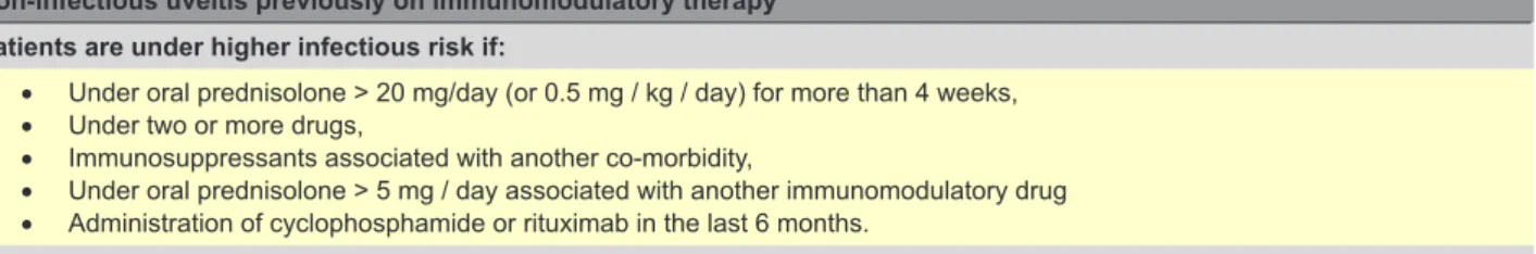
concerning non-infectious uveitis under immunomodulatory treatment can be found in Table 2.
Chloroquine and hydroxychloroquine as treatment of COVID-19: the role of the ophthalmologist
Chloroquine (CQ) and hydroxychloroquine (HCQ) were approved as treatment for COVID-19 by the US Food and Drug Administration (FDA) based on limited in-vitro and an-ecdotal clinical data and is being used as off-label in many countries.55-58 Treatment has been authorized despite
warn-ings from scientific advisers about the lack of randomized controlled trials supporting these drugs’ safety and efficacy in this population.55
An observational study of 1376 patients with COVID-19 who had been admitted to a large medical centre in New York City demonstrated that HCQ administration was not
associated with neither a greatly lowered or increased risk of intubation or death.59
The lack of observed benefit of HCQ associated with in-hospital mortality, following adjustment for pre-existing dis-ease and severity of illness on admission, is consistent with recently reported data from other observational studies.60
Regarding the dose and duration of treatment, there are some variations. For instance, in China, a 10-day course of 500 mg CQ twice daily, or 400 mg HCQ four times daily is being used.61 In France, initially, 600 mg/day of HCQ for 10
days was used.56 In Portugal, although the HCQ as
treat-ment for COVID-19 has been suspended, at the beginning of this pandemic, HCQ was prescribed as 400 mg twice daily for one day, followed by 200 mg twice daily for five or 10 days according to the patient’s disease course.62
Although CQ and HCQ are still used in many countries, taking into account the latest data reported by recent stud-ies that suggested the absence of benefit or even an in-creased risk of mortality associated with HCQ, at the end of May 2020, several European countries, namely France, Belgium, Italy and Portugal have suspended the use of HCQ as treatment for COVID-19.63,64
The AAO recommended a maximum daily HCQ use of ≤ 5.0 mg/kg of real weight, with risk of toxicity being
Table 2 – Recommendations for evaluation and therapy management in non-infectious uveitis on immunomodulatory therapy during COVID-19 pandemics based on Portuguese Group of Ocular Inflammation
Non-infectious uveitis previously on immunomodulatory therapy Patients are under higher infectious risk if:
• Under oral prednisolone > 20 mg/day (or 0.5 mg / kg / day) for more than 4 weeks, • Under two or more drugs,
• Immunosuppressants associated with another co-morbidity,
• Under oral prednisolone > 5 mg / day associated with another immunomodulatory drug • Administration of cyclophosphamide or rituximab in the last 6 months.
How to manage immunomodulatory therapy
• In patients undergoing dose titration, if possible, interrupt dose reduction and maintain the minimum dose previously effective, ideally until the disappearance of pandemic risk and normalization of evaluation conditions for patients;
• Avoid switching or starting new immunomodulatory treatments until that the pandemic is controlled. Analytical study
• Leukocyte count should stay above 4000/μL;
• Repeat an analytical study to monitor pharmacological toxicity whenever necessary, spacing harvest intervals in patients without evidence of toxicity, with leukocytes > 4000/μL and without recent increase in dose drugs;
• Prioritize, if possible, harvests in a location close to the patient. Clinic compatible with COVID-19 infection
• Immunomodulatory therapy can theoretically compromise the response immune system in the early stages of COVID-19. On the other hand, a beneficial effect on the eventual prevention and treatment of ‘Cytokine storm syndrome’ that characterizes stage III of COVID-19 (with emphasis on interferon and tocilizumab) could be seen.
• Asymptomatic patients suspected of having a COVID-19 infection: - Blood count should be monitored;
- To discuss with the doctor responsible for monitoring of SARS-CoV-2 infection the need to reduce or suspension of immunomodulatory therapy;
- Testing will be desirable screening for SARS-CoV-2 infection; • Symptomatic patients with confirmed COVID-19 infection:
- Temporary interruption of immunomodulatory treatment, (conventional or biological) until complete recovery of COVID-19 infection;
- In systemic corticotherapy, the minimum time required for tapering should be guaranteed;
- The maintenance option of interferon or tocilizumab should be mandatorily discussed with the doctor responsible for the treatment of COVID-19 infection.
ARTIGO DE REVISÃO dependent on daily dose and duration of use.65 With this
dosage, retinopathy is rarely seen before 10 or more years of treatment. Nevertheless, in order to treat COVID- 19, the dosages used are higher than those recommended by the AAO, even if for a short period of time.61 Therefore, Marmor
MF suggested that ophthalmic screening is not necessary for COVID-19 patients who take CQ or HCQ for less than two weeks as anti-viral therapy, because the probability of retinal damage is remarkably low even with high dosages.61
Besides, in a time of pandemic with world-wide shortages of medical personnel and equipment, the performance of questionable screening tests would be counterproductive.61
Therefore, ophthalmologists should reassure physicians and the public that retinopathy is not a serious concern re-garding CQ or HCQ usage for COVID-19.61
CONCLUSION
The eye can be affected by this new disease, which is supported by some reports of conjunctivitis in patients with
COVID-19. Moreover, it can be the first organ to show signs of infection, but the frequency of this first manifestation is unknown. The ocular surface can work as an entry point for the virus and the tears can promote the migration of the vi-rus to the respiratory system through the nasolacrimal duct; tears may possibly be a source of infection to other indi-viduals. Given the current scarce evidence, more research is needed to clarify the relationship between SARS-CoV-2 and the eye. Until then, ophthalmologists should continue to use all the recommended protection measures to prevent the possible transmission of SARS-CoV-2 through ocular tissue.
CONFLICTS OF INTEREST
None of the authors has conflict of interest with this sub-mission.
FUNDING SOURCES
No financial support was received for this submission.
REFERENCES
1. Lu H, Stratton CW, Tang YW. Outbreak of pneumonia of unknown etiology in Wuhan, China: The mystery and the miracle. J Med Virol. 2020;92:401-2.
2. Xia J, Tong J, Liu M, Shen Y, Guo D. Evaluation of coronavirus in tears and conjunctival secretions of patients with SARS-CoV-2 infection. J Med Virol. 2020.;92:589-94..
3. Zhang L, Shen FM, Chen F, Lin Z. Origin and evolution of the 2019 novel coronavirus. Clin Infect Dis. 2020 (in press). doi: 10.1093/cid/ciaa112. 4. World Health Organization. Coronavirus disease 2019 (COVID-19)
Situation Report – 122. 2020. [accessed 2020 May 30]. Available from: https://www.who.int/docs/default-source/coronaviruse/situation-reports/20200521-covid-19-sitrep-122.pdf?sfvrsn=24f20e05_2:1-17. 5. Marinho PM, Marcos AA, Romano AC, Nascimento H, Belfort R Jr.
Retinal findings in patients with COVID-19. Lancet. 2020;395:1610. 6. Chen L, Liu M, Zhang Z, Qiao K, Huang T, Chen M, et al. Ocular
manifestations of a hospitalised patient with confirmed 2019 novel coronavirus disease. Br J Ophthalmol. 2020 (in press). doi: 10.1136/ bjophthalmol-2020-316304.
7. Wu P, Duan F, Luo C, Liu Q, Qu X, Liang L, et al. Characteristics of ocular findings of patients with coronavirus disease 2019 (COVID-19) in Hubei Province, China. JAMA Ophthalmol. 2020;138:575-8.
8. Lu CW, Liu XF, Jia ZF. 2019-nCoV transmission through the ocular surface must not be ignored. Lancet. 2020;395:e39.
9. Spreading SARS-CoV-2 through ocular fluids. 2020. [accessed 2020 Apr 10]. Available from: https://www.cebm.net/covid-19/spreading-sars-cov-2-through-ocular-fluids/.
10. Cui J, Li F, Shi ZL. Origin and evolution of pathogenic coronaviruses. Nat Rev Microbiol. 2019;17:181-92.
11. Zhou P, Fan H, Lan T, Yang XL, Shi WF, Zhang W, et al. Fatal swine acute diarrhoea syndrome caused by an HKU2-related coronavirus of bat origin. Nature. 2018;556:255-8.
12. Rabi FA, Al Zoubi MS, Kasasbeh GA, Salameh DM, Al-Nasser AD. SARS-CoV-2 and coronavirus disease 2019: what we know so far. Pathogens. 2020;9.
13. Peiris JS, Yuen KY, Osterhaus AD, Stöhr K. The severe acute respiratory syndrome. N Engl J Med. 2003;349:2431-41.
14. Marie EK, Holly MB, Claire MM, Susan IG, John TW. Middle East respiratory syndrome coronavirus transmission. Emerg Infect Dis. 2020;26:191.
15. Cook TM. Personal protective equipment during the COVID-19 pandemic - a narrative review. Anaesthesia. 2020; 75:920-7..
16. van Doremalen N, Bushmaker T, Morris DH, Holbrook MG, Gamble A, Williamson BN, et al. Aerosol and surface stability of SARS-CoV-2 as compared with SARS-CoV-1. N Engl J Med. 2020; 382:1564-7.. 17. Holshue ML, DeBolt C, Lindquist S, Lofy KH, Wiesman J, Bruce H, et al.
First case of 2019 novel coronavirus in the United States. N Engl J Med. 2020;382:929-36.
18. Bai Y, Yao L, Wei T, Tian F, Jin DY, Chen L, et al. Presumed asymptomatic carrier transmission of COVID-19. JAMA. 2020; 323:1406-7.
19. Li W, Moore MJ, Vasilieva N, Sui J, Wong SK, Berne MA, et al. Angiotensin-converting enzyme 2 is a functional receptor for the SARS coronavirus. Nature. 2003;426:450-4.
20. Glowacka I, Bertram S, Müller MA, Allen P, Soilleux E, Pfefferle S, et al. Evidence that TMPRSS2 activates the severe acute respiratory syndrome coronavirus spike protein for membrane fusion and reduces viral control by the humoral immune response. J Virol. 2011;85:4122-34. 21. Matsuyama S, Nagata N, Shirato K, Kawase M, Takeda M, Taguchi F.
Efficient activation of the severe acute respiratory syndrome coronavirus spike protein by the transmembrane protease TMPRSS2. J Virol. 2010;84:12658-64.
22. Shulla A, Heald-Sargent T, Subramanya G, Zhao J, Perlman S, Gallagher T. A transmembrane serine protease is linked to the severe acute respiratory syndrome coronavirus receptor and activates virus entry. J Virol. 2011;85:873-82.
23. Hoffmann M, Kleine-Weber H, Schroeder S, Krüger N, Herrler T, Erichsen S, et al. SARS-CoV-2 cell entry depends on ACE2 and TMPRSS2 and is blocked by a clinically proven protease inhibitor. Cell. 2020;181:271-80. e8.
24. Huang C, Wang Y, Li X, Ren L, Zhao J, Hu Y, et al. Clinical features of patients infected with 2019 novel coronavirus in Wuhan, China. Lancet. 2020;395:497-506.
25. Guan WJ, Ni ZY, Hu Y, Liang WH, Ou CQ, He JX, et al. Clinical characteristics of coronavirus disease 2019 in China. N Engl J Med. 2020; 382:1708-20.
26. Vaira LA, Salzano G, Deiana G, De Riu G. Anosmia and ageusia: common findings in COVID-19 patients. Laryngoscope. 2020;13:1787. 27. van der Hoek L, Pyrc K, Jebbink MF, Vermeulen-Oost W, Berkhout RJ,
Wolthers KC, et al. Identification of a new human coronavirus. Nat Med. 2004;10:368-73.
28. Astrid V, Thomas M, Julia D, Lia van der H, Stéphanie G, Joëlle P, et al. Human coronavirus NL63, France. Emerg Infect Dis. 2005;11:1225. 29. Loon SC, Teoh SC, Oon LL, Se-Thoe SY, Ling AE, Leo YS, et al.
The severe acute respiratory syndrome coronavirus in tears. Br J Ophthalmol. 2004;88:861.
30. Seah I, Agrawal R. Can the coronavirus disease 2019 (COVID-19) affect the eyes? A review of coronaviruses and ocular implications in humans and animals. Ocul Immunol Inflamm. 2020;28:391-5.
31. Chan WM, Yuen KS, Fan DS, Lam DS, Chan PK, Sung JJ. Tears and conjunctival scrapings for coronavirus in patients with SARS. Br J Ophthalmol. 2004;88:968.
ARTIGO DE REVISÃO 32. Ma D, Chen CB, Jhanji V, Xu C, Yuan XL, Liang JJ, et al. Expression of SARS-CoV-2 receptor ACE2 and TMPRSS2 in human primary conjunctival and pterygium cell lines and in mouse cornea. Eye. 2020 (in press). doi:10.1038/s41433-020-0939-4.
33. Choudhary R, Kapoor MS, Singh A, Bodakhe SH. Therapeutic targets of renin-angiotensin system in ocular disorders. J Curr Ophthalmol. 2017;29:7-16.
34. Liu Z, Sun CB. Conjunctiva is not a preferred gateway of entry for SARS-CoV-2 to infect respiratory tract. J Med Virol. 2020 (in press). doi: 10.1002/jmv.25859.
35. Yuan Y, Cao D, Zhang Y, Ma J, Qi J, Wang Q, et al. Cryo-EM structures of MERS-CoV and SARS-CoV spike glycoproteins reveal the dynamic receptor binding domains. Nat Commun. 2017;8:15092.
36. Abboud H, Abboud FZ, Kharbouch H, Arkha Y, Abbadi NE, Ouahabi AE. COVID-19 and SARS-Cov-2 infection: pathophysiology and clinical effects on the nervous system. World Neurosurg. 2020; 140:49-53.. 37. Xu XW, Wu XX, Jiang XG, Xu KJ, Ying LJ, Ma CL, et al. Clinical findings
in a group of patients infected with the 2019 novel coronavirus (SARS-Cov-2) outside of Wuhan, China: retrospective case series. BMJ. 2020;368:m606.
38. Romero-Sánchez CM, Díaz-Maroto I, Fernández-Díaz E, Sánchez-Larsen Á, Layos-Romero A, García-García J, et al. Neurologic manifestations in hospitalized patients with COVID-19: The ALBACOVID registry. Neurology. 2020 (in press). doi: 10.1212/ wnl.0000000000009937.
39. Lascano AM, Epiney JB, Coen M, Serratrice J, Bernard-Valnet R, Lalive PH, et al. SARS-CoV-2 and Guillain-Barré syndrome: AIDP variant with favorable outcome. Eur J Neurol. 2020 (in press). doi: 10.1111/ ene.14368.
40. Swanson PA 2nd, McGavern DB. Viral diseases of the central nervous system. Current opinion in virology. 2015;11:44-54.
41. Hwang CS. Olfactory neuropathy in severe acute respiratory syndrome: report of a case. Acta Neurol Taiwan. 2006;15:26-8.
42. Conde Cardona G, Quintana Pájaro LD, Quintero Marzola ID, Ramos Villegas Y, Moscote Salazar LR. Neurotropism of SARS-CoV 2: mechanisms and manifestations. J Neurol Sci. 2020;412:116824. 43. Baig AM, Khaleeq A, Ali U, Syeda H. Evidence of the COVID-19 virus
targeting the CNS: tissue distribution, host-virus interaction, and proposed neurotropic mechanisms. ACS Chem Neurosci. 2020;11:995-8.
44. European Centre for Disease Prevention and Control. Infection prevention and control and preparedness for COVID-19 in healthcare settings - second update 31 march. 2020. [accessed 2020 May 01 ]. Available from: https://www.ecdc.europa.eu/sites/default/files/ documents/Infection-prevention-control-for-the-care-of-patients-with-2019-nCoV-healthcare-settings_update-31-March-2020.pdf.
45. Sommer A. Humans, viruses, and the eye-an early report from the COVID-19 front kine. JAMA Ophthalmol. 2020; 138:578-9.
46. Minocha A, Sim SY, Than J, Vakros G. Survey of ophthalmology practitioners in A&E on current COVID-19 guidance at three major UK eye hospitals. Eye. 2020 (in press). doi: 10.1038/s41433-020-0857-5. 47. Aleci C. COVID-19 and opthalmologists. Neuro Ophthalmol Vis
Neurosci. 2020;5:1-1.
48. Romano MR, Montericcio A, Montalbano C, Raimondi R, Allegrini D, Ricciardelli G, et al. Facing COVID-19 in Ophthalmology department. Curr Eye Res. 2020;45:653-8.
49. Centers for Disease Control and Prevention. NIOSH-Approved particulate filtering facepiece respirators. 2020. [accessed 2020 Apr 11]. Available from: https://www.cdc.gov/niosh/npptl/topics/respirators/
disp_part/default.html.
50. American Academy of Ophthalmology. Important coronavirus updates for ophthalmologists. 2020. [accessed 2020 Apr 9]. Available from: https://www.aao.org/headline/alert-important-coronavirus-context. 51. Sociedade Portuguesa de Oftalmologia, Colégio de Oftalmologia.
Recomendações do Colégio de Oftalmologia e da SPO perante a situação de risco epidemiológico de infecção por covid-19. Oftalmologia. 2020. [accessed 2020 May 30]. Available from: https://revistas.rcaap.pt/ index.php/oftalmologia/article/view/19831.
52. Jun IS, Hui KK, Songbo PZ. Perspectives on coronavirus disease 2019 control measures for ophthalmology clinics based on a Singapore center experience. JAMA Ophthalmol. 2020;138:435-6.
53. List N: Disinfectants for use against SARS-CoV-2. United States Environment Protection Agency. [accessed 2020 Apr 9]. Available from: https://www.epa.gov/pesticide-registration/list-n-disinfectants-use-against-sars-cov-2.
54. Grupo Português de Inflamação Ocular. Orientações para a abordagem do doente com inflamação ocular em contexto da pandemia covid-19. Oftalmologia. 2020. [accessed 2020 May 1]. Available from: https:// revistas.rcaap.pt/index.php/oftalmologia/article/view/19830.
55. Lenzer J. Covid-19: US gives emergency approval to hydroxychloroquine despite lack of evidence. BMJ. 2020;369:m1335.
56. Gautret P, Lagier JC, Parola P, Hoang VT, Meddeb L, Mailhe M, et al. Hydroxychloroquine and azithromycin as a treatment of COVID-19: results of an open-label non-randomized clinical trial. Int J Antimicrob Agents. 2020 (in press). doi: 10.1016/j.ijantimicag.2020.105949:105949. 57. Perinel S, Launay M, Botelho-Nevers E, Diconne E, Louf-Durier A,
Lachand R, et al. Towards optimization of hydroxychloroquine dosing in intensive care unit COVID-19 patients. Clin Infect Dis. 2020 (in press). doi: 10.1093/cid/ciaa394.
58. Gao J, Tian Z, Yang X. Breakthrough: chloroquine phosphate has shown apparent efficacy in treatment of COVID-19 associated pneumonia in clinical studies. Biosci Trends. 2020;14:72-3.
59. Geleris J, Sun Y, Platt J, Zucker J, Baldwin M, Hripcsak G, et al. Observational study of hydroxychloroquine in hospitalized patients with covid-19. N Engl J Med. 2020 (in press). doi: 10.1056/NEJMoa2012410. 60. Rosenberg ES, Dufort EM, Udo T, Wilberschied LA, Kumar J, Tesoriero
J, et al. Association of treatment with hydroxychloroquine or azithromycin with in-hospital mortality in patients with COVID-19 in New York state. JAMA. 2020 (in press). doi: 10.1001/jama.2020.8630.
61. Marmor MF. COVID-19 and chloroquine/hydroxychloroquine: is there ophthalmological concern? Am J Ophthalmol. 2020;213:A3-4. 62. Serviço de Doenças Infeciosas do Centro Hospitalar de São João.
Protocolo do CHUSJ de tratamento antivírico da COVID19. Porto: Centro Hospitalar Universitário de São João; 2020.
63. Marmor MF, Kellner U, Lai TY, Melles RB, Mieler WF. Recommendations on screening for chloroquine and hydroxychloroquine retinopathy (2016 revision). Ophthalmology. 2016;123:1386-94.
64. INFARMED. Infarmed e DGS recomendam suspensão do uso de hidroxicloroquina em doentes com COVID-19. 2020. [acessed 2020 jun 21]. Available from: https://www.infarmed.pt/documents/15786/3584909/ Comunicado+de+Imprensa+-+Infarmed+e+DGS+recomendam+suspe ns%C3%A3o+do+uso+de+hidroxicloroquina+em+doentes+com+COV ID-19/1254453b-5943-1668-12cc-828e2bee3ab1:1.
65. Global report: EU countries block hydroxychloroquine, South Korea fears new spike. The Guardian, 2020. [accessed 2020 june 21]. Available from: https://www.theguardian.com/world/2020/may/27/global-report- european-countries-act-against-use-hydroxychloroquine-infections-up-south-koreaat.