Clinical dysphagia risk predictors after prolonged
orotracheal intubation
Gisele Chagas de Medeiros,IFernanda Chiarion Sassi,ILaura Davison Mangilli,IBruno Zilberstein,IIClaudia Regina Furquim de AndradeI
IFaculdade de Medicina da Universidade de Sa˜o Paulo, Physiotherapy, Speech-Language and Hearing Sciences, and Occupational Therapy, Sa˜o Paulo/SP,
Brazil.IIFaculdade de Medicina da Universidade de Sa˜o Paulo, Department of Gastroenterology, Sa˜o Paulo/SP, Brazil.
OBJECTIVES:To elucidate independent risk factors for dysphagia after prolonged orotracheal intubation. METHODS: The participants were 148 consecutive patients who underwent clinical bedside swallowing assessments from September 2009 to September 2011. All patients had received prolonged orotracheal intubations and were admitted to one of several intensive care units of a large Brazilian school hospital. The correlations between the conducted water swallow test results and dysphagia risk levels were analyzed for statistical significance.
RESULTS:Of the 148 patients included in the study, 91 were male and 57 were female (mean age, 53.64 years). The univariate analysis results indicated that specific variables, including extraoral loss, multiple swallows, cervical auscultation, vocal quality, cough, choking, and other signs, were possible significant high-risk indicators of dysphagia onset. The multivariate analysis results indicated that cervical auscultation and coughing were independent predictive variables for high dysphagia risk.
CONCLUSIONS:Patients displaying extraoral loss, multiple swallows, cervical auscultation, vocal quality, cough, choking and other signs should benefit from early swallowing evaluations. Additionally, early post-extubation dysfunction recognition is paramount in reducing the morbidity rate in this high-risk population.
KEYWORDS: Deglutition; Deglutition Disorders; Orotracheal Intubation; Clinical/Bedside Assessment.
Medeiros GC, Sassi FC, Mangilli LD, Zilberstein B, Andrade CR. Clinical dysphagia risk predictors after prolonged orotracheal intubation. Clinics. 2014;69(1):8-14.
Received for publication onJune 11, 2013;First review completed onJune 25, 2013;Accepted for publication onJuly 7, 2013 E-mail: [email protected]
Tel.: 55 11 3091-8406
& INTRODUCTION
Swallowing is a complex process that requires the precise timing and coordination of more than 25 muscles (1), including multiple oral-facial, pharyngeal, laryngeal, res-piratory, and esophageal muscles (2), as well as 6 cranial nerves and frontal lobes (3). Alterations in this process, or dysphagia, can result in profound morbidity and can increase the probability of aspiration and delay proper oral nutrition administration (1). To prevent aspiration, a bolus of food or fluid reaching the posterior oral cavity stimulates neuroreceptors that trigger respiratory muscles to halt respiration, usually during exhalation (2-4).
It is no surprise that orotracheal tubes can disturb these intricately choreographed events and cause post-extubation dysphagia (2). Prolonged intubation, typically defined as an
intubation lasting longer than 48 hours (3,5,6), is thought to contribute to swallowing dysfunction. The development of post-extubation swallowing dysfunction is well documented in the literature and occurs with a high prevalence, with 44 to 87% of these patients developing the condition (5,7). Factors that lead to post-extubation swallowing dysfunction are multifactorial and include oropharyngeal muscle inactivity, glottis injury, mucosal inflammation leading to the loss of tissue architecture, and vocal cord ulcerations. Additionally, the lingering effects of narcotics and anxiolytics can blunt protective airway reflexes (6,8). The clinical significance of post-extubation dysfunction is profound, as it can result in increased morbidity and mortality. Specific risk factors for these outcomes, however, have not been described for intensive care unit (ICU) patients who have received prolonged orotracheal intubation.
Various techniques have been developed to assess swallowing functions, including manometry, manofluoro-graphy, scintimanofluoro-graphy, electromyomanofluoro-graphy, pH monitoring, and ultrasound analyses (5). Traditionally, videofluoro-scopy has been considered the gold standard for swallow-ing evaluations (5,9,10). The clinical utility of this test is compromised, however, by the need to transport moder-ately ill patients to the radiology department, as well as the
Copyrightß2014CLINICS– This is an Open Access article distributed under the terms of the Creative Commons Attribution Non-Commercial License (http:// creativecommons.org/licenses/by-nc/3.0/) which permits unrestricted non-commercial use, distribution, and reproduction in any medium, provided the original work is properly cited.
No potential conflict of interest was reported.
requirement of specialized equipment and personnel that are not readily available in many hospitals (11). Thus, screening protocols that are designed to identify patients at high risk for developing dysphagia are needed. These clinical screening procedures should be effective, based on the presence of specific symptoms, in determining which patients should undergo a more specific form of assessment. Speech-language pathologists are trained to evaluate and treat oral motor function disorders objectively, manage facial and cervical muscle rehabilitation, and advise physicians regarding tube changes and the reintroduction of oral food intake (12,13). The aim of the participation of these professionals in multidisciplinary teams is to prevent and reduce complications resulting from oral motor func-tion alterafunc-tions (12,14,15), thereby reducing the length of hospital stay and readmission rates due to complications (16). Previous studies have already addressed the effective-ness of clinical swallowing assessment protocols (17). The clinical assessment sensitivity for predicting aspirations is still limited, however, because it remains difficult to detect all silent aspirations; therefore, speech pathologists must have reliable instruments when first evaluating post-orotracheal extubation patients (11).
The objective of this study was to elucidate the indepen-dent factors that predict dysphagia risk after prolonged orotracheal intubation (OTI) in ICU patients. Our hypoth-esis, based on the existing literature, was that clinical dysphagia predictors would include multiple swallows per bolus, limited laryngeal elevation during swallowing, and alterations in vocal quality after swallowing.
& MATERIALS AND METHODS
Using the medical records from the Hospital das Clinicas da Faculdade de Medicina da Universidade de Sa˜o Paulo, Brazil, we conducted a retrospective, observational cohort study on extubated ICU patients who had undergone a
bedside swallow evaluation (BSE) by a speech pathologist. The study was approved by the Scientific and Ethics Committee of the Institution (CAPPEsq HCFMUSP 0224/ 10). Additionally, this study was approved as a retro-spective document review; therefore, patient consent was not required.
Patient Population
Patients were eligible if they met all of the following criteria: (a) the patient was admitted to an ICU (Instituto Central do Hospital das Clı´nicas da Faculdade de Medicina da Universidade de Sa˜o Paulo) between September 2009 and September 2011; (b) the patient received prolonged intubation (.48 hours); (c) BSE was administered by a speech pathologist 24 to 48 hours following extubation; and (d) the patient was older than 18 years of age, (e) had clinical and respiratory stability, and (f) had a Glasgow Coma Scale score that was.14 points. The decision to consult a speech pathologist was left to the primary treating physician’s discretion. Patients were excluded if they (a) were using a tracheostomy tube, (b) presented with neurological diseases, (c) presented with esophageal dysphagia, or (d) had undergone head or neck surgical procedures.
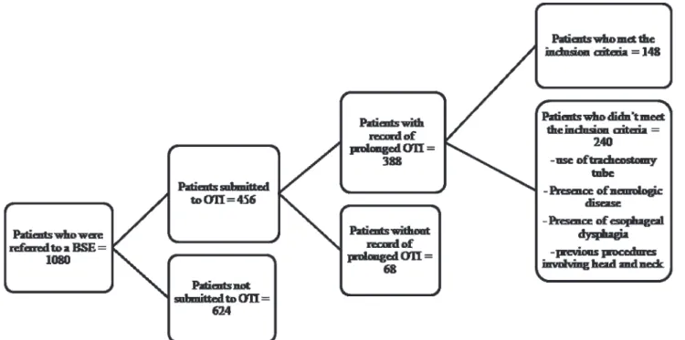
Of the 1,080 ICU patients who received a BSE, 456 had been subjected to an OTI; however, only 85% (388) had records of a prolonged OTI. Of the remaining patients, 148 met the inclusion criteria (Figure 1).
In our hospital, the weaning and discontinuation of ventilatory support protocol are based on the American Association for Respiratory Care and the American College of Critical Care Medicine guidelines (18). These criteria are as follows: (a) evidence for some reversal of the underlying cause of respiratory failure; (b) adequate oxygenation (PaO2/Fio2 ratio .150 to 200, requiring positive end-expiratory pressure [PEEP] #5 to 8 cm H2O; Fio2 #0.4 to 0.5) and a pH$7.25; (c) hemodynamic stability, as defined
by the absence of active myocardial ischemia and clinically significant hypotension (i.e., a condition requiring no vasopressor therapy or a low-dose vasopressor therapy, such as,5m/kg/min of dopamine or dobutamine); and (d)
the capability to initiate an inspiratory effort.
Measurements: Clinical Swallowing Assessment
The BSE included the application of the Dysphagia Risk Evaluation Protocol (DREP) (19), followed by the classifica-tion of the funcclassifica-tional swallowing level according to the American Speech-Language-Hearing Association National Outcome Measurement System (ASHA NOMS) (20).
DREP (19) is a Brazilian bedside assessment protocol designed for early dysphagia risk detection. In our hospital, this is the standard protocol used to assess swallowing dysfunction in patients. DREP has already been validated (21) and includes items previously described as being effective in identifying high-risk dysphagia patients (13,22,23). It includes the controlled administration of water and puree/solid volumes. DREP determines whether a patient should receive larger volumes and different textures of food and liquids, as well as the amount of monitoring necessary for safe feeding. The protocol is divided into 2 sections, a water swallow test and a puree/solid swallow test, and the results are marked as either pass or fail for each of the observed items. As determined by the authors of the protocol, patient swallowing was assessed during the administration of 5 ml of water (via a syringe); 3, 5, and 10 ml of fruit puree (from a spoon); and half a piece of bread. The tests were repeated, if necessary, up to 3 times to confirm the results. The assessment procedures consisted of 11 items for the water swallow test and 12 items for the puree/solid swallow test. Patients were placed in the upright position so that their sitting position would not interfere with the research results (24). The assessed items and the criteria used to interpret the results are described below.
A. The Water Swallow Test (5 ml of water administered from a syringe)
a. Extraoral loss: Pass - Water does not escape from the patient’s lips, and the patient manages the bolus adequately. Fail - The patient has difficulty mana-ging the bolus, which causes drooling/spillage from the mouth.
b. Oral transit time: Pass - The patient swallows the bolus within 4 seconds. Fail - Patient takes longer than 4 seconds to swallow the bolus or does not swallow it.
c. Nasal reflux: Pass - Water does not escape from the patient’s nasal cavities. Fail - Water comes out of the patient’s nasal cavities.
d. Multiple swallows per bolus: Pass - The patient only needs 1 swallow per bolus. Fail - The patient needs more than 1 swallow per bolus, which causes drooling/spillage from the mouth. Additionally, the patient needs cues to complete the task. e. Laryngeal elevation (monitored by positioning the
index and middle fingers over the hyoid bone and the thyroid cartilage): Pass - The patient reaches an average elevation of 2 fingers. Fail - The patient
does not present laryngeal elevation or presents an average elevation of less than 2 fingers.
f. Cervical auscultation (a stethoscope is placed at the lateral aspects above the cricoid cartilage and in front of the sternocleidomastoid muscle and large vessels): Pass - The patient presents the 3 char-acteristic sounds (two clicks followed by an expiratory sound), indicating that the bolus has gone through the pharynx. Fail - The patient does not present any sound or presents sounds other than those described above.
g. Oxygen saturation (baseline oxygen saturation is registered prior to the swallow test using a monitor or pulse oximetry): Pass - The patient does not present oxygen saturation changes of more than 4 units. Fail - The patient presents oxygen saturation changes of more than 4 units.
h. Voice quality: Pass - The patient does not present any alterations within the first minute after swal-lowing. Fail - The patient’s voice becomes gurgly (‘‘wet’’) within the first minute after swallowing. i. Cough: Pass - The patient does not cough within
the first minute after swallowing. Fail – The patient coughs (voluntary or not) with or without throat clearing within the first minute after swallowing. j. Choking: Pass - The patient does not choke after
swallowing. Fail - The patient chokes during and/ or after swallowing.
k. Other signs (cardiac and respiratory frequencies): Pass - The patient does not present significantly increased cardiac (60-100 beats per minute) and respiratory frequency (12-20 breaths per minute) changes. Fail - The patient presents signs of cyanosis, bronchospasm, and significant vital sign alterations.
B. The puree/solid swallow test (3, 5, and 10 ml of fruit puree offered from a spoon along with a half a piece of bread)
a. Extraoral loss: Pass - Water does not escape from the patient’s lips, and the patient manages the bolus adequately. Fail - The patient has difficulty mana-ging the bolus, which causes drooling/spillage from the mouth.
b. Oral transit time: Pass - The patient swallows the bolus within 20 seconds. Fail - Patient takes longer than 20 seconds to swallow bolus or does not swallow it.
c. Nasal reflux: The same as above.
d. Oral residue: Pass - An absence or up to 25% of the bolus residue is observed in the patient’s oral cavity. Fail - More than 25% of the bolus residue is observed in the patient’s oral cavity.
e. Multiple swallows per bolus: Pass - The patient requires 1-3 swallows per bolus. Fail - The patient requires more than 3 swallows per bolus, presents with drooling/spillage from the mouth, and needs cues to complete the task.
i. Voice quality: The same as above. j. Cough: The same as above. k. Choking: The same as above. l. Other signs: The same as above.
The ASHA NOMS swallowing level scale is a multi-dimensional tool designed to measure both the supervision and diet levels required by assigning a single number between 1 and 7 (Table 1). For this study, the patients’ specific diets and supervision levels were used to calculate each patient’s ASHA NOMS swallowing scale score. The speech-language pathologist was certified in assigning the ASHA NOMS swallowing levels. The ASHA NOMS level was determined based on the DREP results.
Statistical Analysis
Only the results obtained from the water swallow test were used for the analyses. The statistical analysis included assessment of the correlation between the water swallow test and the dysphagia risk level (i.e., ASHA NOMS). The purpose of this analysis was to identify which items (including extraoral loss, oral transit time, nasal reflux, multiple swallows per bolus, laryngeal elevation, cervical auscultation, oxygen saturation, coughing, choking, and other signs) were the most significant predictors of high dysphagia risk in the investigated population.
The variables were descriptively presented in contingency tables containing the absolute (n) and relative (%) frequen-cies. A logistic regression model was used to examine the
relationships between the independent dysphagia risk variables. All variables were analyzed using a univariate model to determine statistical significance (p#0.10). All significant variables and the interactions between them were used to obtain selections for a multivariate model (p#0.05), according to the ‘‘enter’’ procedure. The variables that remained in the model were independent predictor variables.
& RESULTS
The selected sample consisted of 91 males and 57 females, with a mean age of 53.64 years (range: 18-90 years). Patients presented an average of 1.08 OTIs (range: 1-2 OTIs) and an average of 182.4 hours of intubation (range: 48-720 hours). The average overall time required to perform the swallow-ing assessment followswallow-ing extubation was 36.0 hours (range: 24-48 hours).
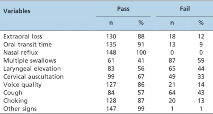
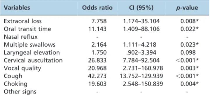
Tables 2 and 3 display the water swallow test results and the distribution of patients according to their ASHA NOMS levels, respectively. In the present study, the oxygen saturation and vital sign monitoring were recategorized as ‘‘other signs’’. Additionally, for statistical purposes, the ASHA NOMS values were also recategorized, whereby L1 represented Levels 1 to 4 and L2 represented Levels 5 to 7. Table 4 presents the logistic regression model (univariate analysis) results, which were based on the independent dysphagia risk variables. For this analysis, ASHA NOMS Level 1 patients were considered a high dysphagia risk population; however, ASHA NOMS Level 2 patients were considered a low dysphagia risk population. The univariate analysis results indicated that the extraoral loss, multiple swallows, cervical auscultation, vocal quality, cough, and choking variables were possible significant high-risk indi-cators of dysphagia. Nasal reflux was not considered in this analysis because none of the patients failed the nasal reflux test. The ‘‘other signs’’ variable was also not considered because only 1 patient failed this item.
Table 2 -The water swallow test results.
Variables Pass Fail
n % n %
Extraoral loss 130 88 18 12
Oral transit time 135 91 13 9
Nasal reflux 148 100 0 0
Multiple swallows 61 41 87 59
Laryngeal elevation 83 56 65 44
Cervical auscultation 99 67 49 33
Voice quality 127 86 21 14
Cough 84 57 64 43
Choking 128 87 20 13
Other signs 147 99 1 1
n – number of patients, % of patients.
Table 3 -The ASHA NOMS results.
L n %
1 72 49
2 76 51
L – level, n – number of patients, % of patients. Table 1 -ASHA NOMS swallowing level scale.
Level 1 The individual is not able to swallow safely with their mouth. All nutrition and hydration is received through non-oral means (i.e., a nasogastric tube).
Level 2 The individual is not able to swallow safely for nutritional and hydration purposes but may achieve some consistency with consistent maximal cues during therapy sessions only. An alternative feeding method is required.
Level 3 An alternative feeding method is required, as the individual receives less than 50% of his/her nutrition and hydration by mouth, swallowing is safe with the consistent use of moderate cues to utilize compensatory strategies, and/or the patient requires maximum diet restriction.
Level 4 Swallowing is safe but usually requires moderate cues to use compensatory strategies, the individual has moderate diet restrictions, and/or the patient still requires tube feeding and/or oral supplements.
Level 5 Swallowing is safe with minimal diet restrictions and/or the patient occasionally requires minimal cues to use compensatory strategies. The patient may occasionally self-cue. All nutrition and hydration needs are met by mouth at mealtime. Level 6 Swallowing is safe, the individual eats and drinks independently, and the individual may rarely require minimal cueing. The
individual usually self-cues when difficulty occurs and may need to avoid or requires additional time (due to dysphagia) to consume specific food items (e.g., popcorn and nuts).
Table 5 presents the logistic regression model (multi-variate analysis) results of the independent variables associated with dysphagia risk. According to these results, cervical auscultation and cough were independent predic-tive variables of high dysphagia risk.
& DISCUSSION
This study represents the largest prolonged orotracheal intubation Brazilian patient group that has been screened for possible signs of dysphagia. To our knowledge, this is one of the few studies that have investigated possible dysphagia risk predictors based on clinical symptoms in ICU patients. Early post-extubation dysfunction recognition is paramount in reducing the rate of morbidity in this high-risk population.
Extended intubation durations have been correlated with dysphagia (5,25-27) and have also been reported to be independent predictors of dysphagia severity (1,28). Post-extubation, a higher dysphagia risk was reported in patients with Glasgow Coma Scale scores #14 (6) and in patients aged$55 years (1,6). In contrast, another study found that neither age nor intubation duration was correlated with increased swallowing dysfunction in post-orotracheal intu-bation patients (29). Prolonged intuintu-bation swallowing disorders extend the time before oral myofunctional/ swallowing assessments can begin and the time to return to normal oral feeding while also delaying subsequent hospital discharges (27,28,30).
Screening procedures are generally designed to be quick (,15 minutes), to be relatively non-invasive, and to provide
little risk to the patient while identifying the dysphagia symptoms that may require an in-depth diagnostic assess-ment (23). In developing countries, the prolonged intensive medical and nursing care that is required by many patients places additional demands on already stretched healthcare
budgets (31). Moreover, routine post-extubation swallowing studies can lead to additional, and possibly unnecessary, imaging, increasing healthcare resource use. Our results indicated that altered cervical auscultation and coughing during water swallow tests increased the likelihood of dysphagia in patients who underwent prolonged orotra-cheal intubation (11).
It is critical that health professionals distinguish between screenings and diagnoses. A screening does not define the nature of a patient’s problem; it simply identifies the patient as being at risk for a significant problem/disorder (in this case, dysphagia) (23). The variables that were strong predictors of high dysphagia risk in our study are also considered to be variables associated with the possible presence of an aspiration (23,32,33). The reasons for aspirations have been discussed frequently in the literature, in which multiple causes have been postulated. It is known that alterations in the chemo- and/or mechanoreceptors (located in the pharyngeal and laryngeal mucosae) that are involved in the swallowing reflex are altered by the presence of an orotracheal tube (1). Inhibition of the sensory larynx abilities led to the absence of coughing or any other behavioral aspiration signs in patients following liquid bolus ingestion. Furthermore, this effect was observed immediately and 4 hours following extubation, and the detrimental effects were significantly reduced within 8 hours post-extubation (34). Additionally, most mucosal lesions caused by orotracheal tubes are healed 3 days following extubation (35).
Swallowing dysfunctions may persist, however, despite the removal of orotracheal tubes and the necessary spontaneous recovery periods. The swallowing dysfunction mechanisms following an extubation are thought to be a combination of muscle ‘‘freezing’’ (which may be attribu-table to the lack of swallowing while intubated) and the loss of proprioception (which may be attributable to mucosal lesions) (6).
Cervical auscultation is increasingly being used to supplement clinical swallowing assessments. The sounds associated with swallowing have been investigated using accelerometers and microphones to identify acoustic char-acteristics (36). Additionally, these sounds may also predict aspiration onset (37). The use of cervical auscultation varies with respect to its reported reliability (38) and validity compared with videofluoroscopic swallow studies (VFSS) (39,40). VFSS itself, however, has poor intra- and inter-rater reliability (41-43). According to Lazareck and Moussavi (37), swallowing sound assessments have great potential to reduce the need for VFSS and to assist in overall clinical swallowing assessments. It is clear from the literature that, despite the methods used to assess swallowing, providing the necessary training is indispensable.
A study by Bordon et al. (1) analyzed swallowing dysfunction risk factors after prolonged intubation in trauma patients. The authors also used a simple bedside speech pathology assessment (specifically, their swallowing failure indicators included coughing when drinking liquids and the presence of multiple swallows). Patients with and without post-extubation dysphagia were then compared by univariate analysis with respect to their age, the number of ventilator and ICU days, the presence or absence of pneumonia, and other variables. The authors suggested that older patients (above 55 years of age) with extended ICU stays and ventilator requirements may benefit from
Table 4 -Logistic regression (univariate analysis) results based on independent dysphagia risk variables.
Variables Odds ratio CI (95%) p-value
Extraoral loss 7.758 1.174–35.104 0.008*
Oral transit time 11.143 1.409–88.106 0.022*
Nasal reflux - -
-Multiple swallows 2.164 1.111–4.218 0.023*
Laryngeal elevation 1.750 .902–3.394 0.098 Cervical auscultation 26.833 7.784–92.504 ,0.001*
Vocal quality 20.968 2.731–160.978 0.003*
Cough 42.273 13.752–129.939 ,0.001*
Choking 19.603 2.548–150.839 0.004*
Other signs - -
-CI – confidence interval, *significant results.
Table 5 -A logistic regression (multivariate analysis) of the independent variables associated with dysphagia risk.
Variables Odds ratio CI (95%) p-value
Extra oral loss 1.837 0.192–17.574 0.598
Multiple swallows 2.056 0.698–6.059 0.191
Cervical auscultation 12.709 2.940–54.931 0.001*
Vocal quality 9.115 0.935–88.853 0.057
Cough 14.817 3.444–63.740 ,0.001*
Choking 2.489 0.194–31.958 0.484
early swallowing evaluations. Similarly, Leder et al. (25) investigated aspiration incidence following extubation in critically ill trauma patients using a bedside transnasal fiberoptic endoscopic swallowing evaluation. The authors noted that aspiration was identified in 45% of the subjects, 44% of whom were silent aspirators. The authors argued that aspiration identification may reduce the likelihood of pulmonary complications following an extubation.
Although there are a few potential limitations of our study (e.g., it was a single-institution investigation and thus may only reflect local patient characteristics, and the study did not include any confirmatory fluoroscopic imaging to document silent or subclinical aspirations), our findings demonstrated that the overall swallowing deficit rates in patients subjected to prolonged orotracheal intubations (i.e., ASHA NOMS Level 1) were comparable to those that have been previously published. Studies performed with a direct laryngoscope indicated that approximately 56% of the observed critically ill patients displayed evidence of swallowing dysfunction (5,8). Similarly, studies performed with fiberoptic endoscopic evaluations (FEES) demon-strated that swallowing dysfunction occurred in approxi-mately 52% of patients after prolonged intubation (29).
In the literature, there is no clear definition of which patients are at risk for dysphagia. The results of our study indicate that if patients who are subjected to prolonged orotracheal intubation present with altered cervical auscul-tation and coughing during a water swallow test, these patients should be given an early and more detailed swallowing assessment. Moreover, these assessments should possibly include imaging examinations before restarting oral nutrition. A similar study, performed with trauma patients subjected to mechanical ventilation (3), investigated whether a BSE could identify swallowing dysfunction in this patient group. One of the main findings of this study was that the patients who failed the BSE required longer mechanical ventilation than those who passed the BSE. Additionally, 78% of the patients intubated for more than 72 hours failed the BSE. All patients who passed the BSE, however, were discharged from the hospital without a clinical aspiration event. The authors also identified independent risk factors for BSE failures, which included tracheostomy, older age, prolonged mechanical ventilation, delirium tremens, traumatic brain injury, and spine fracture.
Finally, we would like to state that we did not study the long-term consequences of post-extubation dysphagia in our cohort because our end point was an evaluation performed within 48 hours of the observed extubations. Future studies at our institution will attempt to answer this and other questions.
& AUTHOR CONTRIBUTIONS
Medeiros GC was responsible for the data collection and analysis, interpretation of the results, and manuscript writing. Sassi FC organized and conducted the statistical analyses, interpreted the results, and wrote a major portion of the manuscript. Mangilli LD participated in the data collection and analyses and organized and conducted the statistical analyses. Zilberstein B was responsible for the medical criteria adopted in the experimental design and contributed to the data analysis and manuscript preparation. Andrade CR was responsible for the research and experimental design and contributed to the data analysis and manuscript preparation.
& REFERENCES
1. Bordon A, Bokhari R, Sperry J, Testa D, Feinstein A, Ghaemmaghami V. Swallowing dysfunction after prolonged intubation: analysis of risk factors in trauma patients. Am J Surg. 2011;202(6):679-82.
2. Heffner JE. Swallowing complications after endotracheal extubation: moving from ‘‘wether’’ to ‘‘how’’. Chest. 2010;137(3):509-10, http://dx. doi.org/10.1378/chest.09-2477.
3. Brown CVR, Hejl K, Mandaville AD, Chaney PE, Stevenson G, Smith C. Swallowing dysfunction after mechanical ventilation in trauma patients. J Crit Care. 2011;26(1):108.e9-13.
4. Martin-Harris B, Brodsky MB, Price CC, Michel Y, Walters B. Temporal coordination of pharyngeal and laryngeal dynamics with breathing during swallowing: single liquid swallows. J Appl Physiol. 2003; 94(5):1735-43.
5. Ajemian MS, Nirmul GB, Anderson MT, Zirlen DM, Kwasnik EM. Routine fiberoptic endoscopic evaluation of swallowing following prolonged intubation implications for management. Arch. Surg. 2001;136(4):434-7, http://dx.doi.org/10.1001/archsurg.136.4.434. 6. Barquist E, Brown M, Cohn S, Lundy D, Jackowski J. Postextubation
fiberoptic endoscopic evaluation of swallowing after prolonged endo-tracheal intubation: a randomized, prospective trial. Crit Care Med. 2001;29(9):1710-3, http://dx.doi.org/10.1097/00003246-200109000-00009. 7. Skoretz SA, Flowers HL, Martino R. The incidence of dysphagia following endotracheal intubation: a systematic review. Chest. 2010; 137(3):665-73, http://dx.doi.org/10.1378/chest.09-1823.
8. Tolep K, Getch CL, Criner GJ. Swallowing dysfunction in patients receiving prolonged mechanical ventilation. Chest. 1996;109(1):167-72, http://dx.doi.org/10.1378/chest.109.1.167.
9. Bastian RW. Videoendoscopic evaluation of patients with dysphagia: an adjunctto the modified barium swallow. Otolaryngol Head Neck Surg. 1991;104(3):339-50.
10. Langmore SE, Schatz MA, Olsen N. Endoscopic and videofluoroscopic evaluations of swallowing and aspiration. Ann Otol Rhinol Laryngol. 1991;100(8):678-81.
11. Mangilli LD, Sassi FC, Santos SS, Andrade CRF. Oral sensorimotor function for feeding in patients with tetanus. Acta Tropic. 2009; 111(3):316-20, http://dx.doi.org/10.1016/j.actatropica.2009.05.015. 12. Otto DJ. Observer variation in evaluation of videofluoroscopic
swallow-ing studies: a continuswallow-ing problem. Dysphagia. 1998;13(3):148-50. 13. Hinchey JA, Shepard T, Furie K, Smith D, Wang D, Tonn S. Formal
dysphagia screening protocols prevent pneumonia. Stroke. 2005; 36(9):1972-6, http://dx.doi.org/10.1161/01.STR.0000177529.86868.8d. 14. Hinds NP, Wiles CM. Assessment of swallowing and referral to speech
and language therapists in acute stroke. Q J Med. 1998;91(12):829-35. 15. ASHA. American Speech-Language-Hearing Association. Model
Medical Review Guidelines for Dysphagia Services 2004. Available from: http://www.asha.org/uploadedfiles/practice/reimbursement/med icare/DunCorpDysphHCE.pdf. Accessed 03 July 2012.
16. Hammond CAS, Goldstein LB. Cough and aspiration of food and liquids due to oral-pharyngeal dysphagia – ACCP evidence-based clinical practice guidelines. Chest. 2006;129(1 Suppl):154-68, http://dx.doi.org/ 10.1378/chest.129.1_suppl.154S.
17. Nishiwaki K, Tsuji T, Liu M, Hase K, Tanaka N, Fujiwara T. Identification of a simple screening tool for dysphagia in patients with stroke using factor analysis of multiple dysphagia variables. J Rehabil Med. 2005;37(4):247-251, http://dx.doi.org/10.1080/16501970510026999. 18. Maclntyre NR. Evidence-based guidelines for weaning and discontinu-ing ventilatory support: a collective task force facilitated by a the American College of Chest Phsysicians; the American Association for Respiratory Care; and the American College of Critical Care Medicine. Chest. 2001;120(6 Suppl):375-96, http://dx.doi.org/10.1378/chest.120.6_ suppl.375S.
19. Padovani AR, Moraes DP, Mangilli LD, Andrade CRF. Protocolo fonoaudiolo´gico de avaliac¸a˜o do risco para disfagia (PARD). Rev Soc Bras Fonoaudiol. 2007;12(3):199-205, http://dx.doi.org/10.1590/S1516-80342007000300007.
20. Rockville MD. American Speech-Language-Hearing Association National Outcome Measurement System (NOMS). Adult Speech-Language Pathology training manual, ASHA, 1998.
21. Padovani AR, Moraes DP, Mangilli LD, Andrade CRF. Protocolo de avaliac¸a˜o fonoaudiolo´gica de risco para disfagia (PARD). In: Andrade CRF, Limongi SCO (Eds). Disfagia – pra´tica baseada em evideˆncias, 1 ed. Sa˜o Paulo: Sarvier, 2012. p.62-73.
22. Smith Hammond CA, Goldstein LB. Cough and aspiration of food and liquids due to oral-pharyngeal dysphagia: ACCP evidence-based clinical practice guidelines. Chest. 2006;129(1 Suppl):154S-168S, http://dx.doi.org/ 10.1378/chest.129.1_suppl.154S.
23. Logemann JA, Veis S, Colangelo L. A screening procedure for oropharyngeal dysphagia. Dysphagia. 1999;14(1):44-51, http://dx.doi. org/10.1007/PL00009583.
Clin. 2000;38(3):219-42, http://dx.doi.org/10.1097/00004311-200007000-00013.
25. Leder SB, Cohn SM, Moller BA. Fiberoptic endoscopic documentation of the high incidence of aspiration following extubation in critically ill trauma patients. Dysphagia. 1998;13(4):208-12, http://dx.doi.org/10. 1007/PL00009573.
26. Postma G, McGuirt F, Butle SG, Ress CJ, Crandall HL, Tansavatdi K. Abnormalities in hospitalized patients with dysphagia. Laryngoscope. 2007;117(10):1720-2, http://dx.doi.org/10.1097/MLG.0b013e31811ff906. 27. Rumbach AF, Ward EC, Cornwell PL, Bassett LV, Muller JM. Clinical
progression and outcome of dysphagia following thermal burn injury: a prospective cohort study. J Burn Care Res. 2012;33(3):336-46, http://dx. doi.org/10.1097/BCR.0b013e3182356143.
28. Barker J, Martino R, Reichart B, Hickey EJ, Ralph-Edwards A. Incidence and impact of dysphagia in patients receiving prolonged endotracheal intubation after cardiac surgery. Can J Surg. 2009;52(2):119-24. 29. El Solh A, Okada M, Bhat A, Pietrantoni C. Swallowing disorders post
orotracheal intubation in the elderly. Intensive Care Med. 2003; 29(9):1451-5, http://dx.doi.org/10.1007/s00134-003-1870-4.
30. Ward EC, Green K, Morton AL. Patterns and predictors of swallowing resolution following adult traumatic brain injury. J Head Trauma Rehabil. 2007;22(3):184-91, http://dx.doi.org/10.1097/01.HTR.0000271119.96780.f5. 31. Thwaites CL, Farrar JJ. Preventing and treating tetanus. BMJ. 2003;
326(7381):117-8, http://dx.doi.org/10.1136/bmj.326.7381.117.
32. Smith-Hammond CA, Goldstein LB, Horner RD, Ying J, Gray L, Gonzales-Rothi L, Bolser DC. Prediciting aspiration in patients with ischemic stroke: comparison of clinical signs and aerodynamic measures of voluntary cough. Chest. 2009;135(3):769-77, http://dx.doi.org/10.1378/chest.08-1122. 33. Camargo FP, Ono J, Park M, Caruso, Carvalho CRR. An evaluation of respiration and swallowing interaction after orotracheal intubation. Clinics. 2010;65(9):919-22, http://dx.doi.org/10.1590/S1807-59322010000900015.
34. Burgess GE, Cooper JR, Marino RJ, Peuler MJ, Warriner RA. Laryngeal competence after tracheal extubation. Anesthesiology. 1979;51(1):73-7, http://dx.doi.org/10.1097/00000542-197907000-00016.
35. Stauffer JL, Olson DE, Petty TL. Complications and consequences of endotracheal intubation and tracheotomy. Am J Med. 1981;70(1):65-76. 36. Cichero JAY, Murdoch BE. Acoustic signature of the normal swallow:
characterization by age, gender, and bolus volume. Ann Otol Rhinol Laryngol. 2002;111(7 Pt 1):623-32.
37. Lazareck LJ, Moussavi ZMK. Classification of normal and dysphagic swallows by acoustical means. IEEE Trans Biomed Eng. 2004;51(12):2103-112, http://dx.doi.org/10.1109/TBME.2004.836504.
38. Borr C, Hielscher-Fastabend M, Phil D, Lucking A. Reliability and validity of cervical auscultation. Dysphagia. 2007;22(3):225-34, http://dx. doi.org/10.1007/s00455-007-9078-3.
39. Mann G, Hankey GJ. Initial clinical and demographic predictors of swallowing impairment following acute stroke. Dysphagia. 2001; 16(3):208-5, http://dx.doi.org/10.1007/s00455-001-0069-5.
40. Mari F, Matei M, Ceravolo MG, Pisani A, Montesi A, Provinciali L. Predictive value of clinical indices in detecting aspiration in patients with neurological disorders. J Neurol Neurosurg Psychiatry. 1997; 63(4):456-60, http://dx.doi.org/10.1136/jnnp.63.4.456.
41. Kuhlemeier KV, Yates P, Palmer JB. Intra- and interrater variation in the evaluation of videofluorographic swallowing studies. Dysphagia. 1998; 13(3):142-7, http://dx.doi.org/10.1007/PL00009564.
42. McCullough GH, Wertz RT, Rosenbek JC, Millis RH, Ebb WG, Ross KB. Inter- and intrajudge reliability for videofluoroscopic swallowing evaluation measures. Dysphagia. 2001;16(2):110-1, http://dx.doi.org/ 10.1007/s004550010004.