653
CLINICS 2007;62(5):653-6
LETTER TO THE EDITOR
1. Nephrology Department - Sao Paulo University Medical School – São Paulo, SP/Brazil
2. Division of Pathology - Sao Paulo University Medical School– São Paulo, SP/Brazil
3. Hospital das Clinicas School of Medicine– São Paulo, SP/Brazil Email: [email protected]
HIV INFECTION AND ACUTE GLOMERULONEPHRITIS
Silvia M. O. Titan1, Leonardo Testagrossa2, Luiz Balthazar Saldanha2, Rui Toledo Barros3, Viktória Woronik1
INTRODUCTION
Human immunodeficiency virus (HIV) infection is known to be associated with various types of glomerulone-phritis. Human immunodeficiency HIV – associated kidney diseases have been classified into 3 different groups: HIV-associated nephropathy (HIVAN), also known as HIV-asso-ciated focal and segmental glomerulosclerosis (HIV-FSGS), hemolytic-uremic syndrome (HIV-HUS), and HIV-associated immune complex renal diseases.1,2 Previously thought to be
rare, these forms of glomerulonephritis have become more frequent, and it is important that the medical community learns more about its diagnosis and treatment.
Case description
A Caucasian 34-year-old man was referred to our clinic due to an acute loss of kidney function. A diagnosis of HIV infection was made 3 years before the patient had been un-der regular follow-up at the Infectious Desease Department since then. The last CD4 count (3 months before consulta-tion) was 374 cells/µL, and the HIV viral load was 1350 000 copies/mL (6.1 log). He was not on anti-retroviral therapy and had never presented any opportunistic infec-tion. There was no history of diabetes, hypertension, kid-ney disease, or any other remarkable comorbidity. There was also no history of intravenous drug use or exposure to any nephrotoxic drugs.
Four to five weeks before consultation, he started to have asthenia. He reported no other symptoms, except for foamy urine. One week before consultation, he presented fever and dry cough, with a normal chest X-ray, and was started on azitromycin 500 mg/day and prophylaxis with sulfamethoxazole-trimethoprim. His laboratory test results on this day revealed a creatinine level of 1.8 mg/dL,
dis-crete anemia, hematuria, and nephrotic-range proteinuria (Table 1). Creatinine and urinalysis 3 months before were normal.
On the day he was first seen in our clinic, physical ex-amination revealed a healthy appearance, a blood pressure of 120 x 70 mm Hg, normal temperature, pulse of 66 beats per minute, and no signs of edema. Chest, heart, and ab-dominal examination showed no abnormalities. Cough and fever had vanished, and the patient was feeling well. His new laboratory results demonstrated a rapid worsening in renal function, with a creatinine value of 3.2 mg/dL (an interval of 1 week after the first test results, Table 1). The 24-hour urine collection confirmed a nephrotic range pro-teinuria of 4.5 g/day. Serum albumin was 2.2 mg/dL; total cholesterol was 192 mg/dL; LDL-cholesterol, 52 mg/dL; HDL-cholesterol, 78 mg/dL; and triglycerides, 309 mg/dL. A discrete anemia was noticed, with a normal mean cor-puscular volume, serum iron concentration of 90 µg/dL, and ferritin of 1882 µg/dL. Despite a high LDH, not confirmed Table 1 - Results of laboratory tests.
One week before On admission
Urea (mg/dL) 89 91
Creatinine (mg/dL) 1.8 3.2
Sodium (mEq/L) 141
Potassium (mEq/L) 4.7 6.8
Hemoglobin (g/dL) / Hematocrit (%) 10.6 / 32 10.2 /32
White cell count (per mm3) 4860 4250
Platelet count (per mm3) 225 000 207 000
Lactate dehydrogenase (U/L) 922 927
Albumin / Globulins (mg/dL) 2.2 / 3.1
Glucose (mg/dL) 94
Urinalysis
Leukocytes (per field) 2 2
Erythrocytes (per field) 10 12
Protein (g/L) > 1 > 1
24h-proteinuria g/day 4.52
Erythrocyte sedimentation rate (mm) 114
C reactive protein (mcg/mL) 2.42
C4 (mg/dL) 24
C3 (mg/dL) 103
654
CLINICS 2007;62(5):653-6 HIV infection and acute glomerulonephritis
Titan SMO et al.
subsequently, there was no other laboratory evidence of thrombotic microangiopathy, since there was no sign of schistocytes in the blood smear and the haptoglobin level was normal. Hepatitis B serology revealed positive anti-Hbe and total antiHbc antibodies, with negative results for AgHbs, AgHbe, anti-Hbs, and IgM anti-Hbc. Hepatitis C serology and tests for antinuclear and anti-neutrophil cy-toplasm antibodies were negative, and the complement lev-els were normal.
He was admitted in the nephrology ward and underwent a kidney biopsy.
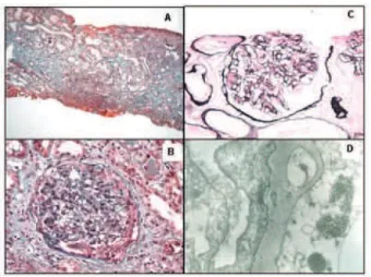
Kidney biopsy
Light microscopic analysis of a specimen containing 5 glomeruli showed mesangial hypercellularity, an epithelial crescent in 1 glomerulus, tubular focal atrophy with thick-ening and shrinking of the tubular basal membrane, and fibrosis on 40% of the interstitial space. Immunofluores-cence demonstrated granular deposits of IgM and C3 in the mesangium. Electron microscopy revealed expansion of the mensagial matrix, hypertrophy and cytoplasmatic vacuoli-zation of mensagial cells, thickening and segmental split-ting of the basal membrane, and hypertrophy and vacualization of podocytes, with diffuse fusion of podocyte foot processes. Electron-dense deposits were noted on the mesangium and subendothelial spaces, and abundant tubuloreticular inclusions were present in the cytoplasm of endothelial cells. Immunohistochemistry for hepatitis B was negative.
Diagnosis: HIV-associated immune complex-mediated glomerulonephritis with a membranoproliferative pattern.
DISCUSSION
Among HIV-associated kidney diseases, HIV-FSGS seems to be the most common form of HIV glomerulopathy. In the United States, this disease is increas-ing in prevalence, and there is a higher risk among patients of the black race. Recent reports suggest that the same pat-tern is not observed in various European countries, with a higher proportion of cases attributed to HIV-HUS and im-mune-mediated glomerulonephritis.3,4 Although there is
lack of epidemiological data concerning other parts of the world, the incidence of HIV-FSGS in African descendents outside the US does not seem to be so high.5
HIV-associated nephropathy can occur in any phase of the HIV infection, including acute seroconversion, but is usually seen later in the disease, when viral load is higher. The clinical picture is that of massive nephrotic syndrome, although edema and hypertension are generally absent. Pa-tients can also present non-nephrotic range proteinuria as-sociated with a variable loss of kidney function. Kidney biopsy usually shows a severe form of FSGS, frequently with characteristics of the collapsing form (severe shrink-ing of the glomerulus accompanied by microcystic tubular dilation). Tubuloreticular inclusions are universally seen, usually located in the cytoplasm of endothelial cells. HIV-associated focal and segmental glomerulosclerosis carries a poor prognosis and is currently the leading cause of end-stage renal disease due to HIV in US.6 Treatment is based
on viral load control involving highly active antiretroviral therapy (HAART) associated with use of angiotesin-con-verting enzyme (ACE) inhibitors. The beneficial impact of ACE inhibitors in HIV-FSGS was shown in 2 nonrandomized case-control studies,7,8 showing reduction
of proteinuria and improvement in renal function. More controversial is the use of corticosteroids, with lack of evi-dence-based data. Although there are reports of improve-ment of proteinuria and renal function with corticosteroids, a high recurrence rate of nephrotic syndrome and an in-crease in infectious-associated morbidity and mortality are still a concern.
HIV has become an important cause of HUS-TTP worldwide. The clinical picture is that of typical hemolytyc anemia associated with low platelet count and an increase in biomarkers of hemolysis (LHD, indirect bilirrubin, low haptoglobin). A discrete consuming of complement might be seen as a result of microangiopathy. The renal manifes-tation is variable, and the disease can present as an acute glomerulonephritis with sudden loss of kidney function, or as a more indolent nephropathy, characterized mainly by non-nephrotic proteinuria and mild-to-moderate renal in-sufficiency. The kidney pathology is no different from other
655
CLINICS 2007;62(5):653-6 HIV infection and acute glomerulonephritis
Titan SMO et al.
forms of renal microangiopathy: platelet and fibrin thrombi in capillaries and renal arterioles, intimal lesion, fibrinoid necrosis, and mesangiolysis due to ischemia of the glomerulus are all histological features. Typical “onion skin” lesions of microangiopathy might be seen. Immun-ofluorescence is usually positive for fibrin and fibrinogen, but low positivity for complement fractions and IgM might be present. Tubuloreticular inclusions are also seen, mainly in vascular endothelial cells.
Previously thought to be extremely rare, diverse forms of HIV-associated immune complex glomerulonephritis have recently been described more frequently. Typical postinfectious acute glomerulonephritis, membranous glomerulonephritis, membranoproliferative glomerulone-phritis, lupus-like glomeruloneglomerulone-phritis, IgA nephropathy, and fibrillary glomerulonephritis3,4,9-11 have all been described.
Of those, membranoproliferative glomerulonephritis and lupus-like nephritis seem to be the most common forms.
It is possible that the variability in renal histology pat-terns observed is a result of the interaction between host environmental and genetic factors and a broad spectrum of immunogenicity triggered by the virus. The pathophysi-ological mechanisms underlying the renal insult remain largely unknown. Direct cytopathic effect, expression of intracellular antigens and deposition of in situ immune complexes, abnormal activation of the immune system with formation of circulating immune complexes, and subse-quent renal deposition are all possible explanations.12
In this present case, renal pathology showed an acute glomerulonephritis, with signs of splitting of the basement membrane and the appearance of double contours. This his-tological pattern associated with the presence of tubuloreticular inclusions suggests the diagnosis of HIV-associated immune complex glomerulonephritis. Tubuloreticular inclusions have been described in all forms of HIV-nephritis, from HIVAN, to HUS and immune com-plex glomerulonephritis. In fact, these inclusions have been described in many organs affected by HIV, and their ap-pearance seems to have a correlation with viral load and disease progression. Despite the initial assumption that these inclusions were composed of viral particles, later studies13-14 suggest that they are related to interferon
pro-duction. Tubuloreticular inclusions (TRI) are not pathog-nomonic of HIV, although they appear very frequently in
this disease: autoimmune diseases (SLE15,
dermatomyosi-tis), lymphoproliferative disease, certain solid tumors, and other viral diseases (parvovirus, Epstein-Barr) have also been associated with these inclusions, suggesting that TRI are probably an immunological epiphenomenon of several diseases.
This case illustrates very nicely how broad the differ-ential diagnosis of HIV-related nephritis can be. This pa-tient was Caucasian, had a CD4 count of 374, and no his-tory of opportunistic infections or drug exposure, and he developed a severe and aggressive form of acute glomeru-lonephritis. Diagnosis could not be established on clinical and laboratory data, and kidney biopsy was crucial to di-agnosis, treatment decision, and prognosis. This case em-phasizes that although HIVAN and HUS are the most com-mon forms of HIV-related kidney diseases, we need to con-sider the immune complex glomerulonephritis as alterna-tive diagnosis.
It might be argued that hepatitis B could be responsi-ble for the acute glomerulonephritis, since the patient has not shown seroconversion to anti-AgHbs. It is known that a few patients with serology showing negative results for AgHbs, AgHbe, and anti-Hbs can have a positive viral load for hepatitis B detected by qualitative assays for detecting serum HBV DNA. More importantly, interpretation of hepatitis B serology is more complicated in HIV patients, since a fall in CD4 is associated with a risk of hepatitis B replication, while antiviral treatment for HIV might cause a reduction in HBV viral load. Although we could not per-form a polymerase chain reaction (PCR) test for hepatitis B in this case, immunohistochemistry was negative for hepatitis B, suggesting that hepatitis B virus was not asso-ciated with the appearance of the glomerulonephritis in this case.
Our patient was started on HAART associated with ACE inhibitors. Considering the good clinical condition of the patient and absence of previous or current opportunistic in-fections, we also started oral 1 mg/kg prednisone for 1 month, with full tapering after 3 months. There was a marked improvement in renal function, with a current creatinine of 1.3 mg/dL (creatinine clearance of 60 mL/min), but no re-gression of proteinuria (4.3 g/day) after 3 months of treat-ment. After 1 year of follow-up, creatinine clearance was 73 mL/min and proteinuria was reduced to 1 g/day.
REFERENCES
1. Weiner NJ, Goodman JW, Kimmel PL. The HIV-associated renal diseases: current insight into pathogenesis and treatment. Kidney Int. 2003;63:1618-31.
656
CLINICS 2007;62(5):653-6 HIV infection and acute glomerulonephritis
Titan SMO et al.
3. Nochy D, Glotz D, Dosquet P, Pruna A, Guettier C, Weiss L, et al. Renal disease associated with HIV infection: a multicentric study of 60 patients from Paris hospitals. Nephrol Dial Transplant. 1993;8:11-9. 4. Casanova S, Mazzucco G, Barbiano di Belgiojoso G, Motta M, Boldorini
R, Genderini A, et al. Pattern of glomerular involvement in human immunodeficiency virus-infected patients: an Italian study. Am J Kidney Dis. 1995;26:446-53.
5. Bechar DM, Shlush LI, Maor C, Lorber M, Skorecki K. Absence of HIV-associated nephropathy in Ethiopians. Am J Kidney Dis. 2006;47: 88-94.
6. US Renal Data System (USRDS): USRDS 2001 Annual Data Report. Bethesda MD, The National Institute of Health, National Institute of Diabetes and Digestive and Kidney Diseases, 2001.
7. Burns GC, Paul SK, Toth IR, Sivak SL. Effect of angiotensin-converting enzyme inhibition in HIV-associated nephropathy. J Am Soc Nephrol. 1997;8:1140-6.
8. Kimmel PL, Mishkin GJ, Umana WO. Captopril and renal survival in patients with human immunodeficiency virus nephropathy. Am J Kidney Dis. 1996;28:202-8
9. Haas M, Kaul S, Eustace JA. HIV-associated immune complex glomerulonephritis with “ lupus-like” features: a clinicopathologic study of 14 cases. Kidney Int. 2005;67:1381-90.
10. Enriquez R, Cabezuelo JB, Escolano C, Perez M, Amoros F, Gutierrez-Rodero F, et al. Postinfectious diffuse proliferative glomerulonephritis and acute renal failure in an HIV patient. Clin Nephrol. 2004;61:278-81.
11. Cheng JT, Anderson HL Jr, Markowitz GS, Appel GB, Pogue VA, D’Agati VD. Hepatitis C virus-associated glomerular disease in patients with human immunodeficiency virus coinfection. J Am Soc Nephrol. 1999;10:1566-74.
12. Ross MJ, Klotman PE. Recent progress in HIV-associated nephropathy. J Am Soc Nephrol. 2002;13:2997-3004.
13. Luu JY, Bockus D, Remington F, Bean MA, Hammar SP. Tubuloreticular structures and cylindrical confronting cisternae: a review. Hum Pathol. 1989;20:617-27.
14. Orenstein JM, Preble OT, Kind P, Schulof R. The relationship of serum alpha-interferon and ultrastructural markers in HIV-seropositive individuals. Ultrastruct Pathol. 1987;11:673-9.