Dementia & Neuropsychologia 2007;1(4):356-360
Clinicopathological correlates of Alzheimer’s
disease in a general autopsy series from Brazil
Lea Tenenholz Grinberg
1,2, Renata Eloah de Lucena Ferretti
1,3, Renata E.P. Leite
1,
Jose Marcelo Farfel
1,3, Silmara P. Pacheco
1, Ana Teresa Di Lorenzo Alho
1,2, Renata P. Grisoli
1,2,
Heidy T.M. Matos
1,2, Eliza G. Moreira
1,2, Erika S. Balbino
1,2, Katia C. Oliveira
1,
Sergio Rosemberg
1, Heráclito Barbosa Carvalho
4, Carlos Augusto G. Pasquallucci
1,
Paulo Hilario N. Saldiva
1, Wilson Jacob-Filho
3, Ricardo Nitrini
5Abstract – The current neuropathological staging models of Alzheimer’s disease (AD) have been developed within the last 20 years. Nevertheless, they were mostly tested on Caucasians of Northern European ancestry or on Asians. Objective: To verify which of the accepted neuropathologic criteria best discriminates AD from normal aging in a well characterized Brazilian clinicopathological series. Methods: A random sample consisting of89 sub-jects belonging to the Brazilian Brain Bank of the Aging Brain Study were clinically and neuropathologically fully assessed using immunohistochemistry. Clinical and functional statuses were assessed by interviewing a reliable informant. The Clinical dementia rating scale (CDR) was compared to Braak and Braak stage, the consortium to establish a registry for Alzheimer’s disease (CERAD) score and NIA-Reagan (National Institute of Aging - Reagan Institute) score. Subjects with a neuropathologic diagnosis other then AD were excluded (n=27). Results:The CDR score distribution for the 62 selected subjects was as follows: CDR0=39, CDR0.5=9, CDR≥1=14. There were no differences regarding age, gender and education among the groups. CDR score correlated best with the CERAD score (r=0.5303; p<0.001) . Braak and Braak stage was significantly higher in subjects with higher CDR. Correlation of the NIA-Reagan criteria was partially disrupted because a large proportion of subjects did not fit any of its categories. Conclusions: In this series, CERAD criteria better correlated with the CDR groups. Consistent with earlier studies, some cognitively normal subjects have AD neuropathological diagnosis.
Key words: Alzheimer’s disease, dementia, diagnostic criteria, neuropathological criteria, brain bank.
Correlação clinicopatológica na doença de Alzheimer em casuística de autópsia no Brasil
Resumo – Os modelos de estadiamento neuropatológico da doença de Alzheimer (DA) têm sido desenvolvidos nos últimos 20 anos. Entretanto, têm sido quase exclusivamente testados em caucasianos de ascendência norte-européia ou em asiáticos. Objetivos: verificar quais dos critérios neuropatológicos discrimina melhor entre a DA e o envelhecimento normal em uma casuística clinicopatológica brasileira bem caracterizada. Métodos: uma amostra aleatória de 89 casos do Banco Brasileiro de Encéfalos do Estudo de Envelhecimento Cerebral foi sub-metida à avaliação neuropatológica completa com imunohistoquímica. As condições clínicas e funcionais foram avaliadas mediante entrevista com informante confiável. Os escores na Clinical Dementia Rating Scale (CDR) foram comparados com os escores dos estágios de Braak e Braak, do CERAD (Consortium to Establish a Registry for Alzheimer’s Disease) e do consórcio NIA-Reagan (National Institute of Aging-Reagan Institute). Casos com diagnósticos neuropatológicos diferentes de DA foram excluídos (n=27). Resultados: Os 62 casos foram classifi-cados em: CDR0=39, CDR0,5=9, CDR≥1=14. Não havia diferenças quanto a idade, gênero e escolaridade entre os grupos. Os escores no CERAD correlacionaram-se melhor com os do CDR (r=0,5303; p<0,001). Os escores nos estágios de Braak e Braak foram significativamente mais elevados nos casos com CDR mais altos. A correlação do CDR com os escores dos critérios NIA-Reagan foi parcialmente rompida porque grande proporção de casos não se enquadrava em nenhuma das categorias diagnósticas destes critérios. Conclusões: Nesta casuística, os cri-térios do CERAD correlacionam-se melhor com os do CDR. Como observado por outros estudos, alguns casos de indivíduos cognitivamente normais, preencheram critérios neuropatológicos para o diagnóstico de DA. Palavras-chave: doença de Alzheimer, demência, critérios diagnósticos, critérios neuropatológicos, banco de encéfalos, banco de cérebros.
1Department of Pathology, University of São Paulo Medical School. 2Instituto Israelita de Ensino e Pesquisa Albert Einstein, São Paulo. 3Division of Geriatrics, University of São Paulo Medical School. 4Department of Preventive Medicine, University of São Paulo Medical School . 5Behavioral and Cognitive Neurology Unit, Department of Neurology, and Cognitive Disorders Refeence Center (CEREDIC). Hospital das Clínicas of the University of São Paulo Medical School.
Lea Tenenholz Grinberg – Avenida Dr. Arnaldo, 455 / 1st floor / room 1351 - 01246-903 São Paulo SP - Brazil. E-mail: [email protected]
Dementia prevalence in Brazil is expected to increase
by around 390% between 2001 and 2040.
1A definitive
di-agnosis of Alzheimer’s disease (AD) requires both a clinical
history of dementia and neuropathologic confirmation at
autopsy. Given that age-related changes overlap with early
changes in AD, it is important to adopt
neuropatholog-ic criteria able to reliably distinguish between these two
conditions. The currently used neuropathologic staging
models of AD have been developed in the last 20 years.
While the relationship of the maximum frequency of
neu-ritic plaques (NP) with the age of the subject associated to
the history of dementia are the pivotal constituents of the
Consortium to Establish a Registry for Alzheimer’s Disease
criteria
(CERAD),
2the distribution and burden of
neuro-fibrillary tangles (NFT) determine each of the seven Braak
and Braak stages.
3Finally, the National Institute on Aging
and the Ronald and Nancy Reagan Research Institute of the
Alzheimer’s Association
4proposed criteria for AD based on
both NFT and NP (hereafter called NIA-Reagan criteria).
The NIA-Reagan criteria stratifies cases by the likelihood
that clinical dementia has been caused by AD lesions in the
brain. Table 1 gives a summary of these criteria.
Although more than 10 years have passed since the last
criteria were published, which of them best correlates with
clinical symptoms remains a matter of debate. In addition,
these criteria were largely tested in Caucasians of Northern
European ancestry or in Asians
5-7and there may be
biologi-cal differences in burden and distribution of AD changes
among different ethnic groups.
8The Brazilian population
is composed mainly of a mixture of Caucasians of
South-ern European ancestry, Africans and Indians. The purpose
of this study was to examine which of the accepted
neu-ropathologic criteria best discriminate AD from clinical
normal aging in a clinicopathological series of 89 subjects
belonging to the Brazilian Brain Bank of the Aging Brain
Study Group (BBBABSG).
Methods
A random sample of 89 retrospective cases
belong-ing to the BBBABSG
9was studied. Brains were obtained
from subjects aged 50 years or older sourced from the Sao
Paulo Autopsy Service. Protocols were approved by the
lo-cal ethics committee and written informed consent forms
obtained.
The subjects’ clinical and functional statuses were
as-sessed through a reliable informant. The protocol included
a series of semi-structured scales and questionnaires that
covered major functional abilities and had been validated
for assessment with an informant elsewhere.
9Clinical
diag-nosis of AD and vascular dementia were based on
DSMIV-R and NINCDS-ADDSMIV-RDA,
10respectively as recommended
by the Brazilian Academy of Neurology
11. The usual criteria
were used for other dementias.
12,13Neuropathological
ex-aminations were carried out based on accepted criteria,
9us-ing immunohistochemistry for
β
-amyloid (4G8),
phospho-tau (PHF-1, gift from Peter Davies),
α
-synuclein (EQV-1,
gift of Kenji Ueda) and where required, ubiquitin-1,
Out of the 89 cases, those having a neurodegenerative
disease other than AD, or having dementia related to
vas-cular changes were excluded.
The 62 remaining cases were divided into three groups
according to the clinical dementia rating (CDR) score:
14CDR0=no cognitive decline; CDR0.5=questionable
de-mentia and CDR
≥
1=dementia. The CDR groups were
compared to the Braak and Braak stage, CERAD score and
NIA-Reagan score.
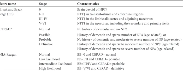
Table 1. Summary of the neuropathologic criteria of Alzheimer’s disease.
Score name Stage Characteristics
Braak and Braak stage (BB)
0 I-II III-IV V-VI
Brain devoid of NFT†
NFT† in transentorhinal and entorhinal regions NFT† in the limbic allocortex and adjoining neocortex
NFT† in the neocortex, including the secondary and primary fields
CERAD* Normal No history of dementia and no NP‡
Possible Probable Definitive
History of dementia and sparse number of NP‡ (age-related), or
No history of dementia and moderate to severe number of NP (age-related) History of dementia and sparse to moderate number of NP‡ (age-related) History of dementia and sparse to severe number of NP‡ (age-related)
NIA-Reagan Normal
Low likelihood Intermediate likelihood High likelihood
BB=0 and CERAD= normal BB=I/II and CERAD= possible BB=III/IV and CERAD= probable BB=V/VI and CERAD= definitive
Correlations were analyzed using the Spearman’s rank
correlation coefficient. The closer r is to one, the stronger
the correlation. The r value was considered statistically
sig-nificant when p<0.05. Statistical analysis was performed
us-ing the software SPSS v. 13 (SPSS, Chicago, Illinois, USA).
Results
The CDR distribution and demographic data of the
62 selected subjects are described in Table 2. There were
no differences regarding age, gender and education among
those groups.
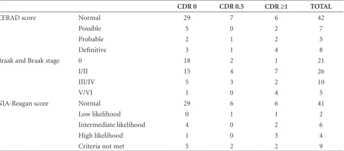
Table 3 depicts the distribution of the cases under each
CDR group by the CERAD score, Braak and Braak stage
and NIA-Reagan score. In 9 subjects, classification
accord-ing to NIA-Reagan score was not possible due to
discrepan-cies between CERAD score and Braak and Braak stage.
Table 4 shows the correlation among CDR score and
neuropathologic criteria. CERAD score correlated best to
the CDR scale (Table 4).
Discussion
In this series, the CERAD criteria correlated to the CDR
score better than Braak and Braak score and NIA-Reagan
criteria, although this moderate correlation was only
slight-ly better than for the Braak and Braak score. Results diverge
in the literature, with McKeel et al. in 2004
15finding NPs
to be the best marker to differentiate normal controls from
questionable dementia subjects, corroborating previous
re-sults,
6,16whereas some studies favor the Braak stage for best
correlation to dementia severity.
17Table 2. Demographic data of the 62 subjects included in this study.
Cognitive status
Number of subjects
Mean age (in years)
Male:female (%)
CDR 0 39 69.7±12.1 61.5:38.5
CDR 0.5 9 71.6±12.3 66.7:33.3
CDR ≥1 14 82.9±4,9 50:50
Total 62 72.9±12.1 59.7:40.3
Table 3. Distribution of the cases in each CDR group by the CERAD score, Braak and Braak stage and NIA-Reagan score.
CDR 0 CDR 0.5 CDR ≥1 TOTAL
CERAD score Normal 29 7 6 42
Possible 5 0 2 7
Probable 2 1 2 5
Definitive 3 1 4 8
Braak and Braak stage 0 18 2 1 21
I/II 15 4 7 26
III/IV 5 3 2 10
V/VI 1 0 4 5
NIA-Reagan score Normal 29 6 6 41
Low likelihood 0 1 1 2
Intermediate likelihood 4 0 2 6
High likelihood 1 0 3 4
Criteria not met 5 2 2 9
*CDR: clinical dementia rating.
Table 4. Clinicopathological correlation by score. The r and p values are depicted on first and second lines, respectively.
Score name CDR* CERAD† Braak and Braak NIA-Reagan
CDR 1.0000
CERAD 0.5303
0.0000
1.0000
Braak and Braak 0.5294
0.0000
0.7563 0.0000
1.0000
NIA-Reagan 0.5076
0.0002
0.9189 0.0000
0.9102 0.0000
1.0000
Despite the criteria, majority of studies comparing AD
neuropathologic criteria have considered neurofibrillary
tangle density and distribution to be directly proportional
to severity of AD.
18-20The present study corroborates these
findings. In our series, 7.7% of the CDR0 subjects met
Braak stages
≥
IV, while 43% of the CDR
≥
1 subjects met
the same stages.
The casuistic used to correlate NIA-Reagan criteria
and CDR was reduced because nine subjects did not meet
the criteria in order to be scored. NIA-Reagan criteria
de-mand a perfect match between CERAD score and Braak
stage. We are not the first to report problems in using the
NIA-Reagan criteria. In 1997, Geddes et al. were only able
to classify 22 out of their 47 cases using the NIA-Reagan
criteria.
21They suggested accommodating discrepant cases
having either a probable CERAD score or a limbic Braak
and Braak stage, into the intermediate likelihood group of
NIA-Reagan criteria. Also, Braak et al. in 1989 published
results showing a mismatch between plaques and tangles
in staging Alzheimer’s pathology.
22Indeed, the r value of
CEARD score and Braak and Braak stage correlation was
0.75. Therefore, the highly specific but low sensitivity
NIA-Reagan criteria are most used for selecting well defined
groups of cases for further analyses. If the prevalence of
dementia of the casuistic is low or if the study focuses
mainly on control subjects, specificity is usually the most
important point and NIA-Reagan criteria are suggested for
subject selection.
Perhaps, the current clinical criteria for AD might not
be able to detect early changes. Corroborating this
hypothe-sis, neuropathological diagnosis of AD was assigned to
con-trol clinical cases in several studies including the present.
23,24On the other hand, NP and NFT may be merely markers of
AD. Giannakopoulus et al. demonstrated, in a large series
of pure AD cases, that more than 50% of CDR scale
vari-ability was not explained by NFT or by amyloid deposits.
7The present study also has limitations.
Informant-based clinicofunctional data might not be reliable enough
for detecting early clinical changes. In order to enhance the
sensitivity of our interview to early clinical changes, the
In-formant Questionnaire on Cognitive Decline in the Elderly
(IQCODE)
25scale is used together with the CDR score. A
study performed in Sao Paulo City verified the IQCODE’s
high sensitivity and specificity for the local population.
26Furthermore, evidence has shown these scales to be reliable
even for subjects having mild cognitive impairment.
27In sum, this series involving Brazilian samples derived
from a general autopsy service corroborates the results
found in other clinicopathological series. These
prelimi-nary results show that our functional assessment has a
good correlation with the neuropathological findings
com-pared to other series. Nevertheless, the clinicopathological
correlation is not yet ideal and further studies may reveal
better markers and criteria.
Grant support
– FAPESP, Albert Einstein Research and
Education Institute – Sao Paulo, CNPq, CAPES,
LIM05-FMUSP, LIM01-FMUSP
References
1. Ferri CP, Prince M, Brayne C, et al. Global prevalence of de-mentia: a Delphi consensus study. Lancet 2005;366:2112-2117. 2. Mirra SS, Heyman A, McKeel D, et al. The Consortium to
Establish a Registry for Alzheimer’s Disease (CERAD). Part II. Standardization of the neuropathologic assessment of Alzheimer’s disease. Neurology 1991;41:479-486.
3. Braak H, Braak E. Neuropathological stageing of Alzheimer-related changes. Acta Neuropathol 1991;82:239-259. 4. The National Institute on Aging and Reagan Institute
Work-ing Group on Diagnostic Criteria for the Neuropathological Assessment of Alzheimer’s Disease. Consensus recommenda-tions for the postmortem diagnosis of Alzheimer’s disease. Neurobiol Aging 1997;18:S1-S2.
5. Bancher C, Jellinger K, Lassmann H, Fischer P, Leblhuber F. Correlations between mental state and quantitative neuropa-thology in the Vienna longitudinal study on dementia. Eur Arch Psychiat Clin Neurosci 1996;246:137-146.
6. Berg L, McKeel DW, Miller JP, et al. Clinicopathologic studies in cognitively healthy aging and Alzheimer disease: Relation of histologic markers to dementia severity, age, sex, and apo-lipoprotein E genotype. Arch Neurol 1998;55:326-335. 7. Giannakopoulos P, Gold G, Kovari E, et al. Assessing the
cog-nitive impact of Alzheimer disease pathology and vascular burden in the aging brain: the Geneva experience. Acta Neu-ropathol 2007;113:1-12.
8. Froehlich TE, Bogardus ST, Jr., Inouye SK. Dementia and race: are there differences between African Americans and Cauca-sians? J Am Geriatr Soc 2001;49:477-484.
9. Grinberg LT, Ferretti RE, Farfel JM, et al. Brain bank of the Bra-zilian aging brain study group - a milestone reached and more than 1,600 collected brains. Cell Tissue Bank 2007;8:151-162. 10. McKhann G, Drachman D, Folstein M, Katzman R, Price D, Stadlan EM. Clinical diagnosis of Alzheimer’s disease: report of the NINCDS- ADRDA Work Group under the auspices of Department of Health and Human Services Task Force on Alzheimer’s Disease. Neurology 1984;34:939-944.
12. Neary D, Snowden JS, Gustafson L et al. Frontotemporal lo-bar degeneration: a consensus on clinical diagnostic criteria. Neurology. 1998;51:1546-1554.
13. McKeith IG, Dickson DW, Lowe J et al. Diagnosis and man-agement of dementia with Lewy bodies - Third report of the DLB consortium. Neurology. 2005;65:1863-1872.
14. Morris JC. The clinical dementia rating (CDR): current ver-sion and scoring rules. Neurology. 1993;43:2412-2414. 15. McKeel DW, Price JL, Miller JP, et al. Neuropathologic
cri-teria for diagnosing Alzheimer disease in persons with pure dementia of Alzheimer type. J Neuropathol Exp Neurol 2004;63:1028-1037.
16. McKeel DW, Ball MJ, Price JL, et al. Interlaboratory histo-pathologic assessment of Alzheimer neuropathology: differ-ent methodologies yield comparable diagnostic results. Alz Dis Assoc Disorder 1993;7:136-151.
17. Grober E, Dickson D, Sliwinski MJ, et al. Memory and mental status correlates of modified Braak staging. Neurobiol Aging 1999;20:573-579.
18. Arriagada PV, Growdon JH, Hedley-Whyte ET, Hyman BT. Neurofibrillary tangles but not senile plaques paral-lel duration and severity of Alzheimer’s disease. Neurology 1992;42:631-639.
19. Bierer LM, Hof PR, Purohit DP, et al. Neocortical neurofibril-lary tangles correlate with dementia severity in Alzheimer’s disease. Arch Neurol 1995;52:81-88.
20. Nagy Z, Esiri MM, Jobst KA et al. Relative roles of plaques
and tangles in the dementia of Alzheimer‘s disease: correla-tions using three sets of neuropathological criteria. Dementia 1995;6:21-31.
21. Geddes JW, Tekirian TL, Soultanian NS, Ashford JW, Davis DG, Markesbery WR. Comparison of neuropathologic crite-ria for the diagnosis of Alzheimer’s disease. Neurobiol Aging 1997;18:S99-S105.
22. Braak H, Braak E, Ohm T, Bohl J. Alzheimer’s disease: mis-match between amyloid plaques and neuritic plaques. Neu-rosci Lett 1989;103:24-28.
23. Morris JC, Storandt M, McKeel DW, et al. Cerebral amyloid deposition and diffuse plaques in ‘’normal’’ aging: evidence for presymptomatic and very mild Alzheimer’s disease. Neu-rology 1996;46:707-719.
24. Crystal H, Dickson D, Fuld P, et al. Clinico-pathologic studies in dementia nondemented subjects with pathologically con-firmed Alzheimer’s disease. Neurology 1988;38:1682-1687. 25. Jorm AF, Jacomb PA. The informant questionnaire on
cog-nitive decline in the elderly (IQCODE): Socio-demographic correlates, reliability, validity and some norms. Psychol Med 1989;19:1015-1022
26. Bustamante SE, Bottino CM, Lopes MA, et al. Combined instruments on the evaluation of dementia in the elderly: preliminary results. Arq Neuropsiquiatr 2003;61:601-606. 27. Isella V, Villa L, Russo A, et al. Discriminative and predictive