Argyrophilic grain disease
An update on a frequent cause of dementia
Lea T. Grinberg
1,2, Helmut Heinsen
2Abstract – Argyrophilic grain disease (AGD) is a sporadic, very late-onset tauopathy, accounting for approximately 4–13% of neurodegenerative dementias. AGD may manifest with a range of symptoms such as cognitive decline and behavioral abnormalities. To date, no study has been able to demonstrate a distinct clinical syndrome associated with AGD. The diagnosis is exclusively based on postmortem findings, the significance of which remains controversial because up to 30% of AGD cases are diagnosed in subjects without any cognitive impairment, while AGD findings often overlap with those of other neurodegenerative processes. Nevertheless, the presence of AGD is likely to have a significant effect on cognitive decline. The neuropathological hallmarks of AGD are argyrophilic grains, pre-neurofibrillary tangles in neurons and coiled bodies in oligodendrocytes found mainly in the entorhinal cortex and hippocampus. This review aims to provide an up-to-date overview of AGD, emphasizing pathological aspects. Additionally, the findings of a Brazilian case series are described.
Key words: pathology, brain, neurology, argyrophilic grain disease, tau.
Doença com grãos argirofílicos: atualização sobre uma causa de demência muito prevalente
Resumo – Doença com grãos argirofílicos (DGA) é uma taupatia, não-familial, de início tardio que corresponde a cerca de 4 a 13% das demências neurodegenerativas. DGA se manifesta com uma gama de sintomas como déficit cognitivo e distúrbios comportamentais. Até o momento, não se conhece nenhuma característica clínica que distinga DGA de outras demências. O diagnóstico é baseado nos achados anatomopatológicos, cujo significado é controverso, quer porque cerca de 30% dos casos de DGA não apresentam sintomas, quer porque DGA é freqüentemente encontrada junto a outras alterações neurodegenerativas. Entretanto, acredita-se que a presença de DGA impacte a cognição. Os marcadores neuropatológicos de DGA são: grãos argirofílicos, pré-emaranhados neurofibrilares e corpúsculos em forma de vibrião em oligodendrócitos. As áreas mais afetadas são o córtex entorrinal, hipocampo e estruturas límbicas. Essa revisão almeja sumarizar o conhecimento mais atual sobre DGA e descrever brevemente achados de uma casuística brasileira.
Palavras-chave: patologia, encéfalo, neurologia, doença com grãos argirofílicos.
1MD, PhD, Department of Pathology, University of São Paulo Medical School, São Paulo, SP, Brazil. 2MD Labor fuer Morphologische Hirnforschung der
Klinik und Poliklinik fuer Psychiatrie und Psychotherapie, University Of Wuerzburg, Wuerzburg, Germany.
Lea Tenenholz Grinberg – Department of Pathology Faculdade de Medicina da Universidade de São Paulo - Avenida Dr. Arnaldo, 455 / 1st floor / room
1353 - 01246-903 São Paulo SP - Brazil.
Disclosure: The authors reports no conflicts of interest.
Received November 1, 2008. Accepted in final form February 5, 2009.
Introduction and historical background
Argyrophilic grain disease (AGD) is a very late-onset
tauopathy, accounting for approximately 4–13% of
neuro-degenerative dementias.
1-5The name AGD stems from the
argyrophilic structures characteristic of this entity.
AGD was first described in 1987 by Braak and
collea-gues as a distinctive degenerative disease characterized by
argyrophilic grains confined to limbic structures affecting
a subset of patients with adult onset dementia.
6Although highly prevalent, to date no study has been
able to demonstrate a distinct clinical syndrome associated
with AGD and only a few series have described clinical
fea-tures that may correlate with the presence of this entity.
7-12The diagnosis is based solely on postmortem findings.
The impact of the grains is controversial for two main
re-asons. Firstly, up to 30% of the AGD cases are diagnosed
in subjects without any cognitive impairment.
8,12Secondly,
Alzheimer’s disease.
8,9,13-16The objective of this review was
to provide an up-to-date overview of AGD and to describe
the findings of a Brazilian case series drawn from the Brain
Bank of the Brazilian Aging Brain Study Group (BBBABSG).
Clinical symptoms
AGD may manifest with a range of symptoms
inclu-ding cognitive decline, dementia
4,7,15,17and behavioral
abnormalities.
7,11,18,19Amnestic cognitive impairment tends to be mild and
non-progressive.
9,20A recent study verified that AGD
pa-tients retain abilities in verbalizing and articulating as well as
problem-solving skills, on average, for approximately 2 years
longer than Alzheimer’s disease (AD) patients. However,
the-re is no distinctive clinical profile for evaluating single cases.
21AGD may occasionally present as frontotemporal
de-mentia, and is considered one of the possible
neuropatho-logical entities underlying frontotemporal dementia.
22,23Although the commonly associated AD pathology
makes it difficult to assign specific clinical symptoms to
AGD, the presence of AGD has a significant effect on
cogni-tive decline; e.g. demented with AGD display considerably
less AD-associated pathology than pure AD would show at
the same clinical stage.
24,25In summary, a precise test for clinical diagnosis of AGD
has yet to be developed.
Neuropathological aspects
Gross examination of the brain shows moderate to
seve-re ceseve-rebral atrophy with average brain weight of 1084±109g
up to 1120g.
16The neuropathological hallmarks of AGD are
argyro-philic grains, pre-neurofibrillary tangles in neurons
(pre-tangle neurons) and coiled bodies in oligodendrocytes.
Given that all of these hallmarks are phospho-tau positive,
AGD is classified as a tauopathy.
Argyrophilic grains (AGs)
The term is derived from their strong staining using
the Gallyas silver iodide method. However, it is noteworthy
that AGs are not stained by all silver methods,
26indicating
that AGs have specific features. AGs are also labeled using
immunohistochemistry against phospho-tau protein, such
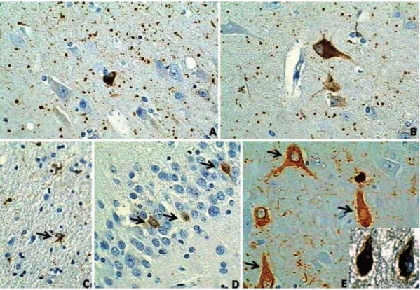
as PHF-1 and AT8 antibodies (Figure 1A,B).
AGs occur mainly in transentorhinal, and entorhinal
cortex, the CA1 area of the hippocampus and
presubi-culum. It is important to notice that these areas are also
affected early by phospho-tau changes in AD. The
ning temporal cortex, orbitofrontal cortex, insular cortex,
basolateral nuclei of the amygdala and hypothalamic
late-ral tubelate-ral nucleus can also be involved.
13,27The source of
AGs probably lies in pre-tangle projection neurons found
in the same location as the AGs
1,4. AGs are predominantly
localized in dendrites and dendritic branches
1,27,28, although
association of AGs with axons has also been reported.
4AGs are small, about 4–8 micrometer, spindle shaped,
rod-like, button-like or round bodies in the neuropil
(Fi-gure 1). Ultrastructurally, AGs contain straight filaments
or tubules measuring 9–25 nm.
13Pre-tangle neurons (Figure 1B,D,E)
Pre-tangle neurons are a constant finding in AGD,
and their regional distribution is the same as that for AD.
They are also found in the dentate gyrus (Figure 1D).
4-6Pre-tangle neurons in AGD do not apparently differ from
pre-tangle neurons in AD
29,30(Figure 1B,D).
Coiled bodies in oligodendrocytes
Although being invariably found in AGD, coiled bodies
are similar to those observed in many other tauopathies
and therefore lack specificity.
31,32(Figure 1C).
Other findings
Tau-containing astrocytes –
Astrocytes containing
phospho-tau show granular immunoreactive cytoplasm
rather than dense inclusions akin to those seen in tufted
astrocytes in progressive supranuclear palsy. Generally, they
appear in clusters, thus being suggestive of plaques seen in
corticobasal degeneration. The presence of tau-containing
astrocytes is variable from one case to another, and when
found are usually confined to the limbic system.
Ballooned neurons –
Α
-
β
-crystallin-positive ballooned
neurons are commonly observed in the amygdala, in the
presubiculum and middle layers of the basal temporal
cor-tex in AGD.
33Yet ballooned neurons are usually interpreted
as non-specific lesions, given these are a common finding
in many familial and sporadic tauopathies and AD.
34,35Tangles and neuropil threads – Variable numbers of
tangles and neuropil threads may be present in the same
regions as in AD. This has caused some confusion about the
borderline between AGD with a few tangles and AGD with
associated AD.
36Most pathologists categorize AD changes
(neurofibrillary tangles and neuropil threads) in AGD
ac-cording to the guidelines of Braak and Braak.
37In their own case series Braak and colleagues classified
most of the AGD cases as having AD ranging from stage
from I to IV.
18However, the apparently small percentage
of AGs in advanced stages of AD must be interpreted with
care, as the substantial phospho-tau-immunoreactive
pa-thology in such cases may incrementally hamper the
vi-sualization of AGs. Recent studies using 4R tau-specific
antibodies which highlight AGs, have shown a higher
pre-valence of AGs in advanced stages of AD.
38Nevertheless,
AGD is usually not accompanied by substantial
β
-amyloid
deposits.
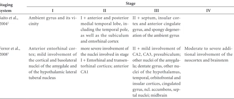
39Staging of AGs –
In 2004, Saito and colleagues proposed
a staging system for AGD based on a refined analysis of a
Table 1. Comparison of the two neuropathological staging systems for argyrophilic grain disease, as proposed by Saito et al. in 2004 and Ferrer et al. in 2008.
Staging system
Stage
I II III IV
Saito et al., 20042
Ambient gyrus and its vi-cinity
I + anterior and posterior medial temporal lobe, in-cluding the temporal pole, as well as the subiculum and entorhinal cortex
II + septum, insular cor-tex and anterior cingulate gyrus, and spongy degener-ation of the ambient gyrus
Ferrer et al., 20085
Anterior entorhinal cor-tex; mild involvement of the cortical and basolateral nuclei of the amygdale and of the hypothalamic lateral tuberal nucleus
more severe involvement of the nuclei involved in stage I + Entorhinal and transen-torhinal cortices; anterior CA1
II + mild involvement of CA2, CA3, presubiculum; other nuclei of the amygda-la; dentate gyrus, other nu-clei of the hypothalamus, temporal, orbitofrontal and insular cortices, cingulated gyrus, ncl. accumbens, sep-tal nuclei; midbrain
large series.
2This system presumes an antero-posterior
pro-gression of the disease. Rare cases have shown widespread
AGs throughout the temporal lobe, limbic system, frontal
cortex and brain stem.
40-42An up-dated staging system was
proposed by Ferrer and colleagues in 2006.
5This recent
systematic staging of AGs does not include accompanying
changes. Table 1 compares the two staging systems.
Biochemistry of tau in AGD
Tau proteins are encoded by the tau gene on
chromo-some 17. Alternative splicing of exons 2, 3 and 10 results in
six isoforms, which in turn give rise to six different mRNAs.
Tau proteins resulting from encoding exon 10 have four
repeat regions (4R tau), whereas those lacking encoding
exon 10 have three repeat regions (3R tau).
43,44The function of tau largely depends on
post-translatio-nal modifications including phosphorylation and
dephos-phorylation, a balanced action between protein kinases and
protein phosphatases. Several kinases have been implicated
in tau phosphorylation.
45-50In contrast to AD, in which 3R tau and 4R tau forms are
found, AGD is characterized by a double band of 68 and 64
kDa similar to that found in progressive supranuclear palsy
and corticobasal degeneration. Therefore, AGD is
conside-red a 4R tauopathy. The use of specific anti-4R antibodies
has corroborated this biochemical observation.
51Interestingly, the occurrence of tangles and pre-tangles
in the hippocampal CA2 area, a very common finding in
AGD, is associated with 4R tauopathy.
52Genetics
AGD appears to be sporadic given that a familial form
has yet to be reported. The tau gene or
microtubule-asso-ciated protein tau (MAPT) locus is located on
chromoso-me 17q21.
53The region is divided into two predominant
haplotypes, H1 and H2. In 2008, a single case with AGD
phenotype was linked to a novel S305I MAPT mutation
and
54there is evidence from one series that the incidence
of MAPT H1 is slightly higher in AD cases with AGD than
in those without AGD.
38However, other genetic studies
have failed to discover a sustained link between AGD and
a particular gene locus. The frequency of apolipoprotein E
e4 (ApoE e4) allele, the most important genetic risk for AD,
proves similar to that of the general population in cases of
AGD.
55Nevertheless, the frequency of ApoE e2 is higher in
AGD than that observed in both AD or controls.
51,56Differential diagnosis
Neuropathological studies have shown frequent
asso-ciation of AGD with other neurodegenerative diseases, the
most common being AD. AGD has also been reported
to-gether with other tauopathies, Creutzfeldt-Jakob disease,
α
-synucleinopathies and hippocampal sclerosis.
8,16,18,57-60AGD in the case series from the Brain Bank
of the Brazilian Aging Brain Study Group
In the BBBABSG series, AGD was diagnosed in 36
(11.5%) out of the first 307 fully analyzed cases. In
accor-dance with other series, AGD was more frequently found in
older subjects (
p
<0.05). No statistically significant
differen-ce was found condifferen-cerning gender, years of schooling,
cogni-tive status, Braak and Braak neurofibrillary stage, presence
of
β
-amyloid plaques or Lewy bodies among the cases with
and without AGD. Most interestingly, AGD was the only
finding in 14.3% of the subjects manifesting moderate or
severe parkinsonism signs. Although AGD is not classically
associated with parkinsonism, we are not the first to report
this association.
61AGD is usually associated with finding
of allocortical neurofibrillary tangles. Accordingly, in our
series only two AGD cases (6.9%) were devoid of tangles.
One of these subjects, a 79-year-old male had no
cogniti-ve decline, whereas the other subject, a 82-year-old female
showed severe dementia, interpreted as being attributed to
the severe burden of microvascular changes and lacunes
rather than the presence of AGs.
Conclusions
AGD is a sporadic and distinct tauopathy often found
in the brain of older subjects. Although linked to cognitive
decline, behavioral problems and even parkinsonism, no
study to date has demonstrated any clinical or laboratory
particularity able to distinguish AGD from other
neurode-generative diseases, while several subjects harboring AGD
appear not to be demented. In recent years, studies based
on well-conducted clinicopathological correlation series
have pointed to older age as the only risk factor for AGD,
and revealed that AGD may lower the threshold for
demen-tia. Neither of these findings was observed in our series.
Several points still remain obscure. What is the origin
of the grains? Is AGD a distinct clinical syndrome? How
can neurofibrillary tangles of Alzheimer disease be
diffe-rentiated from those found in AGD? Is there any hallmark
clinical symptom suggestive of the presence of AGs in the
brain? Additional comprehensive, prospective
clinicopa-thological correlation studies are required to answer many
of these questions.
Acknowledgement –
We would like to thank the
References
1. Tolnay M, Mistl C, Ipsen S, Probst A. Argyrophilic grains of Braak: occurrence in dendrites of neurons containing hyperphospho-rylated tau protein. Neuropathol Appl Neurobiol 1998;24:53-59. 2. Saito Y, Ruberu NN, Sawabe M, et al. Staging of argyrophilic grains: an age-associated tauopathy. J Neuropathol Exp Neu-rol. 2004;63:911-918.
3. Ding ZT, Wang Y, Jiang YP, et al. Argyrophilic grain disease: frequency and neuropathology in centenarians. Acta Neuro-pathol. 2006;111:320-328.
4. Tolnay M, Clavaguera F. Argyrophilic grain disease: a late-onset dementia with distinctive features among tauopathies. Neuropathology 2004;24:269-283.
5. Ferrer I, Santpere G, van Leeuwen FW. Argyrophilic grain disease. Brain 2008;131:1416-1432.
6. Braak H, Braak E. Argyrophilic grains: characteristic pathol-ogy of cerebral cortex in cases of adult onset dementia with-out Alzheimer changes. Neurosci Lett 1987;76:124-127. 7. Ikeda K, Akiyama H, Arai T, Matsushita M, Tsuchiya K,
Mi-yazaki H. Clinical aspects of argyrophilic grain disease. Clin Neuropathol. 2000;19:278-284.
8. Martinez-Lage P, Munoz DG. Prevalence and disease asso-ciations of argyrophilic grains of Braak. J Neuropathol Exp Neurol 1997;56:157-164.
9. Jicha GA, Petersen RC, Knopman DS, et al. Argyrophilic grain disease in demented subjects presenting initially with am-nestic mild cognitive impairment. J Neuropathol Exp Neurol 2006;65:602-609.
10. Botez G, Schultz C, Ghebremedhin E, Bohl J, Braak E, Braak H. Clinical aspects of argyrophilic grain disease. Nervenarzt 2000;71:38-43.
11. Togo T, Isojima D, Akatsu H, et al. Clinical features of argy-rophilic grain disease: a retrospective survey of cases with neuropsychiatric symptoms. Am J Geriatr Psychiatry 2005;13: 1083-1091.
12. Tolnay M, Schwietert M, Monsch AU, Staehelin HB, Langui D, Probst A. Argyrophilic grain disease: distribution of grains in patients with and without dementia. Acta Neuropathol 1997;94:353-358.
13. Braak H, Braak E. Cortical and subcortical argyrophilic grains characterize a disease associated with adult onset dementia. Neuropathol Appl Neurobiol 1989;15:13-26.
14. Knopman DS, Parisi JE, Salviati A, et al. Neuropathology of cognitively normal elderly. J Neuropathol Exp Neurol 2003;62: 1087-1095.
15. Togo T, Cookson N, Dickson DW. Argyrophilic grain dis-ease: neuropathology, frequency in a dementia brain bank and lack of relationship with apolipoprotein E. Brain Pathol 2002;12:45-52.
16. Jellinger KA. Dementia with grains (argyrophilic grain dis-ease). Brain Pathol. 1998;8:377-386.
17. Saito Y, Yamazaki M, Kanazawa I, Murayama S. Severe in-volvement of the ambient gyrus in a case of dementia with argyrophilic grain disease. J Neurol Sci 2002;196:71-75. 18. Braak H, Braak E. Argyrophilic grain disease: frequency of
occurrence in different age categories and neuropathological diagnostic criteria. J Neural Transm 1998;105:801-819. 19. Cairns NJ, Grinberg LT, Lisic R, et al. Expanding the
neu-ropathological spectrum of frontotemporal lobar degenera-tions: review of 833 prospectively assessed dementia cases. J Neural Transm 2006;113:7.
20. Petersen RC, Parisi JE, Dickson DW, et al. Neuropathologic features of amnestic mild cognitive impairment. Arch Neurol 2006;63:665-672.
21. Steuerwald GM, Baumann TP, Taylor KI, et al. Clinical char-acteristics of dementia associated with argyrophilic grain disease. Dement Geriatr Cogn Disord 2007;24:229-234. 22. Kovacs GG, Pittman A, Revesz T, et al. MAPT S305I mutation:
implications for argyrophilic grain disease. Acta Neuropathol 2008;116:103-118.
23. Cairns NJ, Bigio EH, Mackenzie IR, et al. Neuropathologic diagnostic and nosologic criteria for frontotemporal lobar de-generation: consensus of the Consortium for Frontotemporal Lobar Degeneration. Acta Neuropathol 2007;114:5-22. 24. Thal DR, Schultz C, Botez G, et al. The impact of argyrophilic
grain disease on the development of dementia and its rela-tionship to concurrent Alzheimer‘s disease-related pathology. Neuropathol Appl Neurobiol 2005;31:270-279.
25. Josephs KA, Whitwell JL, Parisi JE, et al. Argyrophilic grains: a distinct disease or an additive pathology? Neurobiol Aging 2008;29:566-573.
26. Uchihara T. Silver diagnosis in neuropathology: principles, practice and revised interpretation. Acta Neuropathol 2007; 113:483-499.
27. Schultz C, Koppers D, Sassin I, Braak E, Braak H. Cytoskel-etal alterations in the human tuberal hypothalamus related to argyrophilic grain disease. Acta Neuropathol 1998;96: 596-602.
28. Ikeda K, Akiyama H, Kondo H, Haga C. A study of dementia with argyrophilic grains: possible cytoskeletal abnormality in dendrospinal portion of neurons and oligodendroglia. Acta Neuropathol 1995;89:409-414.
29. Braak E, Braak H, Mandelkow EM. A sequence of cytoskele-ton changes related to the formation of neurofibrillary tangles and neuropil threads. Acta Neuropathol 1994;87:554-567. 30. Tau and ubiquitin immunoreactivity at different stages of
for-mation of Alzheimer neurofibrillary tangles. New York: Alan R. Liss; 1989.
31. Komori T. Tau-positive glial inclusions in progressive supra-nuclear palsy, corticobasal degeneration and Pick’s disease. Brain Pathol 1999;9:663-679.
pathol-ogy in neurodegenerative diseases: their nature and com-parison with neuronal tangles. Neurobiol Aging 1998;19(1 Suppl):S85-91.
33. Tolnay M, Villoz N, Probst A, Miserez AR. Apolipoprotein E genotype in senile dementia with tangles differs from Al-zheimer’s disease. Neuropathol Appl Neurobiol 2003;29: 80-84.
34. Fujino Y, Delucia MW, Davies P, Dickson DW. Ballooned neu-rones in the limbic lobe are associated with Alzheimer type pathology and lack diagnostic specificity. Neuropathol Appl Neurobiol 2004;30:676-682.
35. Togo T, Dickson DW. Ballooned neurons in progressive su-pranuclear palsy are usually due to concurrent argyrophilic grain disease. Acta Neuropathol 2002;104:53-56.
36. Cras P, Perry G. Dementia with argyrophilic grains. Ann Neu-rol 1991;30:853-854.
37. Braak H, Braak E. Neuropathological stageing of Alzheimer-related changes. Acta Neuropathol. 1991;82:239-259. 38. Fujino Y, Wang DS, Thomas N, Espinoza M, Davies P, Dickson
DW. Increased frequency of argyrophilic grain disease in Al-zheimer disease with 4R tau-specific immunohistochemistry. J Neuropathol Exp Neurol 2005;64:209-214.
39. Tolnay M, Calhoun M, Pham HC, Egensperger R, Probst A. Low amyloid (Abeta) plaque load and relative predomi-nance of diffuse plaques distinguish argyrophilic grain dis-ease from Alzheimer’s disdis-ease. Neuropathol Appl Neurobiol 1999;25:295-305.
40. Tsuchiya K, Mitani K, Arai T, et al. Argyrophilic grain disease mimicking temporal Pick‘s disease: a clinical, radiological, and pathological study of an autopsy case with a clinical course of 15 years. Acta Neuropathol 2001;102:195-199. 41. Maurage CA, Sergeant N, Schraen-Maschke S, et al. Diffuse
form of argyrophilic grain disease: a new variant of four-repeat tauopathy different from limbic argyrophilic grain disease. Acta Neuropathol 2003;106:575-583.
42. Ishihara K, Araki S, Ihori N, et al. Argyrophilic grain disease presenting with frontotemporal dementia: a neuropsychologi-cal and pathologineuropsychologi-cal study of an autopsied case with presenile onset. Neuropathology 2005;25:165-170.
43. Goedert M, Spillantini MG, Jakes R, Rutherford D, Crowther RA. Multiple isoforms of human microtubule-associated pro-tein tau: sequences and localization in neurofibrillary tangles of Alzheimer’s disease. Neuron 1989;3:519-526.
44. Himmler A, Drechsel D, Kirschner MW, Martin Jr DW. Tau consists of a set of proteins with repeated C-terminal micro-tubule-binding domains and variable N-terminal domains. Mol Cell Biol 1989;9:1381-1388.
45. Hanger DP, Mann DM, Neary D, Anderton BH. Tau pathol-ogy in a case of familial Alzheimer’s disease with a valine to
glycine mutation at position 717 in the amyloid precursor protein. Neurosci Lett 1992;145:178-180.
46. Mandelkow E, Mandelkow EM. Kinesin motors and disease. Trends Cell Biol. 2002;12:585-591.
47. Goedert M, Spillantini MG, Davies SW. Filamentous nerve cell inclusions in neurodegenerative diseases. Curr Opin Neu-robiol 1998;8:619-632.
48. Lovestone S, Reynolds CH. The phosphorylation of tau: a critical stage in neurodevelopment and neurodegenerative processes. Neuroscience 1997;78:309-324.
49. Reynolds CH, Nebreda AR, Gibb GM, Utton MA, Anderton BH. Reactivating kinase/p38 phosphorylates tau protein in vitro. J Neurochem 1997;69:191-198.
50. Reynolds CH, Utton MA, Gibb GM, Yates A, Anderton BH. Stress-activated protein kinase/c-jun N-terminal kinase phos-phorylates tau protein. J Neurochem 1997;68:1736-1744. 51. Togo T, Sahara N, Yen SH, et al. Argyrophilic grain disease
is a sporadic 4-repeat tauopathy. J Neuropathol Exp Neurol 2002;61:547-556.
52. Ishizawa T, Ko LW, Cookson N, Davias P, Espinoza M, Dick-son DW. Selective neurofibrillary degeneration of the hip-pocampal CA2 sector is associated with four-repeat tauopa-thies. J Neuropathol Exp Neurol 2002;61:1040-1047 53. Andreadis, A, Brown, WM, Kosik, KS. Structure and novel
exons of the human tau gene. Biochemistry (Mosc.). 1992;31: 10626-10633.
54. Kovacs GG, Pittman A, Revesz T, et al. MAPT S305I mutation: implications for argyrophilic grain disease. Acta Neuropathol 2008;116:103-118
55. Tolnay M, Probst A, Monsch AU, Staehelin HB, Egensperger R. Apolipoprotein E allele frequencies in argyrophilic grain disease. Acta Neuropathol 1998;96:225-227.
56. Ghebremedhin E, Schultz C, Botez G, et al. Argyrophilic grain disease is associated with apolipoprotein E epsilon 2 allele. Acta Neuropathol. 1998;96:222-224.
57. Ferrer I, Barrachina M, Tolnay M, et al. Phosphorylated pro-tein kinases associated with neuronal and glial tau deposits in argyrophilic grain disease. Brain Pathol 2003;13:62-78. 58. Masliah E, Hansen LA, Quijada S, et al. Late onset dementia
with argyrophilic grains and subcortical tangles or atypical progressive supranuclear palsy? Ann Neurol 1991;29:389-396. 59. Ikeda K, Akiyama H, Kondo H, et al. Thorn-shaped astro-cytes: possibly secondarily induced tau- positive glial fibrillary tangles. Acta Neuropathol 1995;90:620-625.
60. Beach TG, Sue L, Scott S, et al. Hippocampal sclerosis demen-tia with tauopathy. Brain Pathol 2003;13:263-278.