○ ○ ○ ○ ○ ○ ○ ○ ○ ○ ○ ○ ○ ○ ○ ○ ○ ○ ○
ABSTRACT
○ ○ ○ ○ ○ ○ ○ ○ ○ ○ ○ ○ ○ ○ ○ ○ ○ ○ ○ ○
INTRODUCTION
Central retinal vein thrombosis is typically a disease of older patients, rarely occurring in young adults. It is often associated with either an under-lying vascular disease or a procoagulant state.1 Hypertension, diabetes, vasculitides, and dyslipidemia are the most commonly associated vascular disorders, while the recognized procoagulant states include thrombocytosis, poly-cythemia, macroglobulinemia, the use of oral con-traceptive pill or systemic lupus erythematosus.
Protein S and protein C are vitamin K-de-pendent plasma proteins that modulate coagu-lation. Protein S serves as a cofactor for protein C to inhibit the clotting cascade at the levels of factors V and VIII.2 There are few reports of reti-nal vein thrombosis associated with protein S deficiency,3,4 and these have usually occurred in combination with other thrombophilic condi-tions. This case report describes a young patient with protein S deficiency who developed cen-tral retinal vein prethrombosis. No other prothrombotic risk factors were present.
○ ○ ○ ○ ○ ○ ○ ○ ○ ○ ○ ○ ○ ○ ○ ○ ○ ○ ○ ○
CASE REPORT
A 21-year-old white woman presented with acutely reduced visual acuity and a central scotoma in her left eye. She did not present photophobia, ocular pain, fever or any other local or systemic complaint. The patient had no personal or family history of eye disease, systemic hypertension, diabetes mellitus, thrombophilia, malignancy or use of oral con-traceptive pills. Her best-corrected visual acu-ity was 20/15 in the right eye and 20/25 in the left. The anterior segment examination was
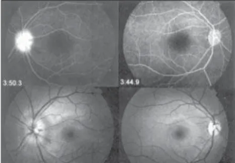
unremarkable, without papillary defects, and the intraocular pressures were 12 mmHg in the right eye and 11 mmHg in the left eye. Ophthalmoscopy of the left eye showed rare dot and blot hemorrhages and blurring of the optic disc. The retinal venules appeared mod-erately tortuous but undilated. In the right eye, the ophthalmoscopy was normal. Fluorescein angiography of the left eye revealed normal ar-terial filling but delayed arteriovenous filling, and there was late disc hyperfluorescence com-patible with prethrombosis (Figure 1). How-ever, the right eye was normal. The results from hematological tests showed an erythrocyte sedi-mentation rate of 18 mm/hour, and the com-plete blood count, prothrombin time, partial thromboplastin time and anticardiolipin bodies were normal. She was negative for anti-nuclear antibodies, rheumatoid factor, lupus anticoagulant antiphospholipid antibodies, and serum protein electrophoresis. The activated protein C resistance, plasma levels of protein C and antithrombin III were normal. Decreased plasma levels were found for both free protein S (6%; normal is 20-40%), and total protein S (36%; normal is 60-140%).
Warfarin therapy was initiated. One month later, the visual acuity of the left eye was 20/20 and complete improvement in ophthalmoscopic findings was noted. Neither parents nor siblings were available for ocular and systemic examination.
○ ○ ○ ○ ○ ○ ○ ○ ○ ○ ○ ○ ○ ○ ○ ○ ○ ○ ○ ○
DISCUSSION
Central retinal vein thrombosis in young adults is considered uncommon and has been difficult to characterize because the cause is
• Paulo de Tarso Ponte Pierre-Filho
• Alessandra Maria Mont’Alverne Pierre
• Maurício Abujamra Nascimento
• Ana Maria Marcondes
Central retinal vein
prethrombosis as an initial
manifestation of
protein S deficiency
Department of Ophthalmology, Universidade Estadual de Campinas
(Unicamp), Campinas, São Paulo, Brazil
CONTEXT: Retinal vein thrombosis is most common in old people, and is often associated with systemic vascular disease. One of its rare systemic causes is protein S deficiency.
CASE REPORT: A case of a 21-year-old woman with retinal vein prethrombosis associated only with protein S deficiency is described. She presented with acutely reduced visual acuity and a central scotoma in her left eye. Warfarin therapy was initiated, and complete improvement in ophthalmoscopic findings was subsequently ob-served. This case illustrates that protein S defi-ciency is a factor that should be considered in cases of retinal vein occlusion, particularly in young patients.
KEY WORDS: Retinal vein. Protein S deficiency. Vas-cular diseases. Case reports.
C
ase Repor
t
São Paulo Medical Journal — Revista Paulista de Medicina
135
often unknown. Although there is no consen-sus, it seems appropriate to refer patients with central retinal vein occlusion for throm-bophilia screening if they are younger than 50 years old.5-7
Prince et al.3 reported a patient with sys-temic lupus erythematosus and acquired pro-tein S deficiency causing central retinal vein thrombosis. Bertram et al.7measured protein C, protein S, and antithrombin III concen-trations in ocular vascular occlusive disease, and found a pathologically reduced anti-thrombin III level in one patient with branch venous occlusion, and severely altered protein C and protein S concentrations in two patients with central retinal vein occlusion.
Protein S deficiency, a condition that may be either inherited or acquired, may result in a hypercoagulable state and it predisposes af-fected individuals to thrombotic events. In the inherited form, clinical manifestations usually
occur before the age of 30 years. It is trans-mitted as an autosomal dominant trait. The deficiency can be acquired under conditions like septic shock, disseminated intravascular coagulation, acute respiratory distress syn-drome, postoperative states, liver diseases, pregnancy, oral contraceptive pill use, human immunodeficiency virus infection and chemo-therapy for breast cancer.8 The initial presen-tation of prethrombosis in this patient was related to protein S deficiency.
Recognition of an association of protein S with thrombotic disease may suggest spe-cific therapeutic approaches. In our patient, therapy with oral anticoagulant was initi-ated because of the known risk of the re-currence of thrombosis in cases of protein S deficiency.8
We suggest that protein S deficiency should be considered in the cases of some pa-tients with unexplained retinal vascular
occlu-sions, particularly when occurring in young patients or when there is a positive family his-tory of thrombotic disease.
1. Kohner EM, Cappin JM. Do medical conditions have an influ-ence on central retinal vein occlusions? Proc R Soc Med. 1974;67(10):1052-4.
2. Walker FJ. Regulation of activated protein C by a new protein. A possible function for bovine protein S. J Biol Chem. 1980;255(12):5521-4.
3. Prince HM, Thurlow PJ, Buchanan RC, Ibrahim KM, Neeson PJ. Acquired protein S deficiency in a patient with systemic
○ ○ ○ ○ ○ ○ ○ ○ ○ ○ ○ ○ ○ ○ ○ ○ ○ ○ ○ ○ ○ ○ ○ ○ ○ ○ ○ ○ ○ ○ ○ ○ ○ ○ ○ ○ ○ ○ ○ ○ ○ ○ ○ ○ ○ ○ ○ ○ ○ ○ ○ ○ ○ ○ ○ ○ ○ ○ ○ ○ ○ ○ ○ ○
REFERENCES
lupus erythematosus causing central retinal vein thrombosis. J Clin Pathol. 1995;48(4):387-9
4. Raus P, Stalmans P, Demeuter E, Spileers W, Dralands L. Unu-sual retinal vasculitis in a patient with protein S deficiency and systemic toxoplasmosis: a case report. Bull Soc Belge Ophthalmol. 2001;(279):7-12.
5. Fong AC, Schatz H. Central retinal vein occlusion in young adults. Surv Ophthalmol. 1993;37(6):393-417.
6. Tekeli O, Gursel E, Buyurgan H. Protein C, protein S and an-tithrombin III deficiencies in retinal vein occlusion. Acta Ophthalmol Scand. 1999;77(6):628-30.
7. Bertram B, Remky A, Arend O, Wolf S, Reim M. Protein C, protein S, and antithrombin III in acute ocular occlusive dis-eases. Ger J Ophthalmol. 1995;4(6):332-5.
8. Greaves M. Aging and the pathogenesis of retinal vein throm-bosis. Br J Ophthalmol. 1997;81(10):810-1.
Pré-trombose da veia central da retina como mani-festação inicial da deficiência de proteína S
CONTEXTO: Trombose venosa retiniana é mais comum em pessoas idosas e freqüentemente está associada com doença vascular sistêmica. Uma causa sistêmica rara é a deficiência de proteína S.
RELATO DE CASO: Um caso de paciente branca, de 21 anos, com pré-trombose de veia central da retina associada à deficiência isolada de
pro-○ ○ ○ ○ ○ ○ ○ ○ ○ ○ ○ ○ ○ ○ ○ ○ ○ ○ ○ ○ ○ ○ ○ ○ ○ ○ ○ ○ ○ ○ ○ ○ ○ ○ ○ ○ ○ ○ ○ ○ ○ ○
RESUMO
Paulo de Tarso Ponte Pierre-Filho, MD.Department of Ophthalmology, Universidade Estadual de Campinas, Campinas, São Paulo, Brazil.
Alessandra Maria Mont’Alverne Pierre, MD.
Faculdade de Medicina, Universidade Federal do Ceará, Fortaleza, Ceará, Brazil.
Maurício Abujamra Nascimento, MD. Department of
Ophthalmology, Universidade Estadual de Campinas, Campinas, São Paulo, Brazil.
Ana Maria Marcondes, MD. Department of
Ophthal-mology, Universidade Estadual de Campinas, Campinas, São Paulo, Brazil.
Sources of funding: None
Conflict of interest: None
Date of first submission: October 23, 2003
Last received: December 29, 2003
Accepted:February 5, 2004
Address for correspondence:
Paulo de Tarso Ponte Pierre-Filho. R. Alexandre Fleming, s/n. Departamento de Oftalmologia, Faculdade de Ciências Médicas
Universidade Estadual de Campinas (Unicamp) Campinas /SP — Brasil — CEP 13081-970. Tel./Fax (+55 19) 3788-7936
E-mail: [email protected]
COPYRIGHT © 2004, Associação Paulista de Medicina ○ ○ ○ ○ ○ ○ ○ ○ ○ ○ ○ ○ ○ ○ ○ ○ ○ ○ ○ ○
Publishing information
teína S é descrito. Ela apresentou súbito embaçamento visual e escotoma central em olho esquerdo. Terapia cumarínica foi iniciada e com-pleta remissão dos achados oftalmoscópicos foi observada. Este caso mostra que deficiência de proteína S deve ser um fator que deve ser consi-derado em casos de oclusão venosa retiniana, particularmente em pacientes jovens.
PALAVRAS-CHAVE: Veia retiniana. Deficiên-cia de proteína S. Doenças vasculares. Rela-tos de casos.
Figure 1. Bottom: Photograph showing rare dot and blot hemorrhages and blurring of optic disc in the left eye. The optic disc, macula, and retinal vessels appear normal in the right eye. Top: Fluorescein angiogram showing late optic disc hyperfluorescence; the disc is extremely edematous on the left.