Overgrowth and Ring Gland cAMP/PKA Signaling Defects
in the
Drosophila melanogaster
Neurofibromatosis-1
Growth Deficiency
James A. Walker1,2, Jean Y. Gouzi1¤a, Jennifer B. Long3, Sidong Huang4¤b, Robert C. Maher1, Hongjing Xia1, Kheyal Khalil1, Arjun Ray1, David Van Vactor3, Rene´ Bernards4, Andre´ Bernards1,2¤b* 1Massachusetts General Hospital Center for Cancer Research and Harvard Medical School, Charlestown, Massachusetts, United States of America,2Center for Human Genetic Research, Massachusetts General Hospital, Boston, Massachusetts, United States of America,3Department of Cell Biology, Harvard Medical School, Boston, Massachusetts, United States of America,4Division of Molecular Carcinogenesis, The Netherlands Cancer Institute, Amsterdam, The Netherlands
Abstract
Neurofibromatosis type 1 (NF1), a genetic disease that affects 1 in 3,000, is caused by loss of a large evolutionary conserved protein that serves as a GTPase Activating Protein (GAP) for Ras. AmongDrosophila melanogaster Nf1(dNf1) null mutant phenotypes, learning/memory deficits and reduced overall growth resemble human NF1 symptoms. These and otherdNf1 defects are relatively insensitive to manipulations that reduce Ras signaling strength but are suppressed by increasing signaling through the 39-59 cyclic adenosine monophosphate (cAMP) dependent Protein Kinase A (PKA) pathway, or phenocopied by inhibiting this pathway. However, whetherdNf1affects cAMP/PKA signaling directly or indirectly remains controversial. To shed light on this issue we screened 486 1st and 2nd chromosome deficiencies that uncover.80% of annotated genes for dominant modifiers of thedNf1pupal size defect, identifying responsible genes in crosses with mutant alleles or by tissue-specific RNA interference (RNAi) knockdown. Validating the screen, identified suppressors include the previously implicateddAlktyrosine kinase, its activating ligandjelly belly(jeb), two other genes involved in Ras/ERK signal transduction and several involved in cAMP/PKA signaling. Novel modifiers that implicate synaptic defects in the dNf1 growth deficiency include the intersectin-related synaptic scaffold protein Dap160 and the cholecystokinin receptor-related CCKLR-17D1 drosulfakinin receptor. Providing mechanistic clues, we show that dAlk, jeb and CCKLR-17D1 are among mutants that also suppress a recently identified dNf1 neuromuscular junction (NMJ) overgrowth phenotype and that manipulations that increase cAMP/PKA signaling in adipokinetic hormone (AKH)-producing cells at the base of the neuroendocrine ring gland restore thedNf1growth deficiency. Finally, supporting our previous contention that ALK might be a therapeutic target in NF1, we report that humanALKis expressed in cells that give rise to NF1 tumors and that NF1 regulated ALK/RAS/ERK signaling appears conserved in man.
Citation:Walker JA, Gouzi JY, Long JB, Huang S, Maher RC, et al. (2013) Genetic and Functional Studies Implicate Synaptic Overgrowth and Ring Gland cAMP/PKA Signaling Defects in theDrosophila melanogasterNeurofibromatosis-1 Growth Deficiency. PLoS Genet 9(11): e1003958. doi:10.1371/journal.pgen.1003958
Editor:Gregory P. Copenhaver, The University of North Carolina at Chapel Hill, United States of America
ReceivedDecember 7, 2012;AcceptedOctober 1, 2013;PublishedNovember 21, 2013
Copyright:ß2013 Walker et al. This is an open-access article distributed under the terms of the Creative Commons Attribution License, which permits unrestricted use, distribution, and reproduction in any medium, provided the original author and source are credited.
Funding:JAW, RCM, HX, KK, AR and AB were supported by NIH/NGMS grant 1-R01 GM084220. AB also received support from an anonymous donor. JYG was supported by Young Investigator Award from the Children’s Tumor Foundation. SH and RB were supported by Dutch cancer society grant NKI 2009-4496. The funders had no role in study design, data collection and analysis, decision to publish, or preparation of the manuscript.
Competing Interests:The authors have declared that no competing interests exist.
* E-mail: abernards@helix.mgh.harvard.edu
¤a Current address: Institute of Cellular and Developmental Biology, Biomedical Sciences Research Centre ‘‘Alexander Fleming,’’ Vari, Greece. ¤b Current address: Department of Biochemistry, McIntyre Medical Building, Montreal, Quebec, Canada.
Introduction
RASopathies, caused by mutations that activate Ras/ERK signaling, are a group of related disorders with features that include facial dysmorphism, skeletal, skin and cardiac defects, cognitive deficits, reduced growth and an increased cancer risk [1]. Neurofibromatosis type 1 (NF1; OMIM 162200), caused by loss of a RasGAP, and Noonan syndrome, caused by mutations that alter Ras/ERK pathway proteins SOS1, KRAS, NRAS, RAF1, BRAF, CBL, PTPN11, or SHOC2, are the most common members of this group, affecting 1 in 3,000, or as many as 1 in 1,000 live births, respectively [2,3]. The genetics of these disorders provides a strong argument that excess Ras/ERK signaling underlies
common RASopathy symptoms, and much effort remains focused on attenuating Ras/ERK signaling as a strategy for therapeutic intervention. However, whether life-long pharmacological inhibi-tion of Ras/ERK signaling is a viable strategy to treat the full range of often non-life-threatening, but nonetheless serious symptoms of these chronic disorders, remains an open question. This motivates our work to better understand the molecular and cellular pathways responsible for NF1 symptom development, in the hope this will identify more specific therapeutic targets.
amino acid length [4]. Like human neurofibromin, the Drosophila protein functions as a GAP for conventional (dRas1) and R-Ras-like (dRas2) GTPases [4,5]. This functional conservation made it all the more surprising when both initially identified dNf1
homozygous null mutant phenotypes, a postembryonic growth deficiency and a neuropeptide-elicited NMJ electrophysiological defect, appeared insensitive to genetic manipulations that atten-uate Ras signaling strength, but were suppressed by increasing signaling through the cAMP-dependent PKA pathway [4,6]. The genetic link betweendNf1and cAMP/PKA led to further studies, which demonstrated that similar to many children with NF1 [7], and Nf1+/2 mice [8], dNf12/2flies exhibit specific learning and
memory deficits [9]. Biochemical studies with fly brain extracts further revealed that loss ofdNf1is associated with reduced GTP-cS-stimulated but not basal adenylyl cyclase (AC) activity [9], and with defects in both classical and unconventional AC pathways [10]. Arguing that the cAMP related function of NF1 is evolutionary conserved, GTP-cS-stimulated AC activity and cAMP levels were also reduced in E12.5 Nf12/2 mouse brain [11], and defects in cAMP generation appear to explain the unique sensitivity toNf1heterozygosity of murine central nervous system neurons [12]. Arguing that NF1 may regulate cAMP signaling at least in part in a cell autonomous manner, reduced cAMP levels and AC activity were also found in NF1 deficient human astrocytes [13]. Thus, while there is little doubt that aberrant AC signaling is an evolutionary conserved NF1 pheno-type, we and others have reached conflicting conclusions about the underlying mechanism.
Based on Drosophila phenotypic rescue studies with human
NF1transgenes, others reported that neurofibromin has physically separable functions as a negative regulator of Ras and a positive mediator of AC/PKA signaling. This conclusion followed from findings that NF1-GAP activity was not required to rescuedNf1
size [10] or learning [14] phenotypes, whereas a transgene encoding a C-terminal part of human neurofibromin that did not include the GAP catalytic domain did suppress both defects. In obvious conflict, in similar experiments withdNf1transgenes, we found that neuronal expression of a functional NF1-GAP catalytic segment was necessary and sufficient to suppress the systemic growth defect, and that other protein segments had no effect.
Moreover, thedNf1growth defect was also suppressed by neuronal expression of the Drosophila p120RasGAP ortholog, and although we extended earlier findings by showing that heterozygous loss of
dRas1ordRas2, or of a comprehensive set of Ras effector proteins did not modify the growth defect, these mutations also did not reduce the elevated phospho-ERK level in the dNf1 central nervous system (CNS). However, some Ras/ERK pathway double mutants did suppress both defects, leading us to conclude that excess neuronal Ras/ERK signaling is the proximal cause of the non-cell-autonomousdNf1 growth defect [5]. Further supporting this notion, recent work implicated the neuronal dAlk tyrosine kinase receptor and its activating ligand jelly belly (jeb) as rate-limiting activators ofdNf1regulated Ras/ERK pathways respon-sible for both systemic growth and olfactory learning defects [15]. The above evidence underlies our hypothesis that loss ofdNf1
increases neuronal dAlk/Ras/ERK activity, which in turn causes reduced cAMP/PKA signaling, which may or may not be cell-autonomous. Obviously, identifying additional components of
dNf1-regulated growth controlling pathways followed by functional analysis might help to test this hypothesis. Here we report results of a dNf1 growth deficiency modifier screen, which identified components of tyrosine kinase/Ras/ERK and neuropeptide/ cAMP/PKA pathways in addition to genes involved in synaptic morphogenesis and functioning. Further analysis showed that the requirement for dNf1 and cAMP/PKA in Drosophila growth regulation involves different tissues, withdNf1required broadly in larval neurons, and cAMP/PKA signaling specifically in AKH-producing cells and perhaps in other parts of the neuroendocrine ring gland. These results, and the recent discovery of a noveldNf1
synaptic overgrowth phenotype [16] that is also suppressed by several genes identified in our screen, set the stage for further work to more precisely define how loss ofdNf1 causes Ras/ERK and other signaling defects, the ultimate consequence of which is reduced systemic growth.
Results
Loss ofdNf1Does Not Phenocopy Starvation or Alter Developmental Timing
Animals use elaborate hormonal mechanisms to coordinate nutrient availability and feeding with changes in metabolism and overall growth. Since starvation or crowding during the larval phase of the Drosophila life cycle reduces systemic growth [17], we first examined whether the small size of dNf1 mutants reflected reduced feeding. Arguing against this hypothesis, wild-type and
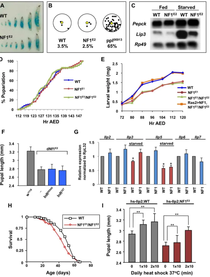
dNf1 larvae ingested similar amounts of dye-stained food throughout their development (Figure 1A). Unlike apumpless(ppl) mutant [18],dNf1larvae also showed no tendency to move away from a food source (Figure 1B). Analysis of the expression of the starvation-inducible Pepck and Lip3 genes [18] provided further evidence that loss of dNf1 does not phenocopy starvation (Figure 1C).
Mechanisms that control Drosophila growth have been the topic of intense study and much has been learned about how an interplay between insulin-like peptide (ILP) controlled growth rate and ecdysone controlled growth duration determines overall growth (see [19] and [20] for reviews). Arguing against an important role for ecdysone or other factors that control the length of the larval growth period, no differences in the expression of canonical ecdysone-regulated genes was found (results not shown) and no difference in developmental timing between wild-type and
dNf1mutants was detected (Figure 1D and S1). Rather, a reduced growth rate throughout larval development results in an
approx-Author Summary
Figure 1. Loss ofdNf1does not phenocopy starvation or alter developmental timing.(A) Wild-type (w1118) anddNf1larvae ingest similar
imately 25% weight reduction ofdNf1pupae relative to isogenic controls (Figure 1E and S1).
Drosophila ILPs control systemic growth, metabolism, longev-ity, and female fecundity [21–24]. Among the eight Drosophila ILP genes,Ilp2,Ilp3andIlp5are co-expressed in bilateral clusters of seven insulin-producing neurosecretory cells (IPCs) in the larval brain [21]. Ablation of these cells causes a severe reduction in overall size, which is rescued by inducing the expression of a hsp70-Ilp2 transgene [22,23]. However, several results argue against a role for ILPs in thedNf1growth defect. Firstly, two hypomorphic insulin receptor alleles, InR05545
and InR327
, did not affectdNf1
pupal size (Figure 1F). Secondly, qRT-PCR analysis of RNA extracted from wandering wild-type and dNf1third instar larvae detected no major differences in the expression ofIlp1(not shown),
Ilp2,Ilp3,Ilp5,Ilp6andIlp7in fed larvae. Among the three IPC expressed ILP genes, the expression ofIlp3andIlp5is reduced in response to starvation [21]. Starved wild-type and dNf1 larvae showed a similar reduction inIlp5expression, whereasIlp3showed a less pronounced response (Figure 1G). Thirdly, while certain insulin receptor or insulin receptor substrate (chico) mutants have an up to 85% increased life span [25,26], the lifespan of dNf1
mutants and isogenic controls was comparable (Figure 1H). We note that others previously reported a reduced life span for the originally identifieddNf1 p-element alleles, generated in a different genetic background [27]. Finally, we previously showed that Ilp2-GAL4drivenUAS-dNf1expression in IPCs did not rescue thedNf1
size defect [5]. Although daily heat shocking ofhsp70-ilp2carrying larvae increased the size ofdNf1pupae, indicating that mutants do not lack the ability to respond to insulin, similar induction of this transgene, as previously noted [21], also substantially increased the size of wild-type controls (Figure 1I). Thus, reduced insulin signaling does not provide an obvious explanation for the slower
dNf1 growth rate, prompting us to perform a screen to identify other genes involved indNf1-mediated systemic growth control.
Screen for Dominant Modifiers ofdNf1Systemic Growth Phenotype
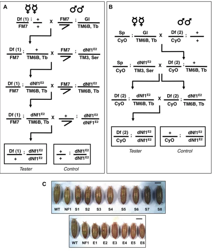
While mostdNf1defects are poorly suited for use in modifier screens, the postembryonic growth defect is robust and readily quantified during the pupal stage [4]. However, using this phenotype in a screen is complicated by the fact that organismal size is sexually dimorphic (females are larger than males) and affected by population density, feeding, environmental factors and genetic background differences. With these confounding factors in mind, we used the crossing schemes outlined in Figure 2 to test collections of isogenic 1st and 2nd chromosome deficiencies for
dNf1E2 pupal size modifier effects or synthetic lethal interactions. For each of 139 1stand 347 2ndchromosome deficiencies from the Exelixis [28], DrosDel [29] or Bloomington Stock Center (BSC) collections, we generated Df(1)/+; Nf1E2/Nf1E2 (Figure 2A) or
Df(2)/+; Nf1E2/Nf1E2(Figure 2B) stocks, respectively. Notably, our work identified only few synthetic lethal interactions, and in all cases tested the synthetic lethality has been specific to the chromosome carrying the Nf1E2
allele, and not observed when the same deficiency was tested in Nf1E2/Nf1E1 null trans-heterozygotes [5]. To guard against size differences caused by inadvertent differences in population density or environmental conditions, each deficiency was scored at least twice using an initial rough caliper measurement of pupae attached to the side of culture vials. For each candidate modifying deficiency thus
identified, microscopy combined with image analysis was used to determine the precise head-to-tail length of at least 40 pupae, which were then allowed to individually eclose in order to establish their sex. Several controls were next performed to eliminate non-specific modifiers or artifactual results. First, for all suppressors the continued presence of theNf1E2nonsense mutation was confirmed by a PCR assay (Figure S2). Secondly, as a critical specificity control, all modifying deficiencies were analyzed in a wild-type background to eliminate those that affect pupal size irrespective of
dNf1 genotype. Further analysis of some of these non-specific modifiers demonstrated that loss ofAct57Bdominantly increases pupal size, whereas heterozygous loss of the glutamate transporter
Eaat1 has the opposite effect. Thirdly, because pupal size is a function of larval growth rate and duration, modifying deficiencies were monitored for obvious changes in developmental timing. Table 1 shows the number of screened chromosome 1, 2L and 2R deficiencies, the fraction of genes uncovered and the number of
dNf1 and wild-type pupal size modifying deficiencies and loci identified. Figure 2C shows the magnitude of the pupal size modification of typical enhancers and suppressors. The number of modifying deficiencies exceeds the number of identified loci, because many modifying deficiencies uncover overlapping geno-mic segments (Figure 3). Not unexpectedly, individual modifying deficiencies increase or decrease dNf1 pupal size to different extents (Figure 4).
Some large non-modifying deficiencies identified in our screen completely overlapped with smaller modifying ones. In such cases, stocks were re-ordered and reanalyzed. If these tests replicated the original results, genetic complementation analysis or PCR amplification using transposon and flanking sequence-specific primers was used to confirm the mapping of the deficiencies in question. This procedure identified several mismapped or mislabeled deficiencies, most of which have since been withdrawn by stock centers. Any suspect or recessive modifying deficiency, or any deficiency that uncovers genes with non-specific size phenotypes, such as Minute loci [30,31], were eliminated from further analysis. Table S1 lists these deficiencies and the reason for their exclusion.
During work to identify genes responsible for observed effects, we prioritized genes uncovered by suppressing deficiencies over those uncovered by enhancers. We also prioritized modifying loci uncovered by more than one deficiency, strong modifiers over weak ones, and genes uncovered by smaller deficiencies over those uncovered by larger ones, reasoning that effects of smaller deficiencies are more likely due to the loss of single genes. Validating the screen, suppressingDf(2R)Exel7144uncovers dAlk
and partially overlapping suppressing Df(2R)BSC199 and
Df(2R)BSC699 each uncover the gene for its activating ligand,
jeb, both previously identified as dominant suppressors ofdNf1size, learning, and neuronal ERK over-activation phenotypes [15]. Other uncovered candidate modifiers, such as PKA catalytic and regulatory subunit genes, were tested in crosses with loss-of-function alleles and/or by tissue-specific knockdown using at least two independent UAS-RNAi transgenes, most of which were obtained from the Vienna Drosophila Stock Center (VDRC) [32]. For deficiencies that lacked obvious candidate modifiers, we used the UAS-RNAi approach to more broadly screen uncovered genes. Figure S3 shows examples of modifiers identified by this latter approach. Although the nutrient sensing fat body and other tissues outside of the CNS play important roles in Drosophila
is not obviously reduced indNf1larvae. H)dNf1adult flies show no altered longevity compared to wild-type controls. (I) Over-expression ofIlp2from ahs-Ilp2transgene indNf1larvae results in a similar increase in size as in wild-type flies.
growth control [33,34], candidate modifiers have only been tested by RNAi knockdown in neurons or glial cells. We focused on these cell types, because neuronal UAS-dNf1 expression sufficed to suppress the growth phenotype [5].
ThedNf1pupal size modifiers identified to date can be classified into three non-exclusive categories, the first of which consists of the
previously implicated dAlk/jeb receptor/ligand pair and two not previously implicated other genes involved in Ras-mediated signal transduction. Another expected category includes genes involved in cAMP/PKA signaling, including the previously reported dnc
cAMP phosphodiesterase suppressor [35], and the newly identified PKA catalytic subunit gene,PKA-C1, which acts as an enhancer. Figure 2. Deficiency screen for dominant modifiers of thedNf1growth defect.Isogenic 1stand 2ndchromosomes deficiencies from the
Exelixis, DrosDel and Bloomington Stock Center collections were tested for their ability to alterdNf1female pupal size. Crossing schemes to generate
Df(1)/+; dNf1E2(A) andDf(2)/CyO; dNf1E2(B) screening stocks. Thetubby-markedTM6B3rdchromosome balancer allowed the selection ofdNf1E2
This group also includes theCCKLR-17D1drosulfakinin receptor, recently implicated as a cAMP-coupled promoter of synaptic growth [36], which is particularly interesting given the recent identification of adNf1 larval NMJ overgrowth phenotype [16]. Finally, our screen also identified multiple genes whose roles in
dNf1 growth control had not been anticipated and whose functional relevance remains to be established. Several genes in this group are predominantly expressed in brain or have known neuronal functions, including genes coding for the aforementioned CCKLR-17D1 receptor, the synaptic scaffold protein Dap160, the neuronal RNA binding protein elav, the neuronal Na,K ATPase interacting protein NKAIN [37], and the larval brain and alimentary channel expressed amino acid transporter NAAT1 [38]. Other genes in this group include CKIIbeta2, encoding a casein kinase regulatory subunit, the endosomal trafficking proteinsdeep-orangeandcarnation, theNotchmodifier heparan sulfate 3-O sulfotransferase Hs3st-B [39], and the ubiquitin E3 ligases
HERC2, which acts as a suppressor, and CUL3, which has the opposite effect. Table 2 lists deficiencies that modifydNf1but not wild-type pupal size, limited to those for which the responsible gene has been identified. Table S2 identifies all analyzed deficiencies, indicates which modifieddNf1 pupal size (providing female pupal sizes as a gauge of modification strength), which also altered wild-type pupal size, and which deficiencies altered developmental timing.
dNf1Pupal Size Modifiers Involved in Jeb/dAlk/Ras/ERK Signaling
We previously reported that thedAlk receptor tyrosine kinase [40] acts as a rate-limiting activator of neuronal Ras/ERK pathways responsible for dNf1 size and learning defects [15]. Therefore, the fact that thedAlkand jebgenes are uncovered by one and two suppressing deficiencies, respectively (Table 2), validates our screen. Others recently reported that Jeb/dAlk signaling allows brain growth to be spared at the expense of other tissues in nutrient restricted Drosophila, and identified a glial cell niche around neuroblasts as the source of Jeb under these conditions [41]. To determine whether glial cells also produce Jeb involved in overall growth control under normal conditions, we used glial and neuronal Gal4 drivers to test the effect of tissue-specific jeb and dAlk knockdown. Arguing that neurons are the main source of Jeb involved in systemic growth control under non-starvation conditions, jeb knockdown with the Ras2-Gal4, C23-Gal4, and n-syb-Gal4 neuronal drivers [5] increaseddNf1E2pupal size (Figure 5A), whereas the Nrv2-Gal4, Eaat1-Gal4 and Gli-Gal4
glial drivers had no effect (data not shown). The only glial driver that gave rise to partial rescue was the pan-glial repo-Gal4 line, although this effect was not enhanced by co-expressingUAS-Dcr2. Control experiments showed that any driver used in these and other experiments had no effect on pupal size in the absence of
UAS transgenes or vice-versa, that UAS transgenes had no effect in the absence of Gal4 drivers (Figure 5A and data not shown). Finally, extending previous findings and further confirming a role forjebas a dominantdNf1size defect suppressor, thejebweli loss-of-function allele [42] dominantly increased dNf1 pupal size (Figure 5B)
Previously, heterozygous mutations affecting RAF/MEK/ERK kinase cascade components Draf (pole hole; phl), Dsor1/dMEK, or ERK/rolled (rl), did not modifydNf1size [5]. In agreement, twophl -uncovering deficiencies,Df(1)ED6574andDf(1)ED11354, did not score as modifiers (Table S2). Norluncovering deficiencies were analyzed, but Df(1)Exel9049, which is among the stronger suppressors identified, deletes Dsor1 and only two other genes, the neurogenic gene almondex (amx), and CG17754, predicting a BTB and Kelch domain protein. Arguing that reduced Ras/ERK signaling upon loss of Dsor1 combined with abnormal neuronal differentiation due to loss of amx may synergistically cause the observed strong effect, Ras2-Gal4 driven UAS-RNAi transgenes targeting either gene, while causing pupal lethality at 25uC, increased dNf1 pupal size at lower temperatures (Figure 5C). Moreover, suppression of the dNf1 pupal size defect was also observed upon individual heterozygous loss of eitherDsor1oramx, although at least with the tested alleles, combined loss of both genes did not have a more pronounced effect (Figure 5B). Previously, we did not observe suppression of thedNf1E2pupal size defect in crosses with the Dsor1S-1221
allele [5]. A potential explanation may be thatDsor1LH110is a null mutant [43], whereas the molecular nature of Dsor1S-1221 is undetermined. Genetic background differences between these Dsor1 alleles are another potential explanation for the discrepant results.
Multiple screens aimed at identifying genes involved in Drosophila tyrosine kinase/Ras signaling have been performed [44–52]. Among the genes identified, several are uncovered by 1st and 2ndchromosome deficiencies that do not modifydNf1 size. SuppressingDf(2R)BSC161uncovers 27 genes including connector enhancer of KSR(cnk), a scaffold protein that functions as a bimodal (both positive and negative) regulator of RAS/MAPK signaling [53,54]. Supporting a role forcnkas adNf1modifier, thecnkXE-385
and cnkE-2083 alleles acted as dominant suppressors (Figure 5B), and suppression was also observed upon RNAi-mediated Cnk knockdown usingRas2-Gal4orP(GawB)C23-Gal4neuronal drivers (Figure 5C). However, Df(2R)BSC154, which uncovers cnk and only nine other genes, did not score as a modifier (Table S2).
dNf1Size Modifiers Involved in cAMP/PKA Signaling
The dNf1 growth defect is suppressed by heat shock-induced expression of a constitutively active murine PKA catalytic subunit transgene, called PKA* [4], or by loss of the dunce (dnc) cAMP phosphodiesterase [35]. Further validating our screen, two dnc
uncovering deficiencies and another that removes the region Table 1.Deficiency screen summary.
Chromosome Number screened % genes uncovered dNf1Modifiers Non-specific modifiers dNf1modifying loci
SUP ENH SUP ENH SUP ENH
1 139 82.1 48 2 5 2 30 2
2L 182 87.7 14 15 1 7 11 10
2R 165 86.9 31 2 4 1 22 1
Indicated are the number of chromosome 1, 2L and 2R deficiencies screened, the fraction of genes uncovered (based on the FB2013_03 FlyBase release), the number of dNf1modifying deficiencies and loci identified, and the number of non-specific modifiers.
immediately upstream of the dnc coding region, all scored as suppressors (Table 2). Moreover, the Pka-R2 gene, encoding a cAMP binding regulatory PKA subunit, whose dissociation from the catalytic subunit activates the latter, is uncovered by two additional suppressing deficiencies, whereas a deficiency that uncovers the major Pka-C1 catalytic subunit gene scored as an enhancer (Table 2).Df(1)ED7261, which uncovers therutabaga(rut) adenylyl cyclase, did not score as a modifier (not shown). Confirmation ofdncand Pka-C1as the genes responsible for the
observed effects was obtained in crosses with threedncand three
Pka-C1 loss-of-function alleles (Table 2). Pka-R2 remains an attractive candidate suppressor, but expressionPka-R2RNAi trans-genes in neurons had no effect and its role as adNf1 modifier remains unconfirmed (results not shown).
NoveldNf1Modifiers
Recently, the cAMP-coupled CCKLR-17D1 drosulfakinin receptor, but not its closely related CCKLR-17D3 paralog, was Figure 3. Cytogenetic locations ofdNf1modifying deficiencies.Locations of modifying deficiencies (drawn to scale) on the 1stand 2nd(2L and 2R) chromosomes. Deficiencies that enhance or suppress are shown in red and green, respectively. Non-specific deficiencies that dominantly affect the size of wild-type pupae are in blue. Many modifying deficiencies uncover overlapping genomic segments.
identified as a positive regulator of synaptic growth [36]. The
CCKLR-17D1gene is uncovered by three suppressing deficiencies, including Df(1)Exel9051, which uncovers only three other genes. The closely linked CCKLR-17D3 paralog is not uncovered by
Df(1)Exel9051, and while Ras2-Gal4 orP(GawB)C23-Gal4 driven
neuronalCCKLR-17D1RNAi expression strongly suppressed the
dNf1pupal size defect, similar suppression ofCCKLR-17D3had no effect (Figure 6A).
BeyondCCKLR-17D1, severaldNf1size modifiers are expressed in brain and/or have neuronal functions. Among these, dynamin-Figure 4. Identified deficiencies increase or decrease pupal size to different extents.Female pupal lengths for the indicated 1, 2L and 2R deficiencies. Control measurements fordNf1E2and wild-type (w1118) are in black. Colors for enhancing, suppressing and non-specific deficiencies are
as in Figure 2. Pupal lengths are shown in mm, error bars denote standard deviations and are based on measurements described in Table S2. All shown deficiencies modifydNf1female pupal size withp-values,0.01.
associated protein 160 (Dap160) is an intersectin-related scaffold implicated in synaptic vesicle exocytosis and neuroblast prolif-eration [55–58].Dap160is uncovered by suppressing deficiencies
Df(2L)Exel6047 and Df(2L)BSC302, whose region of overlap encompasses ten genes. We note that Df(2L)Exel6047 also uncovers the Drosophila Ret tyrosine kinase gene, the human ortholog of which is the receptor for glial-derived neurotrophic factor. Ret initially appeared an especially attractive candidate
suppressor, because activating RET and inactivating NF1
mutations can both lead to human pheochromocytoma [59], and because DrosophilaRetis expressed in larval brain neurons that resemble neuroendocrine cells [60]. However, among multiple lines of evidence that argue against a role for Ret in the dNf1 growth defect, UAS-dNf1 re-expression directed by a newly generated Ret-Gal4 driver that recapitulates the endoge-nous larval brain Ret expression pattern (Figure S4B), or Table 2.Modifying deficiencies and identification of responsible genes.
Deficiency Cytological Breakpoints Modif. Gene Implicated
Modifying allele(s) and/or RNAi
Tyrosine Kinase/Ras signaling
Df(2R)Exel7144 Df(2R)Exel6064
53C8;53D2 53C11;53D11
SUP dAlk dAlk8(lof),dAlk9(lof),v11446, v107083,JF02668
Df(2R)BSC199 Df(2R)BSC699
48C5;48E4 48D7;48E6
SUP Jellybelly (jeb) Jebweli(lof),v103047,v30800
Df(2R)BSC161 54B2;54B17 SUP connector enhancer of ksr (cnk) cnkXE-385(D),cnkE-2083(lof), v107746
Df(1)BSC663 Df(1)Exel9049
8D1;8D5 8D2;8D3
SUP Dsor1andalmondex (amx) Dsor1: Dsor1LH110(amorph), v107276,v40026,HMS00145; amx:amxf06362(hypo),v3296
cAMP/PKA signaling
Df(1)BSC710 Df(1)BSC656 Df(1)BSC834
3B2;3C9 3B3;3D2 3C11;3F3
SUP dunce (dnc) dncM14(amorph),dncML
(amorph),dnc1(hypo)
Df(2L)Exel6024 30C1;30C9 ENH cAMP-dependent protein
kinase 1 (PKA-C1)
PKA-C1BG02142(leth),PKA-C106353
(hypo),PKA-C1B3(leth)
Neuronal Function
Df(1)ED447 Df(1)Exel9051 Df(1)Exel7464
17C1;17F1 17D1;17D3 17D1;17E1
SUP CCK-like receptor at 17D1 (CCKLR-17D1) v100760
Df(2L)BSC302 Df(2L)Exel6047
39A1;39A6 39A2;39B4
SUP Dynamin-associated protein 160 (Dap160) Dap160D1(lof),Dap160D2(lof), v106689,v16158,JF01918
Df(1)Exel6221 Df(1)ED6396
1B4;1B8 1B5;1B8
SUP Embryonic lethal abnormal vision (elav) elavG0031, elav1
Df(2L)BSC216 Df(2L)BSC240 Df(2L)Exel7043 Df(2L)Exel6025
30C6;30E1 30C7;30F2 30D1;30F1 30C9;30E1
ENH Nicotinic Acetylcholine Receptor alpha-30D (nAcRa-30D)
nAcRa-30DDAS1(via) nAcRa-30DDAS2(via)nAcRa-30DKG05852
(via)
Df(2L)Exel8041 37D7;37F2 SUP Rab9 v107192,v36200, HMS02635
Other
Df(1)Exel6254 19C4;19D1 SUP HERC2 v105374
Df(2L)ED800 Df(2L)ED1050 Df(2L)ED1004
35B2;35D1 35B8;35D4 35B10;35D1
ENH Cullin-3 (cul3) cul3gft2(lof)
Df(1)BSC533 Df(1)Exel6290
4F4;4F10 4F7;4F10
SUP Neutral amino acid transporter 1 (NAAT1) v106027,v37380, v50063
Df(1)Exel9068 18B4;18B6 SUP Heparin sulfate 3-O
sulfotransferase-B (Hs3st-B)
v110601
Df(2R)BSC701 56F15;57A9 SUP Casein kinase IIb2 subunit (CKIIb2) v102633, v26915
Df(2R)BSC607 60E4;60E8 SUP Na,K-ATPase Interacting (NKAIN) v105893, v102018
Df(1)BSC275 18C8;18D3 SUP Vps33/carnation (car) car1(hypo),carD146(lof), v110756
Df(1)BSC719 Df(1)Exel8196 Df(1)BSC589
2A3;2B13 2B1;2B5 2B3;2B9
SUP Vps18/deep orange (dor) dor8(leth),v107053,v105330
Modifying deficiencies for which the responsibledNf1interacting gene has been identified. The cytological location, and the dominant effect ondNf1pupal size (SUP -suppressor, ENH – enhancer) of each deficiency is given. The responsible genes for each modifying deficiency are shown with the mutant alleles, VDRC and TRiP RNAi lines used in their identification. Expression of RNAi transgenes was induced with theRas2-Gal4,elav-Gal4,n-syb-Gal4and/or C23-Gal4drivers. Abbreviations: hypo: hypomorphic; leth: lethal; lof: loss-of-function; amorph: amorphic;D: deletion; via: viable.
RNAi-mediated Ret inhibition, did not modifydNf1pupal size, nor did expression of a UAS-Ret K805A kinase dead transgene. Moreover, Ret-Gal4 driven expression of UAS-Ret transgenes carrying the activating C695R mutation, which mimics a mutation found in multiple endocrine neoplasia type 2 did not phenocopy the
dNf1reduced growth phenotype, although the same transgene did produce the previously described rough eye phenotype when driven byGMR-Gal4[60]; Figure S4C]. Further arguing against a role in
dNf1 growth control, Ret is uncovered by non-modifying
Df(2L)BSC312. By contrast, Dap160 loss-of-function alleles (Dap160D1and Dap160D2; [56]), orDap160RNAi expression driven by three neuronal Gal4 drivers, suppressed the dNf1 pupal size defect, identifying it as the responsible modifier (Figure 6B).
The gene for the neuronal RNA binding protein elav is uncovered by suppressing Df(1)Exel6221 and Df(1)ED6396 whose region of overlap includes just three other genes. Identifying elav as the responsible modifier, elav1 andelavG0031 alleles strongly suppressed (Figure 6C).Rab9is a modifier uncovered by suppressing deficiency
Df(2L)Exel8041. Neuronal but not glialRab9RNAiexpression increases
dNf1pupal size, and the same result is seen upon neuronal expression of a Rab9 dominant negative [61] mutant (Figure 6D).
NAAT1, coding for a larval gut and brain expressed amino acid transporter with a unique affinity for D-amino acids [38], is uncovered by suppressingDf(1)Exel6290andDf(1)BSC533whose region of overlap includes only four other genes. Identifying
NAAT1as the responsible suppressor, three neuronal Gal4 lines driving the expression of threeNAAT1targeting RNAi transgenes suppressed the dNf1 size defect, whereas Repo-Gal4 driven glial expression had no effect (Figure 7A and Table 2).
Mammalian E3 ubiquitin ligase HERC2 controls the ubiquitin-dependent assembly of DNA repair proteins on damaged chromosomes [62]. DrosophilaHERC2is uncovered by suppress-ing deficiency Df(1)Exel6254, which also uncovers the syx16, coding for syntaxin 16. No HERC2 alleles exist, but Ras2-Gal4
driven expression of a UAS-HERC2RNAi transgene (v105374) strongly suppressed thedNf1pupal size defect (Figure 7A), whereas similar knockdown ofSyx16had no statistically significant effect (not shown). The gene for another E3 ligase component,Cul-3, is Figure 5. Validation ofdNf1modifiers involved in Jeb/dAlk/Ras/ERK and cAMP signaling.(A) Neuronal expression ofdAlkRNAi using
Ras2-Gal4,Ras2-Gal4+UAS-Dcr-2,c23-Gal4orn-syb-Gal4drivers suppresses thedNf1size defect. Expression ofjebRNAi with the same neuronal drivers
also suppresses. Weaker suppression is observed whenjebRNAi expression is controlled by the pan-glialrepo-Gal4driver. Dark grey bars are control measurements of Gal4 drivers in thedNf1background. Light grey bars are sizes of wild-type (w1118) anddNf1E2controls. (B) Suppression of thedNf1
size defect by the indicatedjeb,cnk,Dsor1andamxalleles. (C) Neuronalcnk,Dsor1oramxknockdown suppressed thedNf1size defect. In the case of
Dsor1 v107276andamx, cultures were maintained at 18uC to prevent lethality observed at 25uC. Some RNAi transgene/driver combinations were lethal ({) even at 18uC. (D) Validation ofdncandPka-C1asdNf1modifiers was obtained in crosses withdncM14,dncML,Pka-C16353andPka-C1BG02142
loss-of-function alleles. In this and subsequent figures, * and ** denotep-values,0.05 and,0.01, respectively. doi:10.1371/journal.pgen.1003958.g005
Figure 6. Validation ofdNf1modifiers with neuronal functions.(A)Ras2-Gal4orC23-Gal4driven neuronal RNAi knockdown ofCCKLR-17D1
but notCCKLR-17D3suppressed thedNf1pupal size defect. (B) Identification of dynamin-associated protein 160 (Dap160) as a suppressor ofdNf1
growth. Neuronal RNAi targeting ofDap160increaseddNf1pupal size as did twoDap160loss-of-function alleles. (C) Twoelavalleles dominantly suppress thedNf1size defect. (D) Neuronal expression of a Rab9 RNAi transgene or of a dominant negative Rab9 mutant suppresses thedNf1size defect.
uncovered by three enhancing deficiencies, and a Cul-3 loss-of-function allele or Ras2-Gal4 driven expression of a Cul-3 RNAi transgene both enhanced thedNf1size defect, identifying it as the responsible gene (Table 2).
SuppressingDf(1)Exel9068uncovers only four genes, including one encoding the TORC2 complex subunit Rictor. However, systematic Ras2-Gal4 driven RNAi knockdown of Df(1)Exel9068
uncovered genes identified Hs3st-B, encoding one of two Drosophila heparan sulfate 3-O sulfotransferases, as a potent
dNf1 size defect suppressor (Figure 7A), whereas knockdown of Rictor had no effect (not shown). Others previously identified
Hs3st-Bas a positive regulator of Notch signaling [39]. However, the heparan sulfate proteoglycan substrates ofHs3st-Bbind various growth factors and other ligands and have been implicated in a variety of biological processes. Exactly why loss of Hs3st-B
suppresses thedNf1growth defect remains to be determined. Two functionally related dNf1 growth defect suppressors
carnation (car/Vps33A) anddeep-orange(dor/Vps18), encode subunits
of the Class C Vacuolar Protein Sorting (VPS) complex, required for the delivery of endosomal vesicles to lysosomes [63]; Figure 7B]. TheVps16Agene encodes a third member of this complex [64], but whetherVps16Alocated on the 3rdchromosome also acts as a
dNf1 suppressor, or whether pharmacological inhibition of lysosomal degradation affectsdNf1 pupal size are questions that remain to be answered.
B4/Susi is a coiled-coil protein without obvious orthologs outside of insects. It functions as a negative regulator of Drosophila class I phosphatidylinositol-3 kinase Pi3K92E/Dp110 by binding to its Pi3K21B/dP60 regulatory subunit. HomozygousB4mutants have an increased body size [65], which may explain why Ras2-Gal4-driven RNAi-mediated suppression of B4, uncovered by suppressing deficiency Df(2L)BSC147, increased dNf1 pupal size (not shown). However, whetherB4 is the responsible dominant modifier is doubtful, given that it is also uncovered by
Df(2L)BSC692, a non-modifying deficiency. Moreover, we previ-ously found that heterozygous loss of Pi3K21B, or neuronal Figure 7. Identification of modifying genes with undetermined roles indNf1suppression.(A) Validation ofNAAT1,HERC2andHs3st-BasdNf1
expression of a dominant negative Pi3K92E transgene, did not modify dNf1 pupal size [5]. Beyond B4, dNf1 size modifying deficiencies uncovered no genes involved in the canonical growth regulating pathways mediated by insulin and ecdysone. Indeed, several such genes were uncovered by non-modifying deficiencies. Among these genes, fat body expressed insulin-like growth factor
Ilp6, which regulates larval growth in the post-feeding phase [66,67], is uncovered by two non-modifying deficiencies. A single non-modifying deficiency,Df(2L)BSC206, uncovers both thechico
and pten genes, whose products antagonistically control insulin-stimulated Pi3K92E/Dp110 activity, leading to changes in body, organ, and cell size [68,69]. Among subunits of the cell growth regulating mTORC1 complex, raptor is uncovered by three and
Torby one non-modifying deficiency. Among genes implicated in ecdysone signaling, the ecdysone co-receptor ultraspiracleand the ecdysone-induced growth regulatingDHR4nuclear receptor [70] are each uncovered by non-modifying deficiencies, and two such deficiencies uncoverPtth, coding for prothoracicotropic hormone, which provides developmental timing cues by stimulating the production of ecdysone [71,72]. These results reinforce our conclusion that the canonical growth regulating pathways involving insulin and ecdysone play no obvious roles in dNf1
growth control.
Manipulating cAMP/PKA Signaling in the Ring Gland AffectsdNf1Systemic Growth Non-Cell-Autonomously
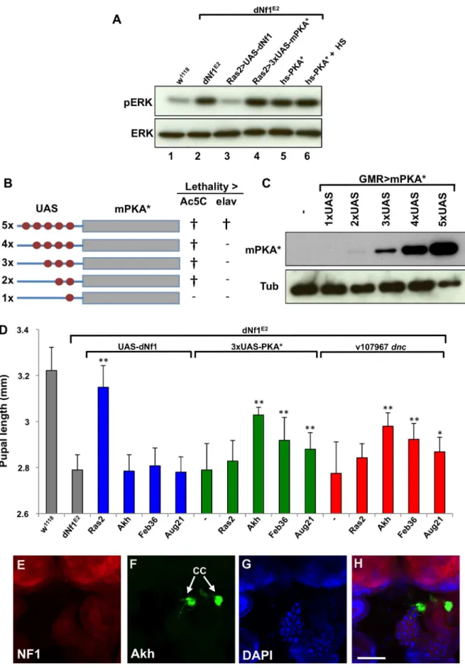
Several results argue that defects in Ras/ERK and cAMP/PKA signaling responsible for the dNf1 growth defect involve non-overlapping cell populations. Firstly, heat shock-induced hsp70 -PKA*, orRas2-Gal4induced attenuatedUAS-PKA*transgene (see below) expression rescued thedNf1pupal size defect, but failed to reduce the elevated larval brain phospho-ERK level (Figure 8A). Moreover, several neuronal RNAi drivers that increasedNf1pupal size when driving UAS-dNf1[5], failed to modify this phenotype when drivingdncRNAitransgenes, even in the presence of the UAS-Dcr-2RNAi enhancer (Table 3). This prompted us to investigate whether genetic manipulation of cAMP/PKA signaling in cells other thandNf1requiring neurons was more effective.
To manipulate cAMP/PKA signaling tissue-specifically we used three UAS-dncRNAi transgenes. We also generated a series of attenuated UAS-PKA* transgenes using vectors with modified Gal4-inducible promoters harboring just 2, 3 or 4 Gal4-binding UAS elements (Figure 8B and C). We made the latter transgenes because a UAS-PKA* expression using the five UAS element containing standard UAS-T vector is lethal in combination with most Gal4 drivers [73]. As reported previously [74], driving UAS-dNf1ubiquitously withAct5C-Gal4, or broadly in neurons withelav
-Gal4,Ras2-Gal4,c23-Gal4, or386Y-Gal4restoreddNf1pupal size, whereas driving the same transgene with more restricted neuronal or non-neuronal drivers had no effect (Figure 8D and Table 3). By contrast, driving the expression ofUAS-dncRNAi
or attenuated UAS-PKA*transgenes with the same set of broadly expressed neuronal drivers was ineffective (Tables 3 and S5). We note that expression of the26UAS-PKA*and36-UAS-PKA*transgenes was generally well tolerated, whereas the46UAS-PKA* and the 56UAS-PKA*
transgenes exhibited increasing levels of lethality (Tables 3 and S5). Arguing that rescue of thedNf1growth defect by manipulating cAMP/PKA signaling or dNf1expression involves different cells, strong pupal size rescue was observed by increasing cAMP/PKA signaling in adipokinetic hormone-producing cells at the base of the neuroendocrine ring gland using the Akh-Gal4 driver (Figure 8D). Rescue was also observed with the Feb36-Gal4and
Aug21-Gal4 ring gland drivers (Figure 8D), which give rise to expression in the corpora allata, the source of juvenile hormone,
but not with the P0206-Gal4or Mai60-Gal4drivers, which express predominantly in the prothoracic gland (Table 3). The tissue specificity of all Gal4 drivers used in this and other experiments was verified by microscopic observation of dissected UAS-GFP
expressing larvae (Table S4 and Figures 8E–H and S5).
dAlk,Jeb,Cnk andCCKLR-17D1Suppress adNf1NMJ Architectural Defect
During larval development, significant expansion of the NMJ arbor must occur, reflecting the steady muscle growth that takes place during larval life. As the NMJ grows, additional branches and boutons are added to the initial synaptic arbor that forms during late embryonic stages upon motor axon contact with its target muscle. As a result, at the wandering third instar stage, wild-type NMJs contain a highly stereotyped, segment specific number of synaptic boutons [75]. Recently, it was reported that
dNf1functions presynaptically to constrain NMJ synaptic growth and neurotransmission [16]. In dNf1 null mutant wandering third instar larvae, while the distribution of major presynaptic proteins is unaffected, increased overall size and synaptic bouton number is apparent at multiple NMJs, supporting a specific role for dNf1 in restricting NMJ expansion [16]. Several dNf1
suppressors that emerged in the current screen have also been linked to synapse morphogenesis, including CCKLR-17D1, which functions as a promoter of NMJ growth [36]. As our screen identified CCKLR-17D1as a dominant dNf1size defect suppressor, we wanted to confirm thedNf1NMJ phenotype and test whether CCKLR-17D1 and other suppressors affected this defect.
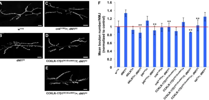
By quantifying bouton number at the NMJ on muscles 6 and 7, we confirmed that dNf1 mutants have a significant increase in mean bouton number (Figure 9A and B). In addition, this analysis confirmed previously published phenotypes for dAlk, jeb and
CCKLR-17D1[36,76]. Importantly, thedNf1synaptic overgrowth phenotype is dominantly suppressed by CCKLR-17D1, dAlk, jeb, andcnkalleles (Figure 9B), arguing that all four genes are epistatic todNf1. As a control we analyzed an allele ofspitz (spi), which encodes an EGF-like growth factor and is uncovered by suppressing Df(2L)Exel8041. However, spi shows no genetic interaction with dNf1, as loss of spi modified neither the pupal size nor the NMJ overgrowth phenotypes (Figure 9B and data not shown).
Human ALK Is Expressed in Schwann Cells and May Serve as a Therapeutic Target in NF1
The identification ofdAlkas a suppressor of all hitherto analyzed
dNf1 defects prompted us to explore whether human ALK represents a therapeutic target in NF1. Given our hypothesis that NF1 negatively regulates ALK stimulated Ras/ERK signaling, in order to play such a role, ALK and NF1 must be co-expressed in cells that give rise to symptoms. We previously found thatdNf1and
presence or absence of ALK mRNA in neurofibroma-derived
NF12/2Schwann cells andNF1+/2fibroblasts, using RNAs kindly
provided by Drs. Eric Legius and Eline Beert. In these experiments, two different primer sets readily detected ALK mRNA inNF12/2Schwann cells, but not inNF1+/2 fibroblasts
derived from the same tumors (Figure S6).
To test whether functional interactions between NF1 and ALK exist in human cells, we used the SK-SY5Y and Kelly neuroblas-toma cells, both of which harbor constitutively active F1174LALK
alleles, and both of which are highly sensitive to pharmacological ALK inhibition [80]. Compatible with a role for NF1 as a negative regulator of mitogenic ALK/RAS signals, qRT-PCR verifiedNF1
knockdown with two shRNA retroviral vectors increased the resistance of both lines to ALK inhibitors NVP-TAE684 and Crizotinib (Figures 10A, 10C and S7). Compatible with a model in which NF1 negatively regulates ALK/RAS signaling,NF1 knock-down resulted in elevated ERK and AKT activation (Figures 10B). Moreover, expression of activatedKRAS,BRAF, orMEKtransgenes, but not of other Ras effector transgenes, in SH-SY5Y cells conferred similar resistance to ALK inhibition (Figure S8).
Discussion
The work reported here was motivated by the fact that human NF1 is a characteristically variable disease, the severity of which is controlled at least in part by symptom-specific modifier genes [81]. Thus, a genetic analysis in Drosophila might not only reveal molecular pathways controlled by the highly conserved (50% identical) dNf1 protein, but also provide clues to the identity of human modifiers, which by virtue of their rate-limiting roles in symptom development might serve as therapeutic targets. The current work was also motivated by the fact that, for reasons that remain poorly understood, mostdNf1null mutant phenotypes are rescued by increasing, or phenocopied by decreasing, cAMP/PKA
signaling. The identification of genetic modifiers of a cAMP/PKA sensitive defect might reveal how loss of dNf1 affects cAMP/PKA signaling, and help to resolve the long-standing controversy as to whether dNf1 affects cAMP/PKA signaling directly, independent of its role as a Ras regulator [10,27], or indirectly, secondary to a Ras signaling defect [5,15].
While recognizing that none of the thus far identified dNf1
phenotypes are ideally suited for use in modifier screens, we selected the pupal size defect as the phenotype to analyze in our screen for three main reasons. First, pupariation occurs at the end of the larval growth period, and pupal size is readily assessed by inspecting pupae attached to the side of culture vials, making this phenotype amenable to a large-scale screen. Second, the growth defect is among several cAMP/PKA sensitivedNf1 phenotypes. Finally, reduced growth is also a symptom of human NF1 and other RASopathies [1,82]. However, while compelling reasons support the selection of this phenotype, confounding factors include that Drosophila size is a sexually dimorphic phenotype affected by population density, feeding, environmental conditions such as temperature, and genetic background differences. More-over, while heterozygousdNf1mutants are marginally smaller than wild-type pupae [5], the more robust size phenotype (,15% reduction in linear dimensions,,25% reduction in weight) used in our screen is only observed upon homozygous loss ofdNf1. Thus, our screen was not designed to find modifiers that act on the dNf1 protein itself, like the recently identified SPRED proteins [83]. Finally, organism size is a function of growth rate and duration, both of which are regulated by hormonal cascades that involve cross-talk between the larval brain, the neuroendocrine ring gland, the fat body and other tissues [19,84]. Thus, a screen for modifiers ofdNf1-regulated growth may uncover genes involved in various aspects of systemic growth control.
Early attempts to identify dNf1 pupal size modifiers were abandoned when.95% of large X-ray induced 2ndchromosome
lethality of these transgenes when driven with eitherAc5C-Gal4orelav-Gal4is indicated by{whereas (2) indicates viable offspring. (C) Western blot of adult head lysates showing relative expression ofGMR-Gal4-driven transgenic PKA*. Tubulin is used as a loading control. (D) Expression of PKA* or knockdown ofdncby shRNAi in the ring gland rescues thedNf1pupal size defect. In contrast,UAS-dNf1expression with the same ring gland drivers fails to restore systemic growth. (E–H) Expression pattern ofAkh-Gal4drivingUAS-GFP, co-stained with DAPI and anti-dNF1. GFP expression in the corpora cardiaca (CC) is indicated. Scale bar = 50mm. As previously noted [74], anti-dNf1 staining is strong in the CNS, whereas staining in the ring
gland is close to background. doi:10.1371/journal.pgen.1003958.g008
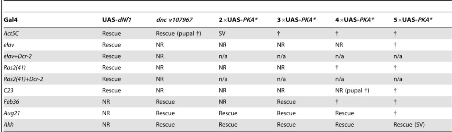
Table 3.Restoration of systemic growth bydNf1and cAMP/PKA involves different tissues.
Gal4 UAS-dNf1 dnc v107967 26UAS-PKA* 36UAS-PKA* 46UAS-PKA* 56UAS-PKA*
Act5C Rescue Rescue (pupal{) SV { { {
elav Rescue NR NR NR NR {
elav+Dcr-2 Rescue NR n/a n/a n/a n/a
Ras2(41) Rescue NR NR NR { {
Ras2(41)+Dcr-2 Rescue NR n/a n/a n/a n/a
C23 Rescue NR NR NR NR (pupal{) {
Feb36 NR Rescue NR Rescue { {
Aug21 NR Rescue Rescue Rescue Rescue {
Akh NR Rescue Rescue Rescue Rescue Rescue (SV)
Act5C-Gal4driven ubiquitousdNf1re-expression, orelav-Gal4andRas2-Gal4driven neuronal re-expression rescues thedNf1pupal size defect, whereasdncRNAi or UAS-PKA* expression controlled by the same drivers is ineffective. By contrast, expressingdNf1in specific parts of the neuroendocrine ring gland with theAkh-Gal4, Feb36-Gal4or Aug21-Gal4drivers fails to rescue, whereas using the same drivers to expressdncRNAi or attenuated UAS-PKA* transgenes does increasedNf1pupal size. All crosses produced viable adults unless otherwise indicated.
{
denotes lethality, SV sub-viable, n/a not applicable, NR non-rescue.
deficiencies were found to be lethal in adNf1background (Glenn Cowley, Iswar Hariharan and A.B., unpublished), or when a pilot chemical mutagenesis screen found the reliable mapping of identified enhancer or suppressor mutations to be impracticable (Suzanne Brill, Iswar Hariharan and A.B., unpublished). Both aborted screens informed the current effort, which used precisely defined small deficiencies, isogenic crossing schemes and exper-imental protocols that guarded against population density differences. In total we analyzed 486 1st and 2nd chromosome deficiencies that together uncover well over 80% of chromosome 1, 2L and 2R genes (Table 1). Among the screened deficiencies, 132 (27.2%) significantly modifieddNf1pupal size (p,0.01; two-tailed Student’st-test). While this is a large number, 20 deficiencies were subsequently eliminated because they also affect wild-type size. Several modifying deficiencies also uncover overlapping genomic segments, further reducing the number ofdNf1modifying loci to 76. During follow-up studies aimed at identifying responsible genes, we prioritized genes uncovered by suppressing deficiencies over those uncovered by enhancing ones, modifiers uncovered by overlapping deficiencies over those uncovered by single deletions, modifiers uncovered by small deficiencies over those uncovered by larger ones and stronger modifiers over weaker ones. We also limited ourselves to genes that function in the nervous system, based on the consideration thatdNf1re-expression in larval neurons is sufficient to suppress the growth defect [5].
We previously reported thatdNf1growth and learning defects are phenocopied by increasing neuronal Jeb/dAlk/ERK signaling, and suppressed by genetic or pharmacological attenuation of this pathway [15]. Validating our screen, deficiencies that uncoverjeb
anddAlkwere identified as dominantdNf1size defect suppressors. Others recently reported that Jeb/dAlk signaling allows brain growth to be spared at the expense of other tissues in nutrient restricted Drosophila and identified a glial cell niche around neuroblasts as the source of Jeb under these conditions [41].
However, Jeb involved in systemic growth appears of mainly neuronal origin, as RNAi-mediated jeb knockdown in neurons increaseddNf1 pupal size, whereas only one of four tested glial drivers produced partial rescue (Figure 5A).
The identification of cAMP/PKA pathway modifiers dnc,
PKA-C1 and tentatively PKA-R2 further validates our screen. Arguing that increased PKA activity doesn’t suppress dNf1
defects by attenuating Ras/Raf/MEK/ERK signaling, hsp70 -PKA* transgene expression, using a daily heat shock regimen that suppresses the dNf1 size defect [4], does not reduce the elevateddNf1larval brain phospho-ERK level, and neither does
Ras2-Gal4 driven neuronal UAS-PKA* expression (Figure 8D). Providing further mechanistic clues, our results demonstrate that
dNf1 and cAMP/PKA both affect systemic growth non-cell-autonomously, but not necessarily in the same cells. Thus, we previously showed that only relatively broadly expressed neuronal Gal4 drivers restored mutant growth when driving
UAS-dNf1, whereas multiple drivers expressed in specific subsets of neurons, including several expressed in the ring gland, lacked the ability to restoredNf1growth [5]. By contrast, using UAS
-dncRNAi or a series of newly generated attenuated UAS-PKA* transgenes that avoid the toxicity associated with high level PKA expression [73], we now show that manipulating cAMP/PKA signaling with broadly expressed neuronal Gal drivers does not affect the dNf1 size phenotype, whereas the same transgenes induced with three ring gland drivers did suppress. Intriguingly, the most potent rescue was observed when UAS-dncRNAi or attenuatedUAS-PKA*transgenes were driven in AKH-producing cells at the base of the ring gland, whereas weaker rescue was also observed with two ring gland drivers that show overlapping expression in the juvenile hormone producing corpora allata. This suggests that thedNf1growth deficiency involves a defect in processes controlled by one or both of these neuroendocrine hormones.
Figure 9. SeveraldNf1pupal size defect suppressors also suppress a NMJ synaptic overgrowth phenotype. (A–E) Representative micrographs of larval muscle 6/7 NMJs of the indicated genotypes. F: Mean bouton number per NMJ normalized to wild-type control. Compared to wild-type (w1118; A),dNf1mutants (dNf1E2; B) have an increased bouton number. While acnkloss-of-function allele had no obvious NMJ phenotype, it dominantly suppressed thedNf1NMJ defect (C). Similarly, thedNf1NMJ phenotype was suppressed inDf(1)Exel9051males that lack CCKLR-17D1 (D), while females heterozygous for CCKLR-17D1 (E) showed a lower level of suppression.Spitz (spi)is uncovered by a modifying deficiency but does not affectdNf1size and was used as a negative control. In panels A–E, scale bars represent 5mm. In panel F, error bars denote standard error of the mean.
As might be expected of a screen that used systemic growth as a read-out, our work identified a diverse set of potential modifiers. Notably, however, among a non-exhaustive set of 18 1st or 2nd chromosome genes implicated in various aspects of Drosophila body, organ, and/or cell size control (dAlk, B4, chico, hpo, Hr4, Ilp6,
jeb, Mer, mir-8, Pi3K21B, Pten, Ptth, SNF1A, sNPF, step, Tor,ush and yki; see Table S3 for details), onlydAlkandjebscored as dominant
dNf1pupal size modifiers, whereas the remaining 16 genes were uncovered by non-modifying deficiencies, or in the case ofPtth, by two deficiencies that altered developmental timing (Table S2). Further explaining this lack of overlap, the previously implicated PI3 kinase regulatorB4act in a recessive manner and several of the above listed genes function outside of the CNS. Our screen excluded such genes, because dNf1 controls growth non-cell-autonomously by regulating neuronal Ras [5]. As previously noted, a special case is provided by insulin pathway components
chico and Pten, which affect growth antagonistically. Both genes map within 5 kb of each other on the 2ndchromosome and are uncovered by the same non-modifying deficiency.
Two newly identified dNf1 growth defect suppressors,Dap160
and CCKLR-17D1, affect synaptic architecture or functioning [36,56,57]. Because dNf1 was recently reported to function downstream of focal adhesion kinase to restrain NMJ synaptic growth and neurotransmission [16], and because the cholecysto-kinin receptor related CCKLR-17D1drosulfakinin receptor stim-ulates NMJ growth [36], we analyzed whether this and three Ras signaling relateddNf1 size defect suppressors also affected NMJ architecture. Our results confirm that dNf1 mutants exhibit synaptic overgrowth, and show that loss of CCKLR-17D1
suppresses this defect. Importantly, loss ofjeb,dAlk, orcnksimilarly suppresses both size and synaptic overgrowth defects, suggesting that both phenotypes may be related.
The results presented here further support our previous conclusion that excess neuronal Jeb/dAlk/Ras/MEK/ERK signaling is the root cause of the cAMP/PKA sensitive dNf1
systemic growth defect. What happens downstream of this primary defect remains less clear, although our demonstration that increasing cAMP/PKA signaling in AKH-producing cells and other parts of the neuroendocrine ring gland suppresses the size defect provides an important new clue, not only about pathways involved in thedNf1growth defect, but also about the likely non-cell-autonomous cause of similar growth defects of PKA-C1 or
dCreb2mutants [85,86]. Other questions that remain to be fully answered concern the role of the NMJ architectural defect in the
dNf1growth deficiency and the role of Jeb/dAlk signaling in the NMJ defect. We note in this respect that that C. elegans ALK ortholog,T10H9.2, has been implicated in synapse formation [87], and that recent work suggests a role for trans-synaptic Jeb/dAlk signaling in the control of neurotransmission and synaptic morphology [88]. However, while thedNf1 growth defect is due to excess dAlk signaling in neurons, NMJ synapse formation has been suggested to involve the release of presynaptic Jeb activating postsynaptic dAlk [88]. Further work will have to establish whether the suppression of thedNf1NMJ overgrowth phenotype byjeb,dAlkandcnkinvolves cell autonomous roles for these genes at synapses, or non-cell-autonomous functions elsewhere in the CNS. Further work is also required to reveal the functional significance and the sites of action of other novel modifiers identified in our screen.
From a clinical perspective, perhaps the most relevant questions raised by our work are whether NF1 regulated ALK/RAS/ERK signaling is evolutionarily conserved and whether excessive ALK/ RAS/ERK signaling contributes to human NF1 symptoms. Much indirect evidence hints at a positive answer to both questions. First, the expression of ALK and NF1 largely overlaps in the murine nervous system [77,78], same as it does in Drosophila [15]. Second, ALK functions as an oncogene and NF1 as a tumor suppressor in neuroblastoma [89–94]. Third, midkine, a ligand that activates mammalian ALK [95], is produced by NF12/2
Figure 10.NF1suppression leads to ERK activation and confers resistance to ALK inhibitors in human neuroblastoma cells.(A)
NF1 knockdown confers resistance to ALK inhibitors in human neuroblastoma cells. SH-SY5Y cells expressing pRS and shGFPcontrol vectors, or shNF1vectors were grown in the absence or presence 50 nM NVP-TAE684 or 250 nM crizotinib. The cells were fixed, stained and photographed after 14 (untreated and crizotinib treated), or 21 (NVP-TAE684 treated) days. (B) Down-regulation ofNF1results in elevated level of phosphorylated p-ERK and p-AKT. Western blot analysis of total lysates of SH-SY5Y cells expressing pRS, shGFPor shNF1vectors. (C) The level ofNF1knockdown by each of the RNAi vectors was measured by examining theNF1mRNA levels by qRT-PCR. Error bars denote standard deviation.
Schwann cells, present at elevated levels in NF1 patient skin and serum, and acts as a mitogen for NF1 tumor cell lines [96–98]. We add to this evidence by showing that shRNA-mediated NF1
knockdown renders two oncogenic ALK-driven human neuro-blastoma cell lines resistant to pharmacological ALK inhibition, and by confirming thatALKmRNA is expressed in neurofibroma-derived NF12/2 human Schwann cells. These findings make a strong case that ALK should be explored as a therapeutic target in NF1, and that loss of NF1expression should be considered as a potential mechanism in cases of acquired resistance to ALK inhibition [99].
Materials and Methods
Fly Stocks and Experiments
The dNf1E1 and dNf1E2 alleles have been described [5]. Exelixis, DrosDel and BSC deficiencies were obtained from the Bloomington Stock Center. Transgenic RNAi lines were obtained from the Vienna Drosophila Research Center (VDRC) and the TRiP Collection at Harvard Medical School. Eaat1SM1 and
Eaat1SM2were provided by D. van Meyel,dALK8andjebweliby R. Palmer,cnkXE-385andcnkE-2083by M. Therrien, andcarD146by H. Kramer, ppl06913by M. Pankratz, hs-Ilp2 transgenic line by E. Rulifson and UAS-Rab9 DN by R. Hiesinger. Flies were maintained on agar-oatmeal-molasses medium at 25uC, unless otherwise indicated.
To assess feeding, larvae at various stages of development were placed on blue food dye-stained yeast paste, removed after 20 min, washed and photographed. To analyze wandering behavior, 100 larvae (age 40–44 hr after egg deposition (AED)) were placed on an agar plate with a central blob of yeast paste, and their position after 24 hr was documented. To assess the expression of starvation-sensitive genes, larvae at 72 h AED were placed in vials with water for 16 hr, after which RNA was prepared and subjected to blot analysis. To determine developmental timing, L1 larvae were collected 24 hr AED using a 2 hr egg collection and reared at 140 animals per vial. The number of larvae that pupariated was scored at hourly intervals. To determine the larval weight, L1 larvae were collected 24 hr AED using a 2 hr egg collection. Larvae were reared at 140 larvae per vial and groups of 10 larvae were weighed at 8 hr intervals. Longevity was assessed by maintaining adult flies under standard conditions and counting the number of dead flies at regular intervals. In each of these assays, genotypes were tested in duplicate. To induce hs-Ilp2
transgene expression, culture vials were placed in a circulating water bath at 37uC for 10 min once or twice a day with an 8 hr interval.
Insulin-Like Protein mRNA Quantification
The 7500 Fast Real-Time PCR System from Applied Biosys-tems was used to determine IlpmRNA levels in RNA prepared from dissected larval brains or from whole wandering stage 3rd instar larvae. Results were normalized to RpL32. The following primers were used: IIp2-Forward, GGCCAGCTCCACAGT-GAAGT,Ilp2-Reverse, TCGCTGTCGGCACCGGGCAT, Ilp3 -Forward, CCAGGCCACCATGAAGTTGT. Ilp3-Reverse, TT-GAAGTTCACGGGGTCCAA, Ilp5-Forward, TCCGCCCAG-GCCGCAAACTC, Ilp5-Reverse, TAATCGAATAGGCCCAA-GGT, Ilp6-Forward, CGATGTATTTCCCAACAGTTTCG,
Ilp6-Reverse, AAATCGGTTACGTTCTGCAAGTC, Ilp7 -For-ward, CAAAAAGAGGACGGGCAATG, Ilp7-Reverse, GCCA-TCAGGTTCCGTGGTT. Expression of the distantly relatedIlp8
and the midgut-expressedIlp4genes [21] was not analyzed.
Genetic Screening, Validation, and Statistical Analysis
The crossing schemes in Figure 2 were used to generatedNf1E2
mutants carrying 1stand 2ndchromosome deficiencies. To avoid crowding, cultures were maintained at 100–200 pupae per culture vial. Initial scoring used calipers set at the length ofdNf1 female pupae, ignoringdNf1heterozygotes recognizable by the presence of the TM6B balancer. Next, the length of individual pupae carrying candidate modifying deficiencies was measured by determining their head-to-tail length using a microscope fitted with NIS-Elements AR 3.0 imaging software. Measured pupae were then placed in 96-well plates (Falcon) to determine their gender and, if necessary, the genotype of eclosed flies. At least 40 pupae were measured for each genotype, and only measurements of female pupae were used to calculate mean values and standard deviations. Statistical significance was assessed with a two-tailed Student’st-test. Throughout this report, single or double asterisks denotep-values,0.05 or,0.01 respectively.
To identify responsible modifiers we used specific alleles or
UAS-RNAiknockdown. Alleles andUAS-RNAilines on the 1stand 2ndchromosomes were crossed into thedNf1E2background. UAS-RNAilines on the 3rdchromosome were recombined withdNf1E2.
UAS-RNAilines in the dNf1E2 background were crossed to Gal4 drivers in the same background. The few deficiencies that gave rise to synthetic lethal interactions were backcrossed withdNf1E1flies to produce Df/+;dNf1E2/dNf1E1progeny.
To test whether genetic suppression reflected the inadvertent introduction of a wild-typedNf1allele, we used fly DNA prepared using DNAzol (Molecular Research Inc.) in a PCR assay with AGTCACATTAATTGATCCTG and GAGATCGTTGATA-AAGAAGT primers. The second primer introduces a penultimate single nucleotide change, which together with the E2 mutation results in the introduction of an RsaI restriction site. RsaI digestion of the PCR product gives rise to 370 and 61 bp fragments for the wild-type allele, and 348, 61 and 22 bp fragments forthe dNf1E2
allele. Digests were run on 8% acrylamide gels using both wild-type (w1118
) anddNf1E2
controls.
Construction ofAkh-Gal4and AttenuatedUAS-PKA* Transgenes
TheAkhpromoter region was amplified withAkh-FORWARD (AGATCTAATCTCCTGAATGCCGCAGCG) and Akh -RE-VERSE (AGATCTATGCTGGTCCACTTCGATTC) primers. The resulting PCR fragment was subcloned into the BamHI site of a GAL4 coding region containing pCaSpeR derivative. The final construct was sequenced to ensure correct orientation of the Akh promoter before being used generate transgenic flies by standard protocols.
reaction with pCR2.1-UAS(26) as template generated pCR2.1-UAS(36) and pCR2.1-UAS(46)). The pCR2.1-UAS clones were sequenced, their inserts excised with PstI and subcloned into PstI-digested p-UAST. Correct insert orientation was verified by sequence analysis, after which the mutationally activated murine PKA* coding region [100] was subcloned into the modified vectors using XbaI and NotI.
Immunofluorescence and Analysis of NMJ Morphology
Wandering third instar larvae were dissected in Ca2+-free saline and fixed in 4% paraformaldehyde for 25 min at room temperature. Following fixation, larval pelts were washed three times in phosphate-buffered saline (PBS) and then blocked for one hour in PBT (PBS+0.1% Triton-X 100)+5% normal goat serum. Larvae were incubated in primary antibody solution for three hours at room temperature. Anti-HRP 568 (1:1000, Invitrogen) was used to visualize neurons and Alexa Fluor 488 phalloidin (1:500, Invitrogen) was used to visualize F-actin in the muscula-ture. Images were collected using a Yokogawa CSU-X1 spinning-disk confocal microscope with the Spectral Applied Research (Richmond Hill, ON, Canada) Borealis modification on a Nikon (Melville, NY) Ti-E inverted microscope using a 606Plan Apo (1.4 NA) objective. The microscope was equipped with a Prior (Rockland, MA) Proscan II motorized stage. Larval samples were excited with 488-nm (for phalloidin) and 561-nm (for HRP) 100-mW solid-state lasers from a Spectral Applied Research LMM-5 laser merge module and was selected and controlled with an acousto-optical tunable filter. Emission was collected with a Semrock (Rochester, NY) quad pass (405/491/561/642 nm) dichroic mirror and 525/50 nm (for phalloidin) and 620/60 nm (for HRP) Chroma (Bellows Falls, VT) emission filters. Images were acquired using a Hamamatsu ORCA-ER-cooled CCD camera. Hardware was controlled with MetaMorph (version 7.7.9) software (Molecular Devices, Sunnyvale, CA.). Five individual animals were imaged for subsequent morphological analysis. Motor nerve terminals of muscles 6 and 7 were imaged in abdominal segments A2 and A3 and Z-stacks (0.25mM between images) and were captured from the top to bottom of each NMJ. Morphological analysis of the NMJ was performed using NIH Image J and was assessed by quantifying the number of synaptic boutons per square micron. The number of synaptic boutons was counted as previously described [16,101] and muscle area covered by the NMJ was quantified by tracing a polygon connecting each terminal branch point [102].
Human NF1 Experiments
The retroviral RNAi vectors targeting human NF1 and expression constructs of active alleles of RAS effectors were as described previously [94]. Crizotinib (S1068) and NVP-TAE648 (S1108) were purchased from Selleck Chemicals. Antibody against NF1 was from Bethyl Laboratories (A300-140A); antibodies against pAKT(S473) and ATK1/2 were from Cell Signalling; antibodies against p-ERK (E-4), ERK1 (C-16), ERK2 (C-14) and CDK4 (C-22) were from Santa Cruz Biotechnology; A mixture of ERK1 and ERK2 antibodies was used for detection of total ERK from human cell lines. Antibody against mouse PKAa-cat (A-2) SC-28315 was from Santa Cruz Biotechnology, b-Tubulin E7 from Developmental Studies Hybridoma Bank.
SH-SY5Y, Kelly and Phoenix cells were cultured in DMEM with 8% heat-inactivated fetal bovine serum, penicillin and streptomycin at 5% CO2. Subclones of each cell line expressing
the murine ecotropic receptor were generated and used for all experiments shown. Phoenix cells were used to produce retroviral
supernatants as described at http://www.stanford.edu/group/ nolan/retroviral_systems/phx.html.
To measure cell proliferation, single cell suspensions were seeded into 6-well plates (1–26104cells/well) and cultured both in the absence and presence of ALK inhibitors. At the indicated endpoints, cells were fixed, stained with crystal violet and photographed. All knockdown and overexpression experiments were done by retroviral infection as described previously [103].
The 7500 Fast Real-Time PCR System from Applied Biosys-tems was used to determine mRNA levels.NF1mRNA expression levels were normalized to expression ofGAPDH. The following primers sequences were used in the SYBR Green master mix (Roche): GAPDH-Forward, AAGGTGAAGGTCGGAGTCAA;
GAPDH-Reverse, AATGAAGGGGTCATTGATGG; NF1 -For-ward, TGTCAGTGCATAACCTCTTGC;NF1-Reverse, AGT-GCCATCACTCTTTTCTGAAG.ALK mRNA levels in neuro-fibroma-derived NF12/2 Schwann cells and NF1+/2 fibroblasts
were analyzed using the following two primer sets: ALK -N-Forward, GGAGTGCAGCTTTGACTTCC; ALK-N-Reverse, TGGAGTCAGCTGAGGTGTTG; ALK-C-Forward, GCAAC-ATCAGCCTGAAGACA; ALK-C-Reverse, GCCTGTTGAGA-GACCAGGAG.
Supporting Information
Figure S1 Loss ofdNf1does not alter developmental timing but reduces larval growth rate. (A) Wild-type,dNf1E1, anddNF1E1/E2
mutants show no altered developmental timing, as judged by their rate of pupariation (also shown in Figure 1D). By contrast, larvae withphm-Gal4drivingUAS-Ras1V12undergo accelerated develop-ment resulting in miniature pupae [104], whereasphm-Gal4driving a dominant negativeUAS-PI3KD954A transgene delayed develop-ment and produced giant pupae [71]. (B) Mouth hook length measurements (inmm) show that dNf1larvae grow at a reduced rate. The marker represents the mean length; the upper box represents the median to Q3 value, the lower box median to Q1 value and the error bars identify the outliers.
(PDF)
Figure S2 PCR/RFLP assay fordNf1E2mutation. (A) To make sure that stocks with putative suppressing deficiencies preserved thedNf1E2C-.T nonsense transition, we used a PCR/Restriction Fragment Length Polymorphism assay. The E2 mutation does not create or destroy a restriction site. Rather, we used a reverse primer with a penultimate A-.C transversion to amplify a 431 genomic fragment as indicated. The mutant primer creates a GTAC RsaI restriction site when E2 genomic DNA is used as a template. (B) RsaI digestion of PCR products gives rise to 370 and 61 bp fragments for the wild-type allele, and 348, 61 and 22 bp fragments fordNf1E2. An example of the assay is shown with both wild-type (w1118) and dNf1E2controls (lanes 2, 3 and 4) and various deficiencies (Df) either in wild-type (Df/CyO;+; lanes 5 and 15),
dNf1 homozygous (Df/CyO; dNf1E2; lanes 6–13) or heterozygous (Df/CyO; dNf1E2/+; lanes 14 and 16) backgrounds.
(PDF)
Figure S3 Systematic identification for dNf1 modifiers. For deficiencies that did not uncover obvious candidate modifier genes, a systematic RNAi approach was used. UAS-RNAi lines targeting genes uncovered by a modifying deficiency were driven byRas2-Gal4in thedNf1E2background and the effect on pupal size determined. (A) Identification of carnation as a dNf1 modifier uncovered by suppressing Df(1)BSC275. (B) Identification of