Homepage:
www.kup.at/
mineralstoffwechsel
Online-Datenbank mit
Autoren- und Stichwortsuche
Indexed in SCOPUS/EMBASE/Excerpta Medica
www.kup.at/mineralstoffwechsel
Österreichische Gesellschaft für Orthopädie und Orthopädische Chirurgie
Österreichische Gesellschaft für Rheumatologie Offizielles Organ der
Österreichischen Gesellschaft zur Erforschung des Knochens und Mineralstoffwechsels
Member of the
Nobember 2008 - Abstracts der
Poster und Vorträge
Journal für M ineralstoffwechsel &
M uskuloskelettale Erkrankungen
PROGRAMMSCHWERPUNKTE
¥ Kollagenosen IPriv.-Doz. Dr. Jochen ZWERINA¥ Die aktinische KeratoseI OÄ Dr. Karin KRENMAYR
¥ Psoriasis Arthritis aus dermatologischer Sicht I Dr. Christine MESSERITSCH-FANTA ¥ Psoriasis Arthritis aus rheumatologischer Sicht I Prim. Doz. Dr. Burkhard LEEB ¥ Morbus Still I ao. Univ.-Prof. DDr. Manfred HEROLD
¥ Die Gicht I tbd
Organisation:
Fischill PR Kochgasse 4/4, 1080 Wien
Tel.: +43 1 408 682 4 - 12
Informationen zur Veranstaltung und Anmeldung:
www.rheuma-days-austria.at
Unter Leitung der Tagungspräsidenten Prim. Doz. Dr. Leeb und Prim. Univ. Prof. Dr. Resch lädt die Fortbildungsinitiative Rheuma Days bereits zum siebten Mal in die Therme Wien Med, um den wissenschaftlichen Austausch anzuregen und den Teilnehmerinnen und Teilnehmern ein thematisches Update im breiten Fach der Rheumatologie zu bieten.
18. März 2017, 09:00 bis ca. 14:30
Therme Wien Med, Kurbadstraße 14, 1100 Wien
Die Veranstaltung wird für das Diplomfortbildungsprogramm (DFP) der Ärztekammer eingereicht.
Programmänderungen vorbehalten, Stand: 23.01.2017
188
J MINER STOFFWECHS 2008; 15 (4)Comparison of SACRAH and M-SACRAH in patients
with rheumatoid arthritis and osteoarthritis
G. Babic, N. Vujasinovic-Stupar, J. Sautner*, D. Petrovic, M. Bukilica, B. F. Leeb*
Clinical Department One, Institute of Rheumatology, Belgrade, Serbia; *Second Medical Department, State Hospital Stockerau
Objectives To quantify the degree of functional impairment, joint stiffness and pain of hand and little finger joints in patients suffering from rheumatoid arthritis (RA) compared with hand osteoarthritis (HOA) and to compare the original SACRAH [1] (Score for the Assess-ment and quantification of Chronic Rheumatoid Affections of the Hands) and the shortened and modified M-SACRAH [2].
Methods The original SACRAH-questionnaire, dating from 2000, consists of 23 questions applying visual analog scales (VAS) covering hand function (17 questions), stiffness (2 questions) and pain (4 ques-tions). The average score of each category is calculated, and the over-all average for the three category scores is then obtained. The overover-all score may therefore range between 0 (without functional impairment, stiffness and pain) and 100 (maximum difficulties) [1]. The M-SACRAH, dating from 2003, is a shortened version of the M-SACRAH, reduced to 12 questions, requiring the same calculation procedure [2].
94 patients suffering from RA completed both questionnaires (83 f/ 11 m; mean age 58 yrs, mean disease duration 114 months, 87 % RF+, mean ESR 1st hour 58 mm, mean morning stiffness 194 minutes). Also
18 patients affected with HOA filled in the questionnaires (mean age 67 yrs, mean disease duration 150 months, mean ESR 1st hour 27 mm).
Results In 14 of 23 questions statistically significant differences were found, in 12 items of the function domain, and in one pain item (during intensive work) and in the item addressing morning stiffness. In the RA group, mean SACRAH amounted to 52 while mean M-SACRAH amounted to 46 (p = 0.96). In the HOA group, mean total SACRAH reached 37 and mean M-SACRAH 36 (p = 0.96).
Conclusion
– Significant differences between RA and HOA patients could be revealed mainly in the function domain mirroring increased func-tional impairment in RA patients.
– This study underlines the prior findings on the highly significant correlation of SACRAH and M-SACRAH, for RA as well as for HOA patients.
References:
1. Leeb BF, Sautner J, Andel I, Rintelen B. SACRAH – A Score for Assessment and Quantification of Chronic Rheumatic Affections of the Hands. Rheuma-tology (Oxford) 2003; 42: 1173–8.
2. Sautner J, Andel I, Rintelen B, Leeb BF. M-SACRAH. A modified, shor-tened version of SACRAH (Score for the Assessment and Quantification of Chronic Rheumatoid Affections of the Hands). Rheumatology (Oxford) 2004; 43: 1409–13.
Titerhöhe und Feinspezifität der
Anti-SS-A/Ro-Antikörper bei Patienten mit Sjögren-Syndrom
M. Bergmann, W. Klotz, A. Österbauer, J. Hermann*, M. Herold Rheumalabor, Universitätsklinik für Innere Medizin I, Medizinische Uni-versität Innsbruck; *Abteilung für Rheumatologie, UniUni-versitätsklinik für Innere Medizin, Medizinische Universität Graz
Einleitung Das Sjögren-Syndrom ist eine chronisch entzündliche autoimmune Erkrankung der exokrinen Drüsen. Die Diagnose eines Sjögren-Syndroms stützt sich gemäß den europäisch-amerikanischen Klassifikationskriterien auf die klinischen Symptome exokrine Insuffizi-enz, den objektiven Nachweis der eingeschränkten Drüsenfunktion, den histologischen Nachweis fokaler Lymphozyteninfiltrate und der spe-zifischen Antikörper gegen SS-A/Ro und SS-B/La.
Ziel Ziel der Diplomarbeit war zu klären, ob sich Patienten mit dia-gnostiziertem Sjögren-Syndrom und Patienten mit anderen Erkran-kungen in der Ro-Titerhöhe unterscheiden. Weiters soll geklärt wer-den, ob bei negativem ANA- (antinukleäre Antikörper) Screening auf der HEp-2-Zelle eine weiterführende Testung mittels ELISA auf Anti-SS-A/Ro-Antikörper zu empfehlen ist. In diesem Zusammenhang wurde untersucht, ob in Seren mit niedrigem ANA-, aber hohem Ro-Titer isoliert Anti-Ro52kD vorliegen.
Methoden 204 Anti-SS-A/Ro-positive Patienten der Universitäts-klinik Innsbruck, davon 61 auf der HEp-2-Zelle ANA-negativ, jedoch im ELISA Anti-SS-A/Ro-positiv, wurden in die Auswertung einbe-zogen. Die Diagnose wurde aus dem aktuellsten Arztbrief entnommen. Die Anti-SS-A/Ro-Titer wurden mit einem kommerziell verfügbaren ELISA (ELiA Ro, Phadia, Freiburg), die ANA-Titer mittels indirekter Immunofluoreszenz (iIF) auf der HEp-2-Zelle ermittelt.
32 Seren, davon 20 Proben (Universitätsklinik Graz) von Patienten mit bioptisch gesichertem Sjögren-Syndrom und 12 ANA-negative, Anti-SS-A/Ro-positive Proben (Universitätsklinik Innsbruck) wurden einer detaillierten immunologischen Analyse unterzogen. Die Seren wurden auf ANA, mit einem auf einem ELISA basierenden Immuno-assayautomaten ELiA auf Anti-SS-A/Ro- und Anti-SS-B/La-Antikör-per sowie mittels ELISA (Pharmacia, Freiburg) auf Anti-Ro52kD-und Anti-Ro60kD-Antikörper getestet. Seren der Universitätsklinik Innsbruck mit diskrepanten Ergebnissen wurden zusätzlich mittels Westernblot und Immunoblot mit rekombinantem Ro52kD und nati-vem Ro60kD-Antigen (Euroimmun, BRD) getestet.
Ergebnisse Sowohl bei den Patienten mit gesichertem Sjögren-Syndrom als auch bei der Gruppe ohne Hinweis auf eine Kollagenose zeigte die Mehrheit der Patienten (33/43 bzw. 59/103) einen Ro-Titer im obersten Messbereich. Seren mit niedrig positiven Ro-Antikör-pertitern stammten überwiegend von Patienten ohne Kollagenose. Von den 20 Patienten mit bioptisch gesichertem Sjögren-Syndrom waren 9 (45 %) iIF-ANA-positiv, 14 (70 %) hatten Anti-SS-A/Ro-Antikörper.
Von 23 iIF-ANA-negativen Proben (Titer < 1:100) waren 6 (26,1 %) isoliert Anti-Ro60kD-positiv (3 von Patienten mit diagnostiziertem Sjögren-Syndrom, 1 von einem Patienten mit systemischem Lupus erythematodes), 3 Proben (13,0 %) waren isoliert Anti-Ro52kD-positiv (davon 2 von Patienten mit diagnostiziertem Sjögren-Syndrom) und 3 Proben (13,0 %) waren Anti-Ro52kD- und anti-Ro60kD-positiv (alle 3 Proben stammten von Patienten mit diagnostiziertem Sjögren-Syndrom).
Schlussfolgerung Anti-SS-A/Ro-Antikörper sind ein wichtiges Diagnosekriterium des Sjögren-Syndroms, deren Vorliegen aber keine absolute Bedingung für die Diagnose darstellt. Der Antikörpernachweis erleichtert die Diagnosestellung aufgrund des hohen Anteils an
Pati-Jahrestagung der ÖGR
28. und 29. November 2008
Abstracts der Poster und Vorträge
*
enten mit SS-A/Ro-Antikörpern, aber ohne Hinweis für eine Kolla-genose ist eine Testung auf Anti-SS-A/Ro-Antikörper nur in Zusam-menhang mit der entsprechenden klinischen Symptomatik sinnvoll. Entgegen unseren Erwartungen finden sich in der Gruppe der iIF-ANA-negativen Patienten sowohl Antikörper mit isolierter Aktivität gegen das 52kD- oder 60kD-Antigen als auch Seren, die Antikörper gegen beide Antigene enthalten. Eine weiterführende Untersuchung an einem größeren Kollektiv iIF-ANA-negativer Seren ist nötig, um die diagnostische Sinnhaftigkeit einer SS-A/Ro-Bestimmung in iIF-ANA-negativen Seren abzuklären.
Pten regulates osteoclast biology and inflammatory
arthritis
S. Blüml, G. Schabbauer*, B. Niederreiter, J. Smolen, K. Redlich Division of Rheumatology; *Dep. of Vascular Biology and Thrombosis Research, Medical University of Vienna
Pten is a lipid phosphatase, whose substrate is phosphatidylinositol 3,4,5-trisphosphate. Therefore, pten is one of the main antagonists of the PI3-kinase, which plays a major role in many important cellular functions, such as proliferation, migration or response to inflamma-tory stimuli.
Here we investigated the role of pten in osteoclast biology as well as in collagen induced arthritis.
We show that in vitro osteoclastogenesis in mice with a conditional, monocyte/macrophage-specific pten knock-out (LysMCrePtenflox/-) is
increased by a factor of two compared to control wild-type mice. This phenotype was also detectable in vivo, where analysis of tibiae of female mice revealed a significant increase in numbers of osteoclasts per bone perimeter (N.Oc/B.Pm) and osteoclast surface per bone sur-face (Oc.S/BS). However, enhanced osteoclastogenesis did not result in systemic bone loss, since no differences in bone volume per tissue volume (bv/tv), trabecular thickness and trabecular separation could be detected, suggesting impaired function of osteoclasts lacking pten. In collagen induced arthritis, LysMCrePtenflox/-mice showed a
signi-ficant reduction in clinical severity of arthritis compared to wild-type animals. Interestingly, total collagen antibodies as well as anti-collagen IgGs were identical in both groups. Flow cytometric analy-sis revealed that dendritic cell and macrophage recruitment to the draining lymph node was not impaired, suggesting regular recruit-ment of antigen presenting cells. Histological analysis of CIA, Lys-MCrePtenflox/-mice displayed significantly reduced erosive bone
de-struction. There was also a strong tendency towards reduced numbers of osteoclasts, which did not reach statistical significance. These data point to a regulatory role of pten in osteoclast biology and arthritis development.
HO-1 end products biliverdin and carbon monoxide
ameliorate murine collagen induced arthritis
M. Bonelli, A. Rapp, A. Savitskaya, E. Rath, F. H. Bach*, J. S. Smolen, C. Scheinecker
Division of Rheumatology, Internal Medicine III, General Hospital of Vienna, Medical University of Vienna; *Beth Israel Deaconess Medical Center, Harvard Medical School, Boston, MA, USA
Background Heme oxygenase 1 (HO-1), which degrades heme to free iron, biliverdin and carbon monoxide (CO), plays an important role in inflammation. Beneficial effects of HO-1 overexpression have been shown in various inflammatory conditions. There are, however, conflicting results regarding the role of HO-1 in rheumatoid arthritis (RA).
Objectives We therefore set up experiments in order to analyze the therapeutic potential of the end products of HO-1 activity, CO and biliverdin, in the collagen induced arthritis (CIA) model of RA.
Methods For the induction of CIA DBA/1 mice were immunized with type II collagen (CII) and boosted after 10–14 days. Upon the onset of arthritis the animals were scored for clinical signs of arthritis including paw swelling and grip strength. Anti-CII antibody levels were determined by ELISA. Animals were randomized into treatment
groups after they developed the first clinical signs of arthritis. CO was administered for one hour at a concentration of 250 ppm. Bili-verdin (35 mg/kg) was injected i. p. twice daily and control animals were injected with PBS. All animals were treated for 14 days. After another 5 days all animals were sacrificed and paraffin sections of the affected joints were analyzed for histomorphological signs of inflam-mation as well as cartilage and bone destruction.
Results All animals immunized with CII developed serum anti-CII antibodies. Antibody levels were slightly decreased in biliverdin and in CO-treated animals. Both, biliverdin and CO treatment significantly improved clinical disease activity scores as compared to control ani-mals. In line with this, histomorphological analysis of joint sections in CO and biliverdin treated animals as compared to controls revealed a decrease in inflammation, area of erosion, osteoid area and num-bers of osteoclasts.
Conclusion Our data demonstrate a beneficial therapeutic effect of treatment with HO-1 end-products CO and biliverdin in the CIA model of RA. Future experiments will have to determine the mechanisms of how CO and biliverdin interfere with disease progression in CIA.
Analyses of a novel regulatory T cell subset in
patients with systemic lupus erythematosus (SLE)
M. Bonelli, A. Savitskaya, E. Rath, J. S. Smolen, C. Scheinecker Division of Rheumatology, Internal Medicine III, General Hospital of Vienna, Medical University of Vienna
Introduction CD4+CD25+ regulatory T cells (Treg) that specialize
in the suppression of immune responses might be critically involved in the pathogenesis of autoimmune diseases. Little, however, is known about the phenotype and functional properties of CD4+CD25-Foxp3+
T cells. We therefore performed phenotypic and functional analyses of CD4+CD25-Foxp3+ T cells in patients with autoimmune disease
and healthy controls (HC).
Methods The phenotype analyses of peripheral blood CD4+CD25
-Foxp3+ T cells was determined using flow cytometry in SLE patients,
patients with systemic sclerosis (SSc), rheumatoid arthritis (RA) and HC. The percentage of CD4+CD25-Foxp3+ T cells was correlated with
clinical data, the daily cortisone dose and the SLE disease activity index (SLEDAI). In addition we isolated CD4+CD127–CD25– T cells
from HC and SLE patients by Flourescence Activated Cell Sorting (FACS) and cocultured them with responder T cells (Tresp).
Results Proportions of CD4+CD25-Foxp3+ T cells are increased in
patients with SLE as compared to patients with SSc, RA or HC. They do not have an activated phenotype. We tried to correlate the CD4+CD25–Foxp3+ T cells with the clinical disease activity and the
daily cortisone dose and we found a significant correlation for the SLEDAI score and the daily cortisone dose. Finally we isolated CD4+
CD127-CD25+ T cells and CD25– T cells, which both expressed Foxp3,
cocultured them with Tresp and found a suppressive capacity in both populations.
In conclusion one might speculate that this population represents a countermechanism in active SLE patients.
Childhood stroke as the presentation of Takayasu’s
arteritis: Diagnostic delay can cause catastrophic
complications
J. Brunner1, 2, D. Armstrong3, B. M. Feldman4, R. Schneider1, S. Benseler1 1Division of Pediatric Rheumatology Neurology, The Hospital for Sick Children, Toronto, Ontario, Canada; 2Pediatric Rheumatology, Depart-ment of Pediatrics, Medical University of Innsbruck; 3Diagnostic and In-terventional Paediatric Neuroradiology, Department of Diagnostic Imaging, The Hospital for Sick Children, Toronto, Canada; 4Health Policy Management & Evaluation, and Public Health Sciences, University of Toronto, Division of Pediatric Rheumatology, The Hospital for Sick Child-ren, Toronto, Canada
Introduction Takayasu’s arteritis (TA) is a granulomatous inflam-mation of the aorta and its major branches. Clinical presentation in adults is characterized by symptoms due to stenosis or aneurysms of the involved vessels.
The studies about TA in childhood are rare. Symptoms are either ty-pical as hypertension and pulselessness, but a more unspecific pre-sentation may also indicate TA. Children may present with organ specific or with nonspecific systemic features. Therefore connecting the symptoms with the diagnosis may be a challenge in pediatric pa-tients. The disease can develop tremendous complications, when the diagnosis is missed and the consecutive treatment starts with a delay.
Case report We present a 9½ year old girl who developed 8 epi-sodes of stroke in 2006. The last stroke before diagnosis developed in August 2006 going along with 3 episodes of binocular diplopia and unsteadiness lasting 3 minutes. Laboratory examination was normal. The MRI showed new infarcts in the left caudate nucleus and hypo-thalamus. The MRA showed evidence of flow disturbance at the origin of the brachiocephalic artery. The chest CT revealed a filling defect in the left CCA. According to the EULAR/PRES consensus criteria the diagnosis of TA was established. Proper anti-inflammatory treat-ment was initiated.
Conclusion TA in children has a different presentation from adults and may cause catastrophic complications. Therefore TA has to be considered in children presenting with stroke.
Analysis of the classical, alternative, and MBL
pathways of the complement system in juvenile
idiopathic arthritis
J. Brunner, M. Sailer, E. Binder, L.-B. Zimmerhackl, M. Prelog Department of Pediatrics, Medical University of Innsbruck
Introduction The complement system is involved in the host defence by recognition and elimination of potentially harmful exogenous and endogenous structures from the human body. Activation of comple-ment may also promote inflammatory reactions and cause tissue da-mage if adequate control is not provided by the complement regula-tory proteins. Significant amounts of biologically active products arising from complement activation have been detected in patients with rheumatoid arthritis.
Objective To investigate the role of complement cascade in juvenile idiopathic arthritis.
Method 12 serum samples and 2 samples from synovial fluid were obtained from 2 individuals with juvenile idiopathic arthritis (JIA). The complement kit for assessment of classical, alternative and MBL pathway activity was developed by the EU consortium and prepared centrally at Wieslab (Sweden).
Results The samples of synovial fluid showed a deficiency in the classical, alternative and MBL pathway of the complement system. The results in the sera were normal.
Conclusion Complement system might play a major role in the development of joint effusion in JIA.
Chitotriosidase activity in juvenile idiopathic
arthritis
J. Brunner, M. Sailer, E. Binder, L.-B. Zimmerhackl, M. Prelog Department of Pediatrics, Medical University of Innsbruck
Background Juvenile idiopathic arthritis (JIA) is an inflammatory joint disease of unknown aetiology. The pathogenesis is driven by T-and B-cells. The role of macrophages remains unclear. Chitotriosida-se belongs to the chitinaChitotriosida-se protein family and is Chitotriosida-secreted by activated macrophages. The chitinases are able to catalyze the hydrolysis of chitin or chitin-like substrates such as 4-methylumbelliferyl chitotrioside.
Methods Chitotriosidase activity was determined using the substrate 4-methylumbelliferylb-DNN’N’’-triacetylchitotriosiose (MUbGlc-Nac, SIGMA Chemical Co). The substrate and serum was incubated with the serum in a citrate/phosphate buffer. The reaction was stopped
by adding glycina buffer. The fluorescence of 4-methylumbelliferone was evaluated by fluorimeter at excitation 365 nm and emission 430 nm.
Result We report about chitotriosidase measurements in patients with JIA. The chitotriosidase level in synovial fluid was up to 2000 nmol/ h/ml at disease onset before therapy. The level in sera was below 600 nmol/h/ml.
Conclusion Serum chitotriosidase levels could represent the activity of macrophages in the synovial fluid in JIA.
Rituximab is a therapeutical option for juvenile
microscopic polyangiitis
J. Brunner, M. Sailer, E. Binder, L.-B. Zimmerhackl, M. Prelog Department of Pediatrics, Medical University of Innsbruck
Introduction Microscopic polyangiitis is characterized by anti-neu-trophil cytoplasmic antibodies (ANCA) and small vessel vasculitis. The diseases usually respond to cyclophosphamide (Cyc) but some patients are therapy resistant. Rituximab (RIT) is a chimeric antibo-dy to CD20 causing lysis of B-lymphocytes and used for treatment of lymphomas. RIT has also been successfully used in patients with rheu-matoid arthritis (RA), idiopathic thrombocytopaenic purpura, auto-immune haemolytic anaemia, cold agglutinin disease, systemic lupus erythematosus and vasculitides.
Case report A 16 year old female patient with hemoptysis, nephritis, dermatitis was diagnosed as a myeloperoxidase-ANCA-positive microscopic polyangiitis. The patient was resistant to conventional therapy or had relapsed repeatedly after cessation of cyclophospha-mide (Cyc). The patient was treated with intravenous infusions of rituximab (RIT).
Conclusion RIT seems promising and safe in pediatric microscopic polyangiitis. Controlled studies should be conducted.
Omega-3 fatty acids – a therapeutic option in
secondary fibromyalgia?
C. Deutsch, T. Nothnagl, P. M. Haindl, B. Rintelen, I. Andel, B. F. Leeb II. Medizinische Abteilung, NÖ Zentrum für Rheumatologie, NÖ Landes-klinikum Weinviertel Stockerau, Karl-Landsteiner-Institut für klinische Rheumatologie, Stockerau
Background Secondary fibromyalgia (FMS) may be prevalent in up to 20 % of patients with systemic lupus erythematosus, in up to 15 % of rheumatoid arthritis (RA) patients, as well as in patients with Sjoegren’s syndrome, and chronic inflammatory bowel disease. Often, severe pain is present in contrast to low inflammatory activity of the underlying disease. So far, only a minority of patients responds well to standard therapy. Omega-3 fatty acids have been shown to somewhat improve the course of RA [1].
Objective To study whether omega-3 fatty acids could constitute a treatment option in patients with secondary FMS accompanying RA.
Patients and methods 10 patients (9 females, 1 male; mean age 47.5 yrs.) were included in this prospective open study. 9 patients suffered from seronegative RA, and 1 patient from rheumatoid factor positive RA. All patients met the ARA classification for RA (1987) and the 1990 ACR classification criteria for FMS at the time of inclu-sion into the study. Patients received omega-3 fatty acids intravenously on 5 consecutive days once a month at a single dose of 2 ml/kg/d. This medication was continued over 12 weeks. Outcome parameter included number of positive tender points, pain perception on a visu-al anvisu-alogue scvisu-ale (0–10 cm), C-reactive protein (CRP, mg/dl) and ery-throcyte sedimentation rate ESR (first hour). Morning stiffness and tender and swollen joint count (TJC, SJC) were documented as well as the DAS 28 calculated.
chan-ges could be seen with respect to ESR and CRP levels and morning stiffness. Treatment was generally well tolerated.
Conclusion Treatment with intravenously administered omega-3 fatty acids resulted in some improvement in RA patients suffering from secondary FMS. The results obtained here could give the basis to justify further controlled studies which are of course warranted to prove efficacy and tolerability.
References:
1. Leeb BF, Sautner J, Andel I, Rintelen B. Intravenous application of omega-3 fatty acids in patients with active rheumatoid arthritis. The ORA-1 trial. An open pilot study. Lipids 2006; 41: 29–34.
Treatment patterns, disease activity, and quality of life
of patients with ankylosing spondylitis in Central and
Eastern Europe: a cross-sectional survey
W. Ebner, T. Palotai, C. Codreanu, P. Géher, A. Pahor, K. Pavelka, J. Smolen, J. Szechi´nski, M. Z`´lnay
Medical Department II, Rheumatology, Hospital Hietzing, Vienna
Objective We sought to obtain information on the profile of pati-ents with ankylosing spondylitis (AS), disease activity, previous and current treatments, and the proportion and profile of patients treated with conventional medications but considered eligible for anti-tumour necrosis factor (TNF) therapy.
Methods Rheumatologists from 7 Central and Eastern European countries who were experts in treating patients with AS participated in the study. Physicians were to include 3–5 patients who had never received anti-TNF therapy and were asked to decide whether they considered their patients candidates for anti-TNF therapy.
Results A total of 1506 patients were analyzed. 61 % of AS patients who had never received anti-TNF therapy until the time of the survey qualified for anti-TNF therapy based on the clinical judgment of their rheumatologists, with this proportion ranging from 36 % in Slovakia to 84 % in Romania. Overall, candidates had higher levels of disease activity and functional impairment, and they were more likely to report a lower quality of life. 38 % of candidates fulfilled the Assessment in Ankylosing Spondylitis (ASAS) recommendations with respect to a Bath Ankylosing Spondylitis Disease Activity Index (BASDAI) of at least 4 and previous use of at least 2 non-steroidal anti-inflammatory drugs (NSAIDs), ranging from 18 % in Poland to 57 % in Hungary.
Conclusion In summary, more than half of the AS patients currently treated with other medications may be eligible for anti-TNF therapy, even though there were considerable discrepancies between the coun-tries. Also, rheumatologists regarded disease activity as the determining factor for starting anti-TNF drugs but their decision did not always fully comply with the ASAS recommendations, confirming the need for continued exchange among the medical community to increase awa-reness of the ASAS recommendations.
Quantitative variables of salivary gland scintigraphy
differ between parotid and submandibular gland in
primary and secondary Sjögren’s syndrome and
non-Sjögren’s sicca syndrome
S. F. Egger1, I. Hurtl2, B. Ibi2, P. Petera3, I. Virgolini2, J. S. Smolen3 1Klinikum Malcherhof, Baden; 2Institute of Nuclear Medicine, Hospital Hietzing, Vienna, 32nd Dept. of Internal Medicine and Centre for Diagnosis and Treatment of Rheumatic Diseases, Vienna
Background Sjögren’s syndrome (SS)is an immune mediated dis-order of exocrine glands, predominantly affecting lacrimal and sali-vary glands. SS can occur as primary disorder (pSS) or secondary to other established connective tissue diseases (sSS). Other conditions in which lacrimal and salivary gland functions are diminished, but without any evidence of underlying inflammatory or immune mediated processes, have been summarized as non-Sjögren’s sicca syndrome (nSS-sicca). Salivary scintigraphy is a method that allows to evaluate both parotid and submandibular glands. Quantification of the paren-chymal function of accumulation as uptake (UT) and of the stimulated secretory function as ejection fraction (EF) have been proved reliable and accurate reflections of salivary gland functions. It is currently unknown, if patients of the different diagnostic groups, pSS, sSS, and nSS-sicca, differ in these quantitative variable of parotid and sub-mandibular gland scintigraphy.
Objectives The aim of this study was to compare quantitative vari-ables of both parotid and submandibular gland scintigraphy in diffe-rent diagnostic groups, pSS, sSS and nSS-sicca.
Methods 106 suffering from pSS (n = 31), sSS (n = 46), or nSS-sicca (n = 29) who underwent sequential salivary gland scintigraphy with secretory stimulus by ascorbic acid were analysed retrospec-tively. After the injection of 185 MBq 99mTc-sodium pertechnetate, scintigraphic percentage uptake (UT) and, after stimulus, ejection fraction (EF in %) were calculated for parotid and submandibular gland in all patients. Results were compared between the different diag-nostic groups using the Mann-Whitney U test.
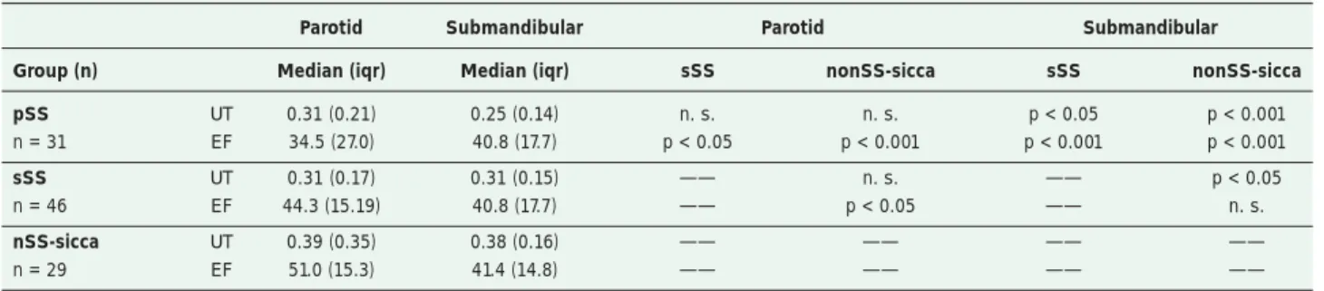
Results In the parotid gland there were no statistically significant differences in UT between any of the diagnostic groups. However, in the pSS group EF was significantly decreased when compared to sSS and nonSS-sicca (p < 0.05 and p < 0,001, respectively; Table 1). In addition, EF was significantly lower in sSS when compared to nonSS-sicca (p < 0.05). In the submandibular gland significant differences in UT were shown between pSS, sSS and nonSS-sicca group, with UT being lowest in pSS, intermediate in sSS and highest in nonSS-sicca. Furthermore, highly significantly decreased values of EF were observed in the pSS group compared to sSS and nonSS-sicca. EF was not significantly decreased in the sSS group compared to nonSS-sicca.
Conclusion By using quantitative analysis in salivary gland scinti-graphy, EF of the parotid gland and UT of the submandibular gland may allow differentiation of pSS from other forms of sicca syndrome, especially nonSS-sicca.
Table 1: Egger SF et al. Differences between diagnostic groups in UT and EF of the parotid and submandibular gland
Parotid Submandibular Parotid Submandibular Group (n) Median (iqr) Median (iqr) sSS nonSS-sicca sSS nonSS-sicca pSS UT 0.31 (0.21) 0.25 (0.14) n. s. n. s. p < 0.05 p < 0.001 n = 31 EF 34.5 (27.0) 40.8 (17.7) p < 0.05 p < 0.001 p < 0.001 p < 0.001
sSS UT 0.31 (0.17) 0.31 (0.15) —— n. s. —— p < 0.05 n = 46 EF 44.3 (15.19) 40.8 (17.7) —— p < 0.05 —— n. s.
The integrin linked kinase is upregulated in the
rheumatoid synovial membrane
J. Grisar, L. Amoyo, B. Niederreiter, K. Redlich, J. S. Smolen
Division of Rheumatology, Department of Internal Medicine III, Medical University of Vienna
Background Integrin linked kinase (ILK) is a β1-integrin cytoplas-mic domain interacting protein. Its activity is stimulated by adhesion to the extracellular matrix and by growth factors in a phosphatidyli-nositol 3-kinase dependent manner. In the tissue of several tumors, this kinase, which is located upstream of NFkappa B, is upregulated. ILK plays a pivotal role in angiogenesis, invasion and proliferation and since these features are also hallmarks of rheumatoid arthritis (RA), we assessed if ILK would also be upregulated in the rheumatoid synovial membrane.
Methods To determine the expression of ILK in RA, we performed stainings with an antibody against ILK in paraffin embedded 2 µm hind limb sections of arthritic joints of TNF transgenic (tg) mice, a well established TNF driven arthritis model. Hind limb sections of wild type (WT) mice served as controls. Furthermore, immunohisto-chemistry was also performed on paraffin embedded sections of syn-ovial tissue of patients with RA and patients with osteoarthritis (OA) that underwent joint replacement.
Moreover, we analyzed the extracts of synovial fibroblasts (SF) from RA and OA patients and in WT and TNF tg mice by western blot detecting the expression for ILK. Furthermore, SF of RA patients were also stimulated with TNF at a dose of 10 ng/ml for 24, 48 and 48 hours and ILK-expression was assessed in the same manner as de-scribed above and compared with SF cultured in medium alone.
Results The expression of ILK was very pronounced in the synovial membrane of arthritic joints of TNF transgenic mice. ILK was espe-cially expressed in the synovial infiltrates and at the pannus-bone junction. Furthermore, some, but not all chondrocytes and osteoclasts were found to stain for ILK. In contrast, in WT mice immunohisto-chemistry showed only little ILK expression, mainly in chondrocy-tes and the synovial membrane. In OA, we also observed cells ex-pressing ILK but to a lesser extent than in RA.
Western blot analyses showed that ILK is expressed in both OA- and RA-SF. After stimulation with TNF, RA-SF showed an increasing ILK expression with increasing incubation period, amounting to 1.4 fold after 96 h stimulation with TNF. This suggests that also other pathways lead to the upregulation of ILK in human RA.
The expression of ILK analysed out of the SF isolated from TNF tg mice was clearly higher than in WT mice.
Conclusion Our results show that the kinase ILK is overexpressed in cells of the synovial membrane of arthritic mice and RA patients. This overexpression is partly TNF-dependent. These findings sug-gest that ILK might be an interesting candidate for interfering with the course of synovial inflammation in RA.
Gout: Old disease – new therapy
J. Gruber
Universitätsklinik für Innere Medizin I, Medizinische Universität Innsbruck
Introduction Gout is an old malady known in ancient cultures and is one of the first diseases recognized as a clinical entity. It is a com-mon chronic arthritis that can lead to significant disability. Gouty arthritis is often self limited and is characterized as an inflammatory reaction to monosodium urate (MSU) crystals in the joints and in the adjacent structures. So far the influence of MSU crystals on the im-mune system was hardly noticed and the crystals were regarded as “innocent bystander”. Recently, the role of the MSU crystals in the induction of an inflammatory process, mediated through an intracel-lular sensor, i. e. the NALP3 Inflammasome, and consecutively inter-leukin-1 (IL-1) was recognized. A similar mechanism may be respon-sible for the development of pseudogout. Therefore, new agents in the management of gout and pseudogout have emerged.
Patients and methods 6 patients with gout and 1 patient with pseudogout were treated on an open label basis with 100 mg anakinra subcutaneously usually for 3 days. All our patients either failed con-ventional treatment with NSAIDs, colchicine or glucocorticoids or
had contraindications for these drugs. Patients with an active untrea-ted infection, with uncontrolled diabetes, with uncontrolled heart or respiratory failure were not eligible. All patients consented to receive anakinra. Clinical efficacy was assessed by clinical examination of the swollen and tender joints as well as the patient’s evaluation of the efficacy of the treatment in terms of reduction of pain and symptoms with the use of a visual analog scale (0–100 mm scale).
Results All patients with gouty arthritis responded within 48 hours. 1 patient with involvement of the Achilles tendon responded later and needed 4 injections. 1 patient had a relapse, probable due to an excessive alcohol intake, and had to be treated with a second series of 3 injections. The response after the second series was also very swift and occurred again within 48 hours. No treatment related side effects were observed during therapy and there were no infectious complica-tions.
1 patient suffered from a pseudogout attack of the first carpometacar-pal joint of the left hand. The aspirate showed clearly calcium pyro-phosphate dihydrate (CPPD) crystals. She was treated with 100 mg anakinra on three consecutive days and responded with 72 hours with marked reduction of pain and swelling.
Conclusions With the use of anakinra a new therapeutic principle for the treatment of gout and pseudogout has emerged and, if proven in further studies, could be used as a promising agent in patients where glucocorticoids, colchicine or NSAIDs are contraindicated or could even be used as a first line therapy.
Wirksamkeit und Kosteneffektivität eines ambulanten
multimodalen Rehabilitationsprogramms bei
Patienten mit chronischen Kreuzschmerzen
W. Habelsberger, C. Grunt-Göschl Rehamed, OÖGKK, Linz
Einleitung Rund eine Million Österreicher im Alter von 15 und mehr Jahren leidet an chronisch rezidivierenden Kreuzschmerzen. Der Großteil der verursachten direkten und indirekten Krankheitskosten ist der Patientengruppe mit chronischen Kreuzschmerzen zuzuord-nen. Der Frage nach Wirksamkeit und Ökonomie verfügbarer Reha-bilitationsmaßnahmen kommt daher eine besondere Priorität zu. Im Rahmen dieser Studie wurde eine retrospektive Untersuchung ver-schiedener subjektiver Parameter (Schmerz, Depressivität, Befinden, Schmerzbeeinträchtigung), der „return to work“-Rate (Arbeitsfähig-keit, Invaliditäts- bzw. Berufsunfähigkeitspensionierung) und der direkten Gesundheitskosten vor und nach Teilnahme an einem am-bulanten Rehabilitationsprogramm von Patienten mit chronischen Kreuzschmerzen durchgeführt.
Methoden 43 Patienten (22 w, 21 m; Altersmedian 43,0 Jahre) mit unspezifischen chronischen Kreuzschmerzen und einer schmerz-bedingten Krankenstandsdauer von mehr als 3 Monaten (Median der Arbeitsunfähigkeitsdauer 109,5 Tage) nahmen im Zeitraum Juli 2005 und Dezember 2006 an einem 4-wöchigen ambulanten Rehabilitati-onsprogramm teil. Entsprechend den Empfehlungen der European Guidelines for the Management of Chronic Nonspecific Low Back Pain (2004) absolvierten die Patienten in einem Gruppensetting 5 Tage pro Woche und 6 Stunden täglich eine multimodale Rehabilitation mit den Inhalten Bewegungs- und Trainingstherapie, „work harde-ning“ bzw. ergonomisches Training sowie Verhaltenstherapie. Als primäres Interventionsziel wurde einerseits die Wiederherstellung der für den Beruf erforderlichen funktionellen Fähigkeiten und die nach-haltige Reintegration ins Erwerbsleben evaluiert – anhand des Er-werbsstatus zu den Zeitpunkten 1, 3, 6 und 12 Monate nach Ende der Rehabilitation – und andererseits die Verminderung der direkten Gesundheitskosten, gemessen anhand der Ausgaben der Krankenver-sicherung für extramurale Krankenbehandlung, Heil- und Hilfsmit-tel und Tagsätze für Krankenhausbehandlungen im Jahr vor und nach Beendigung der Rehabilitation, definiert. Als sekundäres Ziel wurde eine Verbesserung der subjektiven Wahrnehmung in den Dimensio-nen Schmerz (NRS, SES), Depression (ADS) und Schmerzbeeinträch-tigung (PDI) zwischen Rehabilitationsbeginn und -ende festgelegt.
Symptome). 1 Monat nach Ende der Rehabilitation waren 53 % der Rehabilitanden arbeitsfähig und erwerbstätig, nach 3 Monaten betrug der Prozentsatz 63 %, nach 6 Monaten 75 % und nach 12 Monaten 78 %. Nach einem Jahr waren nur 5 % der Patienten vorzeitig auf-grund ihrer funktionellen Einschränkungen pensioniert. Die durch-schnittlichen direkten Gesundheitsausgaben pro Person betrugen im Jahr vor der Rehabilitation 2.748 Euro, im Jahr danach reduzierten sie sich um 29,7 % auf durchschnittlich 1.932 Euro pro Person. Die zu Beginn und am Ende der Rehabilitation evaluierten subjektiven Parameter für Schmerz, gemessen mittels einer NRS und der Schmerz-empfindungsskala, verbesserten sich von 68,4 auf 45,8 (p = 0,01) bzw. von 72 auf 53 (p = 0,01), depressive Verstimmtheit, gemessen an-hand der ADS, von 24,6 auf 15,4 (p = 0,05) und Schmerzbeeinträch-tigung, gemessen anhand des PDI, von 36 auf 27.
Pulmonary toxicity of methotrexate in rheumatoid
patients with pre-existent lung disorders
P. M. Haindl1, G. Eberl2, H.-P. Brezinschek3, B. Rintelen1, B. F. Leeb1 1II. Medizinische Abteilung, NÖ Zentrum für Rheumatologie, NÖ Landes-klinikum Weinviertel Stockerau, Karl-Landsteiner-Institut für klinische Rheumatologie, Stockerau; 2Klinikum Malcherhof, Baden/Wien; 3 Abtei-lung für Rheumatologie, Universitätsklinik für Innere Medizin, Medizini-sche Universität Graz
Background Methotrexate (MTX) is only used cautiously in pati-ents with lung disease. This review evaluates the evidence of MTX and its potential pulmonary toxicity in rheumatoid arthritis (RA) pa-tients with pre-existent lung disorders.
Method We performed a systematic literature research using the keywords “RA”, “MTX”, “Lung Toxicity or Pulmonary Function”, “Asthma”, “COPD”, “Pneumonitis”, “Sarcoidosis”, “Rheumatoid Lung”, “Fibrosis” and “Interstitial Lung Disease”. Databases searched in-cluded Medline, Cochrane Clinical Trials, EULAR and ACR abstracts 2005–2007. According to defined inclusion and exclusion criteria, two investigators selected 27 manuscripts for final analysis. Their Oxford level of evidence ranged from 2b to 4.
Results Asthma: No prospective or retrospective study nor even a case report could be found on RA and asthma. However, data exists on MTX as a steroid sparing agent in adult asthma. COPD: 1 rando-mised controlled trial dealing with COPD as co-morbidity of RA con-cluded that neither DMARD therapy nor pulmonary disease was as-sociated with increase in serious adverse events. MTX pneumonitis: 11 studies were found on PLD as risk factor for MTX induced pneu-monitis, although the majority (6) stated no definite statistical results. Yet 3 studies concluded that radiologic interstitial abnormalities or pulmonary fibrosis are risk factors for occurrence of this entity. A meta-analysis performed in 2004 calculated an odds ratio of 7.4 for MTX pneumonitis in RA patients with prior lung disease (n = 164) versus no prior lung disease (n = 1660). However, the result is of limited value due to heterogeneity of the study population, as defini-tion of prior lung disease varied considerably in these 6 studies (from history of cough to fibrosis). Lung function: 2 prospective studies on MTX and its long term effect on pulmonary function did not show evidence that rheumatoid patients with pre-existent lung disease are at higher risk for deterioration of pulmonary function. Interstitial lung disease: Adjusted Risk Ratio (ARR) for MTX induced interstitial lung disease (ILD) was 3.1 (95 % CI 1.5–6.4) in RA patients without prior MTX use and no prior pulmonary disease. In comparison, patients previously on MTX and with prior diagnosed ILD had an ARR of 0.4 (95 % CI 0.2–0.9). Sarcoidosis: Sarcoidosis and RA is rare; 3 cases treated with MTX were reported.
Conclusion Evidence for MTX and its pulmonary short and long term effects in patients with asthma, COPD, sarcoidosis and interstitial lung disease is limited. Given the high prevalence of obstructive lung dis-ease and the effectiveness of MTX in combination with TNF alpha blockers in RA treatment, further studies evaluating the effects of MTX on pulmonary function, as well as the occurrence of severe adverse events are necessary.
The study was performed as part of 3e Initiative “MTX in Rheumatic disorders”.
A comparison of the RAPID-3 and the RADAI-5 in
routine care of rheumatoid arthritis patients
P. M. Haindl, J. Sautner, H. T. H. Mai, C. Deutsch, B. Rintelen, B. F. Leeb II. Medizinische Abteilung, NÖ Zentrum für Rheumatologie, NÖ Landes-klinikum Weinviertel Stockerau, Karl-Landsteiner-Institut für klinische Rheumatologie, Stockerau
Objective To evaluate the degree of agreement between the Routine Assessment of Patient Index Data-3 (RAPID-3) and a modified version of the Rheumatoid Arthritis Disease Activity Index (RADAI-5), both completely patient administered rheumatoid arthritis (RA) activity assessment questionnaires refraining from joint counts, in daily rou-tine.
Patients and methods 128 RA out-patients completed the RADAI-5 and the RAPID-3. Simultaneously, the DAS28-ESR, and the CDAI were applied. Cronbach’s alpha as a measure for internal consistency was calculated and factorial analysis for both question-naires was performed. For agreement analysis gamma as an ordinal symmetric measure was calculated.
Results On average patients were in a low disease activity stage. The median RADAI-5 was 2.8 (0–9.2) and the median RAPID-3 3.3 (0–8.6). Alpha amounted to 0.904 for the RADAI-5 and to 0.757 re-spectively for the RAPID-3. In addition, alpha for the Multidimensio-nal Health Assessment Questionnaire (MDHAQ) appeared to be 0.899. Factorial analysis by principal component analysis revealed that both questionnaires constitute mono-dimensional instruments. Gamma for the relationship between the RADAI-5 and the RAPID-3 appeared to be 0.591 (p < 0.001), between RADAI-5 and MDHAQ 0.489 (p < 0.001).
Conclusion The RAPID-3 and the RADAI-5 proved to be in signi-ficant agreement. As all domains of the longer RAPID-3 are also addressed and covered by the shorter RADAI-5, the fewer questions, 5 in comparison to 12, can be seen as an advantage of the RADAI-5.
Does methotrexate (MTX) increase the risk of
infection in rheumatoid arthritis patients?
E. Hartl, P. M. Haindl, B. Rintelen, H. T. H. Mai, C. Deutsch, J. Sautner, B. F. Leeb
II. Medizinische Abteilung, NÖ Zentrum für Rheumatologie, NÖ Landes-klinikum Weinviertel Stockerau, Karl-Landsteiner-Institut für klinische Rheumatologie, Stockerau
Background Rheumatoid arthritis (RA) patients are known to have an “a priori” higher risk of infection. A systematic literature research, performed on the topic of incidence rate (IR) of infection in RA pati-ents on methotrexate (MTX), showed controversial results after ad-justment for age, sex or co-morbidities. While some studies reported a higher risk with hazard ratios up to 2.13, others could not show an association with certain DMARD therapies. Interestingly, glucocor-ticoids (GC) as well as longstanding disease were identified as pre-disposing factors.
Objective To evaluate the prevalence of infection in an unselected RA population and to identify possible risk factors for determination of a high risk group.
Methods We conducted a retrospective patient survey in our out-patient cohort. Occurrence of infection over the last 12 months was assessed by the patients’ self or physician’s report and chart review. We distinguished between non-serious, serious and life threatening diseases. Serious infection was defined by antibiotic treatment and/ or hospitalisation. Further documentation included type of DMARD therapy including biologics, glucocorticoid use, disease activity assessment, co-morbidities and smoking.
hospitalisa-tion due to infechospitalisa-tion. RA patients on other DMARDs including biolo-gics (Leflunomid: n = 21, SSZ: n = 13, Chloroquin: n = 3, TNFalpha inhibitors: n = 27, Rituximab: n = 2, combination: n = 10) showed similar results: 16 (28.6 %) required antibiotic treatment and 3 (5.4 %) were hospitalised.
In our cohort neither long standing RA disease, extraarticular mani-festation nor MTX or GC dose were associated with an increased risk of infection. However, only 4 patients were on GC dosages above 7.5 mg/d. Patients with co-morbidities (diabetes, COPD, hypertension, coronary artery disease, dyslipidaemia, thyroid disorders or smok-ing) did not suffer from more infections.
Conclusion RA patients on MTX may be at increased risk for se-rious infection; nevertheless patients on other DMARDs had similar rates of antibiotic treatment and hospitalisation. No risk factor could be identified.
Erfolgreiche Anwendung von Krallendorn
®bei Patienten mit rheumatoider Arthritis und
begleitenden Arthrosebeschwerden
Th. Haueis, A. Österbauer, M. Bergmann, W. Klotz, M. Herold Rheumaambulanz und Rheumalabor, Universitätsklinik für Innere Medi-zin I, MediMedi-zinische Universität Innsbruck
Einleitung Krallendorn®-Kapseln (Firma Immodal) sind bei
Pati-enten mit rheumatoider Arthritis (RA) als Zusatzbehandlung zu einer antirheumatischen Basistherapie und bedarfsorientierten Schmerz-therapie zugelassen. Basierend auf unseren Erfahrungen haben wir gezielt bei Patienten mit RA und Beschwerden im Rahmen einer gleichzeitig bestehenden Arthrose in einer Anwendungsbeobachtung untersucht, ob eine ergänzende Therapie mit Krallendorn® eine
Ver-besserung in Bezug auf die Parameter SF-SACRAH und HAQ bringt.
Methode Insgesamt 34 Patienten (27 Frauen und 7 Männer) im Alter von 31 bis 85 Jahren (Mittelwert ± SD = 61,3 ± 11,9 Jahre; Median 64 Jahre) erhalten eine Therapie mit Krallendorn® 3 x 1 Kapsel täglich
über einen Zeitraum von 3 Monaten. Zu den Zeitpunkten 0, 1, 2 und 3 Monate nach Therapiebeginn wurde Gelenksstatus gemäß DAS28, SF-SACRAH, HAQ sowie die Einschätzung der Schmerzen sowohl durch den Patienten als auch durch den Arzt anhand einer VAS-Skala erhoben.
Resultate Zum Zeitpunkt der Abstracteinreichung lagen folgende Ergebnisse vor: Nach einem Monat (32 Patienten) zeigten 17 Patienten (53,1 %) eine Verbesserung, 12 Patienten (37,5 %) eine Verschlechte-rung und 3 Patienten (9,4 %) keine VerändeVerschlechte-rung des SF-SACRAH. Nach zwei Monaten (15 Patienten) ergab sich bei 11 Patienten (73,3 %) eine Verbesserung, bei 4 Patienten (26,7 %) eine Verschlechterung im Vergleich zum Therapiebeginn und im Vergleich zwischen 1. und 2. Monat (15 Patienten) bei 10 Patienten (66,7 %) eine Verbesserung, bei 5 Patienten (33,3 %) eine Verschlechterung des SF-SACRAH. Nach dem 3. Monat (1 Patient) zeigte dieser eine Verbesserung zwi-schen Therapiebeginn und Therapieende. 2 Patienten haben aufgrund eines ihrer Einschätzung nach mangelnden Therapieerfolgs diese Anwendungsbeobachtung abgebrochen.
Schlussfolgerung Eine erste Auswertung dieser noch laufenden Anwendungsbeobachtung gibt Hinweise, dass Krallendorn®-Kapseln
regelmäßig eingenommen auch Arthrosebeschwerden lindern. Zum Zeitpunkt der ersten Auswertung sind mit Ausnahme von 2 Patienten alle anderen Teilnehmer an einer Fortführung der Therapie über den Zeitraum von 3 Monaten interessiert, um eventuell eine Einsparung nebenwirkungsreicher NSAR zu erreichen. Nach Angabe des Her-stellers sollte frühestens nach einer dreimonatigen Anwendung ein Therapieerfolg beurteilt werden.
Loss of p53 partially rescues TNF-mediated systemic
bone loss
S. Hayer1, M. Hecking2, D. L. Boyle3, G. S. Firestein3, J. Smolen1, G. Schett1, 4 1Division of Rheumatology, Internal Medicine III, Medical University of Vienna; 2Division of Nephrology, Internal Medicine III, Medical Universi-ty of Vienna; 3Division of Rheumatology, Allergy and Immunology, Rheu-matic Diseases Core Center, University of California, San Diego, USA; 4Internal Medicine 3, University of Erlangen-Nuernberg, Erlangen, Ger-many
Objective To investigate the role of tumor suppressor protein p53 in TNF-mediated arthritis in respect of inflammatory joint destruc-tion and systemic bone loss.
Methods To study the involvement of p53 in TNF-mediated arthri-tis, p53 knock out mice were crossed with hTNFtg mice. Clinical course of arthritis was weekly assessed from p53–/–hTNFtg and
hT-NFtg mice from 4 to 10 weeks of age. Paw sections from 10 weeks old mice were histologically analysed for joint inflammation, sub-chondral bone erosion and cartilage damage using H&E, TRAP and toluidine-blue staining. To characterize osteoblast function, osteo-blasts were isolated from 4 days-old calvarial bones and cultured in presence of ascorbic acid and glycerol-2-phosphate for 20 days. Osteo-blast differentiation and their capability of bone formation was eva-luated by alizarin red staining, quantitative RT-PCR and western blot-ting. In addition, osteoblasts were counted each 3 days for 3 weeks to evaluate the growth rate of osteoblasts.
Results p53 deficiency does not affect the clinical course of TNF-mediated arthritis. Progress of both paw swelling and grip strength was similar in p53–/–hTNFtg in comparison to hTNFtg mice from 4 to
10 weeks of age. The area of pannus inflammation, subchondral bone erosion and cartilage damage did not differ between these 2 geno-types as revealed from histological analysis from hind paws of 10 weeks old mice. However, histomorphological analysis of tibial bo-nes revealed increased osteoblast formation and enhanced trabecular bone mass in p53–/–hTNFtg compared to hTNFtg mice. Interestingly,
loss of p53 demonstrates a dramatically enhanced proliferation in in vitro osteoblast cultures from hTNFtg mice. Depletion of p53 leads to abolished expression of cyclin-dependent kinase inhibitor p21 as observed by western blot analysis. Moreover, p53 deficiency demons-trated a higher osteoblast activity as shown by increased bone nodule formation using alizarin red staining. Enhanced osteoblast differenti-ation demonstrated an increased expression of osteocalcin and bone sialoprotein mRNA as assessed by quantitative real-time PCR. Fur-thermore, increased osteoblast differentiation and bone nodule formation was accompanied by increased activation of MAPkinases p-Akt and p-ERK in p53–/–hTNFtg osteoblasts compared to hTNFtg
osteoblasts as shown by western blots.
Conclusion Human tumor necrosis factor (hTNFtg) transgenic mice develop osteoporosis/osteopenie due to enhanced osteoclastogenesis and reduced bone formation, respectively. Loss of p53 leads to increased activity of bone-forming osteoblasts and counteracts TNF mediated inhibition of osteoblast-dependent bone formation. Thus, depletion of p53 leads to more bone formation and can partially rescue TNF-mediated systemic bone loss.
PI3Kgamma regulates cartilage damage in chronic
inflammatory arthritis
S. Hayer1, N. Pundt2,J. Penninger4, J. S. Smolen1, T. Pap2, G. Schett3 1Division of Rheumatology, Internal Medicine III, Medical University of Vienna; 2Division of Molecular Medicine of Musculoskeletal Tissue, Uni-versity Hospital Muenster, Germany; 3Department of Internal Medicine III, University Erlangen-Nuernberg, Germany; 4Institute of Molecular Bio-technology of the Austrian Academy of Sciences, Vienna
Objective To study the regulatory role of the gamma isoform of the phosphoinositide-3 kinase (PI3Kg) in chronic inflammation and joint destruction in the TNF-dependent arthritis mouse model (hTNFtg) as well as in human patients with rheumatoid arthritis.
tumor necrosis factor transgenic (hTNFtg) mice. Clinical course of arthritis was weekly assessed in hTNFtg and PI3Kg–/–hTNFtg from 4
to 10 weeks of age. Synovial inflammation, subchondral bone erosions and cartilage damage was evaluated in H&E, TRAP and toluidine-blue stainings. Further immunohistochemical stainings were perfor-med for cellular markers (CD3, CD45R, F480, 7/4) and for cartilage neoepitope VDIPEN. MMP-3 expression and MAPkinases were in-vestigated in isolated murine synovial fibroblasts from hTNFtg and PI3Kg–/–hTNFtg mice. Cocultures of chondrocytes with TNF
stimu-lated wildtype or PI3Kg–/–fibroblasts were performed and expression
of MMPs and ADAMTs was analysed by qPCR. Furthermore, PI3Kgamma expression was analyzed in synovial tissue and isolated synovial fibroblasts from patients with RA using immunohistoche-mical staining, western blotting and qPCR. In addition, inhibitor TNF-induced MMP-3 and downstream activated MAPkinases AKT and ERK in presence or absence of a pan PI3K inhibitor or specific PI3K was determined by western blot analysis and by ELISA.
Results The loss of PI3Kg leads to a milder inflammatory arthritis as indicated by slightly reduced paw swelling and higher grip strength in the PI3Kg–/–hTNFtg compared to hTNFtg mice. Histological
ana-lysis demonstrated less severe joint inflammation and bone erosion in 10 weeks old PI3Kg–/–hTNFtg mice as compared to hTNFtg
ani-mals. Interestingly, PI3K deficiency showed significantly less dama-ge of cartiladama-ge, revealed by proteoglycan loss of articular cartiladama-ge and less empty lacunae resulting from chondrocyte death. However, PI3Kg-defect does not alter the recruitment of inflammatory cells, but significantly reduces cartilage damage through reduced expression of MMPs in fibroblasts and chondrocytes. In vitro analyses demons-trate that PI3Kg deficiency decreases the TNFa induced expression of MMP-3 of synovial fibroblasts and the invasiveness of these cells through decreased phosphorylation of AKT and ERK. Using PI3Kg specific inhibitors, these data are confirmed in human synovial fibro-blasts from RA patients that exhibit a disease-specific upregulation of PI3Kg.
Conclusion PI3Kg is an important regulator of fibroblast-induced cartilage destruction during chronic destructive arthritis. Our data indi-cate that in addition to mediating the recruitment of inflammatory cells, PI3Kg is an important regulator of fibroblasts-mediated joint destruc-tion in RA, and suggest that specific inhibitors of PI3Kg will interfere with the activation of RASF and reduce cartilage destruction in RA.
99m
Tc-Infliximab-Szintigraphie zur Beurteilung der
spezifischen Gelenksentzündung bei rheumatoider
Arthritis
J. Hermann1, A. Dunzinger2, G. Schaffler3, H. Kvaternik4, R. Lipp2, W. Graninger1
1Abteilung für Rheumatologie, Universitätsklinik für Innere Medizin, Medizinische Universität Graz; 2Abteilung für Endokrinologie und Nuklear-medizin, Universitätsklinik für Innere Medizin, Medizinische Universität Graz; 3Zentralröntgeninstitut, Medizinische Universität Graz; 4 Depart-ment für Gesundheitsphysik, Austrian Research Centers, Seibersdorf
Einleitung Tumor necrosis factor alpha (TNF-α) ist ein zentraler Mediator der Entzündung bei rheumatoider Arthritis (RA) und die Expression von TNF im entzündeten Gelenk kann szintigraphisch durch radioaktiv markierte Antikörper nachgewiesen werden. Wir haben diese Methode deshalb zur Beurteilung der spezifischen Ge-lenksentzündung bei RA getestet.
Methoden Wir verabreichten 11 TNF-Blocker naiven RA-Patien-ten mit aktiver Erkrankung und einem Disease Activity Score (DAS) 28 von mindestens 5,1 sowie 2 Patienten mit schmerzhafter Arthrose nach schriftlicher Einwilligung der Patienten eine mittlere Dosis von 618 (439–847) MBq 99mTc-markiertem Infliximab intravenös über
einen Zeitraum von 15 Minuten. Vor der Infusion wurden alle Patien-ten klinisch untersucht und es wurde Blut zur Bestimmung der Ent-zündungsparameter abgenommen. Zudem wurden bei allen Patien-ten die peripheren Gelenke sonographisch untersucht. Innerhalb von 24 Stunden wurde zudem die klinisch stärker betroffene Hand von 8 RA-Patienten MR-tomographisch evaluiert und die Aufnahmen von 2 unabhängigen Radiologen ausgewertet.
Resultate Eine spezifische Tracer-Aufnahme war bei 10 von 11 RA-Patienten (91 %), aber nicht bei den 2 untersuchten RA-Patienten mit Arth-rose nachweisbar. Die mittlere „lesion to background ratio“ war in den betroffenen Gelenken schon nach 10 Minuten signifikant höher als in den nicht betroffenen Gelenken (2,1 ± 0,7 vs. 1,3 ± 0,5; p < 0,001) und blieb auch nach 3 Stunden (2,5 ± 0,5 vs. 1,4 ± 0,3; p < 0,001) und nach 24 Stunden (2,3 ± 0,5 vs. 1,5 ± 0,2; p < 0,001) signifikant erhöht. Eine signifikante Tracer-Aufnahme fand sich in 43 % der klinisch geschwollenen Gelenke und die Zahl der Gelenke mit signifikanter
99mTc-Infliximab-Aufnahme korrelierte signifikant mit der Zahl an
geschwollenen Gelenken (r = 0,70; p < 0,05), mit der Zahl an Gelen-ken mit sonographischen Zeichen einer Synovitis (r = 0,67; p < 0,05) und stark mit der Höhe des C-reaktiven Proteins (r = 0,93; p < 0,001; Cronbachs 0,79). Eine spezifische 99mTc-Infliximab-Aufnahme fand
sich auch in 39 % der Fingergelenke mit MR-tomographischen Hin-weisen auf eine Arthritis. Nebenwirkungen waren sowohl während als auch nach der Verabreichung des Tracers nicht aufgetreten.
Schlussfolgerungen Die 99mTc-Infliximab-Szintigraphie ist bei
RA-Patienten eine sichere Methode zum Nachweis der TNFα -asso-ziierten Synovitis in großen und in kleinen Gelenken und könnte Pa-tienten identifizieren, die auf eine Therapie mit TNF-Blocker anspre-chen.
Alpha-fodrin antibodies in the diagnosis of Sjögren’s
syndrome
J. Hermann1, C. Jost1, W. Graninger1, 2, U. Demel2
1Division of Rheumatology, Department of Internal Medicine; 2Clinical Immunology, Medical University Graz
Introduction The relevance of α-fodrin antibodies in the diagno-sis of Sjoegren’s syndrome (SS) has not yet been defined. As the ap-plication of different diagnostic criteria to diagnose SS may have con-tributed to these unequal results [1] we tested only patients with primary SS (pSS) and a positive salivary gland biopsy with a focus score of 3 or higher for the presence of α-fodrin antibodies.
Methods 31 consecutive patients with pSS (pSS) (30 female, 1 male; mean ± SD age 52 ± 12 years) and 22 patients with osteoarthritis (OA) (20 female, 2 male; mean ± SD age 65 ± 11 years) who attended our outpatient clinic were included in the study. All patients with pSS underwent a thorough clinical examination and diagnostic work-up including Schirmer’s test, salivary gland scintigraphy and lip biopsy. In addition, blood was drawn to test for IgG- and IgA-α-fodrin anti-bodies using an immunoassay (AESKU.Diagnostics GmbH, Wendels-heim, Germany) and for the detection of antinuclear antibodies (ANA), Ro and La antibodies using commercially available assays. According to the manufacturers’ recommendation test results of more than 15 IU/ml were considered positive. Patients with signs of secondary SS were excluded in the analysis. Patients with OA were diagnosed on clinical and radiological grounds after a comprehensive medical his-tory, clinical examination and laboratory tests revealed no signs or symptoms of any inflammatory or autoimmune disease.
Results IgG-α-fodrin antibodies were detectable in 4 of 31 patients with pSS and in 2 of 22 OA patients (p > 0.05) (sensitivity 13 %; specificity 91 %). In contrast, IgA-α-fodrin antibodies were found more frequently in 7 of 31 patients with pSS and in 4 of 22 OA pati-ents (p > 0.05) (sensitivity 23 %; specificity 82 %). When results of IgG and IgA antibody tests were pooled together, α-fodrin antibodies could be detected equally frequently in 32 % of patients with pSS and 27 % of OA patients (sensitivity 32 %; specificity 73 %). Interestin-gly, all of the patients with positive test results were either positive for IgG or IgA antibodies. None of the patients investigated tested positive for both antibodies. ANA, Ro- and La antibodies were detec-table in 49 %, 77 %, and 55 % of patients with pSS, whereas only in one OA patient ANA were detectable at low titres.
Conclusion Although the number of patients investigated was low results of our study do not support α-fodrin antibodies as a diagnostic tool in pSS.
New onset psoriasis in a patient receiving abatacept
for rheumatoid arthritis
C. Jost, J. Hermann, C. Laila El-Shabrawi*, W. Graninger
Abteilung für Rheumatologie, Universitätsklinik für Innere Medizin, Me-dizinische Universität Graz; *Universitätsklinik für Dermatologie und Venerologie, Medizinische Universität Graz
Background Abatacept has been recently introduced for the treat-ment of RA after failure of TNF-blocking therapy and considered a treatment option in psoriasis. Here we present a case of a patient with longstanding rheumatoid factor positive RA who developed new onset psoriasis after 4 infusions of abatacept. To our knowledge this is the first description of psoriasis associated with the administration of abatacept.
Case A 51 year old female with rheumatoid factor positive RA was started on abatacept due to insufficient response to all previous thera-pies including TNF-α-blockers and rituximab. After four courses of abatacept she developed a few scattered erythematous plaques on her lower legs. After an additional dose of abatacept more wide spread eruptions with psoriasiform skin lesions occurred. Histological fin-dings from a skin biopsy were consistent with psoriasis. Therefore abatacept was discontinued, the skin lesions improved substantially under topical treatment with steroids and calcipotriol. Two months later the skin lesions had entirely disappeared.
After the termination of abatacept her RA exacerbated. Due to the lack of other treatment options and after having obtained informed consent abatacept was re-established in our patient.
Three weeks later psoriasiform skin lesions reoccurred, the patient required the cessation of this drug and the skin cleared entirely within six weeks.
Discussion Current paradigm indicates a primarily T-lymphocyte-based immunopathogenesis of psoriasis. Hence, the new onset of psoriasis in a patient receiving a T-lymphocyte costimulation modu-lator is an unexpected event. Readministration of abatacept resulted in reoccurence of the skin manifestations suggesting a causal con-nection between the drug exposure and the development of psoriasis. Alternative explanations for the development of psoriasiform skin lesions under abatacept therapy, such as an allergic reaction, under-lying psoriatic arthritis or TNF-blocker induced psoriasis were ruled out. Recently, considerable effort was undertaken to elucidate a pos-sible role for blocking costimulatory signals between the antigen pre-senting cells and the T-cell in the treatment of psoriasis. Blockade of T-lymphocyte costimulation with abatacept (CTLA4Ig) was found to reverse the cellular pathology of psoriatic plaques, including the ac-tivation of keratinocytes, dendritic cells and endothelial cells, there-fore suggesting a potential therapeutic use for this novel immunomo-dulatory approach in T-cell mediated diseases such as psoriasis. A phase I study investigating the effect of CTLA4-Ig in patients with psoriasis showed some clinical improvement. In the light of our fin-dings, psoriasis may be a previously unknown paradoxical adverse effect of abatacept and abatacept may not be a suitable treatment modality for psoriasis.
IFN
γγγγγ
promotes fibroblast-like synoviocyte motility
T. Karonitsch, K. von Dalwigk, I. Radda, B. Niederreiter, R. Byrne, G. Steiner, J. S. Smolen, H. P. Kiener
Division of Rheumatology, Department of Medicine III, Medical University of Vienna
IFNγ is a pleiotropic cytokine that is expressed in the inflamed syno-vium of patients with rheumatoid arthritis (RA). IFNγ is best known for its role in orchestrating immune reactions such as antigen presen-tation as well as differentiation of lymphocytes. Here we demonstra-te that IFNγ promotes fibroblast-like synoviocyte (FLS) motility th-rough a signaling cascade that involves activation of focal adhesion kinase (FAK).
In rheumatoid synovitis, increased FLS motility may contribute to mesenchymal tissue reorganization (e. g. lining layer hyperplasia, pannus formation). To investigate a role for IFNγ in facilitating FLS cellular reorganization in arthritis, we performed migration assays
using modified Boyden chambers. Strikingly, IFNγ stimulated FLS demonstrated a two-fold increased migratory activity when compa-red to unstimulated FLS. The IFNγ induced migratory activity was similar to that of platelet derived growth factor (PDGF). Using im-munofluorescence and confocal microscopy, we further analyzed the FLS cellular response to IFNγ. Cell spreading assays revealed promi-nent membrane protrusions and multiple peripheral focal adhesions connected to longitudinal actin fibers after 15 minutes in culture for FLS that were exposed to IFNγ. By contrast, unstimulated FLS rarely formed membrane protrusions and few focal contacts were enmeshed in cortical actin. Thus, IFNγ stimulation had a profound impact on FLS actin cytoskeletal reorganization and focal adhesion develop-ment. Since actin reorganization, cell-to-matrix adhesion, and cell motility are controlled by FAK, we hypothesized that IFNγ modula-tes FAK activity. Indeed, western blot analysis using a monoclonal antibody to Tyr397-phosphorylated FAK revealed that IFNγ stimula-tion results in FAK activastimula-tion in FLS. FAK phosphorylastimula-tion at Tyr397 increased within 5 minutes and peaked 15 minutes after the addition of IFNγ to the culture medium. As a specificity control, tumor necro-sis factor (TNF) stimulation had no effect on FAK phosphorylation. Together, these data implicate IFNγ in the regulation of actin reorga-nization and cell-to-matrix adhesion of FLS that is critical for their migratory activity.
These studies suggest a role for IFNγ in the mesenchymal tissue re-sponse to inflammation and may provide insight into FLS behavior and function in arthritis, especially rheumatoid arthritis.
Quantitative Messung von Anti-CCP-Antikörpern im
Krankheitsverlauf von Patienten mit chronischer
Polyarthritis (CP)
W. Klotz, C. Bachleitner, A. Österbauer, M. Herold
Rheumalabor, Universitätsklinik für Innere Medizin I, Medizinische Uni-versität Innsbruck
Einleitung Antikörper gegen citrullinierte Peptide (ACPA) haben einen hohen Stellenwert [1] bei der Diagnose der cP (auch rheumato-ide Arthritis, RA). Mit einer Spezifität von annähernd 99 % und einer Sensitivität von etwa 70 % sind sie der verlässlichste laborchemische Parameter zur Diagnose, die Aussagekraft des Antikörper-Titers im Krankeitsverlauf wird aber kontroversiell diskutiert [2–4]. Eine re-trospektive Auswertung der Krankenkarteien aller Patienten, die bisher in unserem Labor mehrfach auf Anti-CCP-Antikörper getestet wur-den, soll zur Klärung beitragen, ob eine wiederholte Bestimmung von ACPA sinnvoll ist.
Methoden 16.947 Seren wurden im Zeitraum vom 14.3.2002 bis 26.8.2008 mittels zweier kommerziell erhältlicher EIA (QUANTA Lite TM anti-CCP IgG ELISA, Fa. INOVA, San Diego, CA; ELIA anti-CCP, Fa. Phadia, Freiburg, BRD) auf Anti-CCP-2-Antikörper untersucht. In 1431 Proben (8,4 %) ließen sich Antikörper gegen ci-trullinierte Proteine (ACPA) nachweisen. Ausgeschlossen wurden Proben von Patienten, welche in diesem Zeitraum nur einmal gemes-sen wurden oder bei denen zwischen 2 Messungen die Methode ge-wechselt wurde. Ausgeschlossen wurden auch Patienten, bei denen keine Diagnose gestellt wurde oder keine Messwerte vorhanden waren, welche Rückschlüsse auf die Krankheitsaktivität erlauben würden (DAS28, C-reaktives Protein). Zur Auswertung gelangten 203 Pati-enten, bei denen klinisch die Diagnose einer chronischen Polyarthri-tis gestellt oder im Arztbrief der dringende Verdacht auf diese Er-krankung geäußert wurde und bei denen mindestens 2 Messwerte im Abstand von 6 Monaten oder mehr vorhanden waren. Die Proben-paare wurden in Gruppen zu je 3 Monaten zusammengefasst und die Änderungen der Anti-CCP-Werte im Vergleich zum ersten Messwert berechnet.
Resultate
1. Ein Vergleich der Anti-CCP-Werte gegenüber DAS28 und CRP aller Patienten zum ersten Zeitpunkt zeigt keine Korrelation (r = 0,02 und < 0,01).