Survival Nomogram for Curatively Resected
Korean Gastric Cancer Patients: Multicenter
Retrospective Analysis with External
Validation
Bang Wool Eom1, Keun Won Ryu1☯*, Byung-Ho Nam2☯*, Yunjin Park2, Hyuk-Joon Lee3, Min Chan Kim4, Gyu Seok Cho5, Chan Young Kim6, Seung Wan Ryu7, Dong Woo Shin8¤a,
Woo Jin Hyung9, Jun Ho Lee1¤b
1Gastric Cancer Branch, National Cancer Center, Goyang, Gyeonggi-do, Republic of Korea,2Biometric Research Branch, Division of Cancer Epidemiology and Prevention, National Cancer Center, Goyang, Gyeonggi-do, Republic of Korea,3Department of Surgery, Seoul National University College of Medicine, Seoul, Korea,4Department of Surgery, Dong-A University College of Medicine, Busan, Korea,
5Department of Surgery, Soonchunhyang University Bucheon Hospital, Bucheon, Gyeonggi-do, Republic of Korea,6Department of Surgery, Chonbuk National University Medical School, Jeonju, Jeollabuk-do, Republic of Korea,7Department of Surgery, Keimyung University School of Medicine, Daegu, Republic of Korea,8Department of Surgery, Bundang Jesaeng Hospital, Seongnam, Gyeonggi-do, Republic of Korea, 9Department of Surgery, Yonsei University College of Medicine, Seoul, Republic of Korea
☯These authors contributed equally to this work.
¤a Current address: Department of Surgery, Hallym University Dongtan Sacred Heart Hospital, hwaseong, Gyeonggi-do, Republic of Korea
¤b Current address: Department of Surgery, Samsung Medical Center, Sungkyunkwan University School of Medicine, Seoul, Republic of Korea
*[email protected](KWR);[email protected](BHN)
Abstract
Background
A small number of nomograms have been previously developed to predict the individual survival of patients who undergo curative resection for gastric cancer. However, all were de-rived from single high-volume centers. The aim of this study was to develop and validate a nomogram for gastric cancer patients using a multicenter database.
Methods
We reviewed the clinicopathological and survival data of 2012 patients who underwent cu-rative resection for gastric cancer between 2001 and 2006 at eight centers. Among these centers, six institutions were randomly assigned to the development set, and the other two centers were assigned to the validation set. Multivariate analysis using the Cox proportional hazard regression model was performed, and discrimination and calibration were evaluated by external validation.
OPEN ACCESS
Citation:Eom BW, Ryu KW, Nam B-H, Park Y, Lee H-J, Kim MC, et al. (2015) Survival Nomogram for Curatively Resected Korean Gastric Cancer Patients: Multicenter Retrospective Analysis with External Validation. PLoS ONE 10(2): e0119671. doi:10.1371/ journal.pone.0119671
Academic Editor:Qing-Yi Wei, Duke Cancer Institute, UNITED STATES
Received:August 28, 2014
Accepted:January 15, 2015
Published:February 27, 2015
Copyright:© 2015 Eom et al. This is an open access article distributed under the terms of the
Creative Commons Attribution License, which permits unrestricted use, distribution, and reproduction in any medium, provided the original author and source are credited.
Data Availability Statement:All relevant data are within the paper and its Supporting Information files.
Funding:This work was supported by the National Cancer Center, Republic of Korea (Grant no.1410140). The funders had no role in study design, data collection and analysis, decision to publish, or preparation of the manuscript.
Results
Multivariate analyses revealed that age, tumor size, lymphovascular invasion, depth of inva-sion, and metastatic lymph nodes were significant prognostic factors for overall survival. In the external validation, the concordance index was 0.831 (95% confidence interval, 0.784–0.878), and Hosmer-Lemeshow chi-square statistic was 3.92 (P= 0.917).
Conclusions
We developed and validated a nomogram to predict 5-year overall survival after curative re-section for gastric cancer based on a multicenter database. This nomogram can be broadly applied even in general hospitals and is useful for counseling patients, and scheduling fol-low-up.
Introduction
Gastric cancer is the fourth most common cancer, and approximately one million new cases
are diagnosed annually worldwide [1]. Although the incidence has decreased substantially,
gas-tric cancer remains the second leading cause of cancer-related deaths in the world and the
most common cancer among Korean males [2,3].
The prognosis of gastric cancer patients is mainly associated with the extent of disease. The American Joint Committee on Cancer (AJCC) has developed a staging system to classify gastric cancer into eight risk groups based on the depth of invasion, the number of metastatic lymph
nodes, and distant metastasis [4]. Generally, this staging system is strongly correlated with
sur-vival; however, different prognoses were also observed among patients at the same pathological stage. These differences may be due to other prognostic factors such as age, sex, tumor size, his-tological type, and adjuvant chemotherapy, all of which could affect overall survival. Therefore, a more refined method for predicting individualized survival of gastric cancer patients is re-quired, and a nomogram is a good method for this purpose.
A nomogram was initially established by Kattan et al.[5] in 2003. They analyzed 1039
pa-tients’clinicopathological data and developed a nomogram predicting 5-year disease-specific
survival after R0 gastric cancer resection at a single US institution. This nomogram showed su-perior discrimination to the American Joint Committee on Cancer (AJCC) stage grouping, and
was validated using two European cohorts [6,7]. However, Strong et al.[8] observed different
survivals between the Unites States and Korea. Even though several different clinical variables were adjusted in the multivariate model, same result was revealed. This result indicated that a different nomogram was required for Eastern gastric cancer patients.
Recently, two nomograms were developed based on the Korean database [9,10]. These
no-mograms are valuable because they were derived from data collected in Eastern countries, and one was validated by an independent data set (Japanese database). However, it is questionable whether these nomograms can be applied to general hospitals because data originating from a single high-volume center may be biased in terms of treatment outcomes and survival.
Materials and Methods
Study cohort and data
Between January 2001 and December 2006, a total of 3,284 patients underwent conventional open gastrectomy for gastric cancer by nine surgeons from eight institutions (Chonbuk Na-tional University Hospital, Dong-A University Hospital, Jaeseng Hospital, Keimyung Universi-ty Hospital, Seoul National UniversiUniversi-ty Hospital, Soonchunhyang UniversiUniversi-ty Hospital, Yonsei University Severance Hospital, and the National Cancer Center). Among these patients, we ex-cluded 352 patients who underwent R1 or R2 resection, 392 patients with missing clinicopatho-logical characteristic data, 432 patients with no information regarding adjuvant chemotherapy, and 96 patients with no survival data. Lastly, 2012 patients were included in this study.
The database reviewed retrospectively consisted of patients’age, sex, pathological
character-istics (size, location, histological type, lymphovascular invasion, number of harvested lymph nodes, depth of invasion, lymph node metastasis), treatment-associated factors (extent of lymph node dissection, adjuvant chemotherapy), and follow-up period with survival status. The tumor size was measured at the widest diameter and grouped as less than 5 cm, from 5 cm; to 9.9 cm, and more than 10 cm. The tumor location was categorized as upper, middle, lower one-third, and overlapping based on the center of the main lesion. Overlapping is defined that can-cer extents over more than two one-third. Regarding histological type, differentiated type in-cluded papillary, well-differentiated and moderately-differentiated tubular adenocarcinoma. Undifferentiated type included poorly-differentiated tubular adenocarcinoma, signet ring cell carcinoma, mucinous adenocarcinoma and other special types such as squamous adenocarcino-ma, and hepatoid carcinoma. The depth of invasion and lymph node metastasis were
catego-rized according to the 7thAJCC tumor-node-metastasis (TNM) classification.4The extent of
lymph node dissection was classified as D1 plus and D2 according to the Japanese treatment
guidelines [11].
After surgery, the patients were followed up regularly with physical examinations, laborato-ry tests (including evaluation of the tumor markers carcinoemblaborato-ryonic antigen and
carbohy-drate antigen 19–9), chest radiography, endoscopy, and computed tomography. These
examinations were performed every 6 months during the first 3 years and annually for the next
2 years [12]. When metastasis was suspected, further evaluations were performed, such as
posi-tron emission tomography, bone scan, endoscopic biopsy, and fine-needle aspiration. The fol-low-up period was calculated from the day of surgery to the last folfol-low-up date, and National Statistical Office data were used for patients who were lost to follow-up.
Development and validation sets
For development of the nomogram and independent external validation, eight institutes were randomly divided into two groups. Six institutions were assigned to the development set (n = 1,579), and the remaining two institutions were assigned to the validation set (n = 433).
Statistical analysis
The Cox proportional hazard regression model was used to estimate the hazard ratio (and cor-responding 95% confidence interval [CI]) for each of the potential risk factors. Three variable selection methods (forward, backward, and stepwise; inclusion and exclusion criteria of type I error = 0.1 based on likelihood ratio tests) were considered in the multivariate model to build the risk prediction model.
Discrimination refers to the ability of a model to correctly distinguish non-events and events,
and it can be quantified by calculating the C-statistic developed for the survival model [13].
The C-statistic is a concordance measure analogous to area under the receiver operating char-acteristic (ROC) curve.
Calibration measures how closely the predicted probabilities agree numerically with the actual
outcomes and a H-L chi-square statistic was used for this purpose [13]. This chi-square statistic
was calculated by first dividing the data into 10 groups (deciles) based on the predicted probabili-ties produced by the model in ascending order. Then, for each decile, the average predicted
prob-abilities were compared to the actual event rate estimated by the Kaplan—Meier approach.
P values were two-sided, and values of<0.05 were considered statistically significant. All
data were analyzed using SAS version 9 (SAS Institute Inc., Cary, NC, USA) and a nomogram was generated based on the multivariate prediction model using R software. All of the results were interpreted by a biostatistics specialist (BH Nam).
Ethic Statement
This study was performed with the approval of the institutional review boards of the 8
institu-tions. (National Cancer Center, NCCNCS-13–830; Seoul National University Hospital,
H-1407–031–592; Dong-A University Hospital, 14–149; Soonchunhyang University Bucheon
Hospital, SCHBC 2014–07–011; Chonbuk National University Hospital, CUH 2012–01–003–
001; Keimyung University Dongsan Medical Center, DSMC 2014–07–058; Bundang Jesaeng
Hospital, 14–01; Yonsei University Severance Hospital, 2014–1194–001). The participants’
in-formed consent was waived by each institutional review boards because this study involved routinely collected medical data that were anonymously managed in all stages, including stages of data cleaning and statistical analyses.
Results
Clinicopathological characteristics of the development and validation
sets
The clinicopathological characteristics of the development and validation sets are shown in
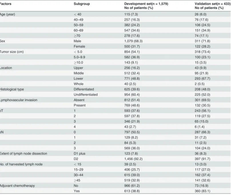
Table 1andS1 Dataset. A majority of the patients underwent D2 lymph node dissection (92.2% and 91.7% in the development and validation sets, respectively) and more than 15 lymph nodes were dissected in most cases (97.5% and 97% in the development and validation sets, respectively). The proportion of patients receiving adjuvant chemotherapy was consider-ably different between the two sets (38.8% vs. 83.1% in the development and validation sets, re-spectively). It was revealed that even early gastric cancer patients received oral chemotherapy agents after the operation in one hospital belonging to the validation set.
Risk factors for overall survival and development of the nomogram
The mean follow-up period for the development set was 51.7 ± 23.5 months (median, 52.0 months), and 351 (22.2%) patients died during the follow-up period.
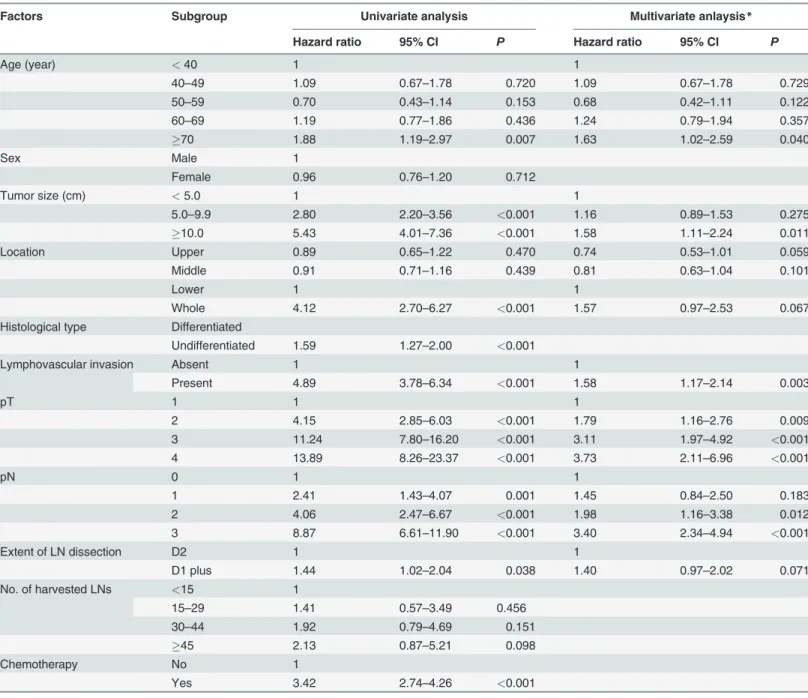
In the univariate analysis, age, tumor size, location, histological type, lymphovascular inva-sion, depth of invainva-sion, lymph node metastasis, extent of lymph node dissection, and adjuvant
chemotherapy were significantly associated with overall survival (Table 2). In contrast, sex and
many metastatic lymph nodes were revealed as significant independent factors for overall survival.
Based on these results, we developed a prediction model and a nomogram predicting 5-year
overall survival was generated (Fig. 1). Location and extent of lymph node dissection had no
significance in the multivariate analysis, however, they were included in the nomogram accord-ing to the exclusion criteria of 0.1. Each clinicopathological factors corresponds to a specific point by drawing a line straight upward to the Points axis. After sum of the points is located on the Total Points axis, the sum represents the probability of 5-year survival by drawing straight down to the 5-year survival axis. For example, a 45-year-old male (35 points) underwent D2 gastrectomy (0 point) for 7cm sized (12 points) gastric cancer located in the lower one-third of stomach (23 points). In the pathological report, the tumor invaded into subserosa (84 points) Table 1. Demographic and Clinicopathological Characteristics of the Development and Validation set.
Factors Subgroup Development set(n = 1,579) Validation set(n = 433)
No of patients (%) No of patients (%)
Age (year) <40 115 (7.3) 26 (6.0)
40–49 257 (16.3) 76 (17.6)
50–59 382 (24.2) 106 (24.5)
60–69 547 (34.6) 151 (34.9)
70 278 (17.6) 74 (17.1)
Sex Male 1,079 (68.3) 311 (71.8)
Female 500 (31.7) 122 (28.2)
Tumor size (cm) <5.0 854 (54.1) 318 (73.4)
5.0–9.9 582 (36.9) 100 (23.1)
10.0 143 (9.1) 15 (3.5)
Location Upper 256 (16.2) 43 (9.9)
Middle 512 (32.4) 95 (21.9)
Lower 771 (48.8) 293 (67.7)
Whole 40 (2.5) 2 (0.5)
Histological type Differentiated 625 (39.6) 208 (48.0)
Undifferentiated 954 (60.4) 225 (52.0)
Lymphovascular invasion Absent 812 (51.4) 301 (69.5)
Present 769 (48.6) 132 (30.5)
pT 1 593 (37.6) 243 (56.1)
2 597 (37.8) 119 (27.5)
3 346 (21.9) 65 (15.0)
4 43 (2.7) 6 (1.4)
pN 0 797 (50.5) 287 (66.3)
1 129 (8.2) 31 (7.2)
2 84 (5.3) 11 (2.5)
3 569 (36.0) 104 (24.0)
Extent of lymph node dissection D1 plus 123 (7.8) 36 (8.3)
D2 1,456 (92.2) 397 (91.7)
No. of harvested lymph node <15 39 (2.5) 13 (3.0)
15–29 406 (25.7) 117 (27.0)
30–44 615 (39.0) 162 (37.4)
45 519 (32.9) 141 (32.6)
Adjuvant chemotherapy No 966 (61.2) 73 (16.9)
Yes 613 (38.8) 360 (83.1)
with lymphovascular invasion (34 points), and there were five metastatic lymph nodes (50 points). For this example, the total point equals 238, and the suspected 5-year survival is ap-proximately 60%. This calculated value could be used in decision making for treatment plans and patient counseling.
Table 2. Risk factors for overall survival according to Cox proportional hazards regression model.
Factors Subgroup Univariate analysis Multivariate anlaysis*
Hazard ratio 95% CI P Hazard ratio 95% CI P
Age (year) <40 1 1
40–49 1.09 0.67–1.78 0.720 1.09 0.67–1.78 0.729
50–59 0.70 0.43–1.14 0.153 0.68 0.42–1.11 0.122
60–69 1.19 0.77–1.86 0.436 1.24 0.79–1.94 0.357
70 1.88 1.19–2.97 0.007 1.63 1.02–2.59 0.040
Sex Male 1
Female 0.96 0.76–1.20 0.712
Tumor size (cm) <5.0 1 1
5.0–9.9 2.80 2.20–3.56 <0.001 1.16 0.89–1.53 0.275
10.0 5.43 4.01–7.36 <0.001 1.58 1.11–2.24 0.011
Location Upper 0.89 0.65–1.22 0.470 0.74 0.53–1.01 0.059
Middle 0.91 0.71–1.16 0.439 0.81 0.63–1.04 0.101
Lower 1 1
Whole 4.12 2.70–6.27 <0.001 1.57 0.97–2.53 0.067
Histological type Differentiated
Undifferentiated 1.59 1.27–2.00 <0.001
Lymphovascular invasion Absent 1 1
Present 4.89 3.78–6.34 <0.001 1.58 1.17–2.14 0.003
pT 1 1 1
2 4.15 2.85–6.03 <0.001 1.79 1.16–2.76 0.009
3 11.24 7.80–16.20 <0.001 3.11 1.97–4.92 <0.001
4 13.89 8.26–23.37 <0.001 3.73 2.11–6.96 <0.001
pN 0 1 1
1 2.41 1.43–4.07 0.001 1.45 0.84–2.50 0.183
2 4.06 2.47–6.67 <0.001 1.98 1.16–3.38 0.012
3 8.87 6.61–11.90 <0.001 3.40 2.34–4.94 <0.001
Extent of LN dissection D2 1 1
D1 plus 1.44 1.02–2.04 0.038 1.40 0.97–2.02 0.071
No. of harvested LNs <15 1
15–29 1.41 0.57–3.49 0.456
30–44 1.92 0.79–4.69 0.151
45 2.13 0.87–5.21 0.098
Chemotherapy No 1
Yes 3.42 2.74–4.26 <0.001
*Backward variable selections methods was conducted with selection criteria of 0.2. Sex, extent of lymph node dissection, No. of harvested lymph nodes were excluded in the multivariate analysis because of no significant effect in the univariate analysis.
CI, confidence interval; LN, lymph node.
External validation set and performance
In the validation set, the mean follow-up period was 47.0 ± 16.3 months (median, 49 months), and 55 (12.7%) patients died during the follow-up period.
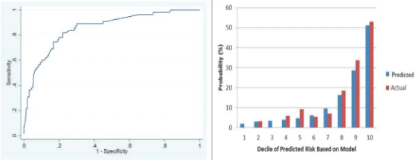
The external validation was performed by evaluating the performance of the model with re-spect to its discrimination and calibration abilities. The C-index, which indicated
discrimina-tion ability, was 0.831 (95% CI, 0.784–0.878), and the receiver operating characteristic curves
are shown inFig. 2a. The H-L chi-square statistic, which revealed calibration ability was 3.92,
and the calibration plot is presented inFig. 2b(P= 0.917).
Discussion
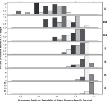
The TNM staging system is the most common method for predicting patient prognosis. How-ever, considerable survival variation has been observed even in patients at the same stage of gastric cancer. In the current study, we developed a nomogram predicting overall survival after gastric cancer surgery based on a multicenter database, and good performance was shown in the external validation. The advantage of this nomogram over AJCC stage grouping is shown inFig. 3, the heterogeneity of overall survival within each stage was seen, particularly in the stage IIIA, IIIB, and IV.
Fig 1. Nomogram to predict 5-year overall survival after curative surgery for gastric cancer.Each clinicopathological factor corresponds to a specific point by drawing a line straight upward to the Points axis. After sum of the points is located on the Total Points axis, the sum represents the probability of 5-year survival by drawing straight down to the 5-year survival axis.
doi:10.1371/journal.pone.0119671.g001
Fig 2. The ROC curves(a) represents the discrimination ability of the model measured by the C-index was 0.831 (95% CI, 0.784–0.878). Calibration plots (b) show the relationship between the predicted probabilities base on the nomogram and actual values of the validation set. The x-axis represents deciles of predicted risk, and the y-axis reveals predicted and actual probability of 5-year survival. The H-L chi-square which measure the calibration was 3.92 (P= 0.917).
As mentioned above, there have been three nomograms predicting survival after
gastrecto-my for gastric cancer. One is initial US nomogram reported by Kattan et al.[5] in 2003, others
were developed by Han et al.[9] and Song et al.[10] based on the Korean database. The main
difference between the previous nomograms and the current nomogram is study cohort. Previ-ous nomograms derived from a single high-volume center data. Memorial Sloan-Kettering
Cancer Center, Seoul National University Hospital, and the Seoul St. Mary’s Hospital had big
database consisted of more than 1000 patients in each hospital. External validation was also performed using other large-volume center database. According to many previous studies, sur-gical outcomes and survival after gastrectomies were significantly associated with hospital
vol-ume [14–17]. Therefore, it is possible that the previous nomograms are limited to tertiary
high-volume hospital.
Another difference is that previous Korean nomograms included only D2 gastrectomies. It is well known that D2 lymphadenectomy has been accepted as a standard procedure for
pa-tients with resectable gastric cancer [18,19]. However, limited lymphadenectomy is also
per-formed in high risk patient or in complicated situation. Moreover, the recent Japanese treatment guideline recommended D1 plus for early gastric cancer, and in Korea, most early
gastric cancer patients undergo D1 plus gastrectomy [11,20–22]. Previous Korean nomograms
could not be adapted to patients who underwent limited lymphadenectomy.
On the other hand, the current nomogram derived from various size of hospital database,
and the nine involved surgeons collected all their patients’data from the initial case. Therefore,
the current nomogram could be broadly adopted by both tertiary hospitals and general hospi-tals, and both experienced surgeons and inexperienced surgeons. Moreover, we included both D2 gastrectomy and limited lymphadenectomy, and the proportion of limited
lymphadenect-omy amounted to 7–8%. Therefore, even patient who underwent limited lymphadenectomy
could use the current nomogram for predicting survival.
Some differences are also observed in prognostic factors between previous nomograms and
the current one. Kattan et al.[5] revealed that the Lauren classification and the number of
nega-tive nodes were significant prognostic factors. Han et al.[9] demonstrated that sex and the
Fig 3. Distribution of nomogram predictions within each AJCC stage grouping.
number of examined lymph nodes had significant prognostic effects on overall survival. Simi-larly, sex, gross type, and Lauren classification were independent factors for overall survival in
a study by Song et al.[10] However, in the current study, and the number of harvested lymph
nodes had no statistical significance even in the univariate analysis. Histological type had a sig-nificant effect in the univariate analysis; however, it was eliminated in the multivariate analysis with the selection method. Instead, tumor size and lymphovascular invasion were independent prognostic factors for overall survival in the current study. Considering that the current study was based on multicenter data, we assumed that the number of harvested lymph nodes would vary depending on the surgeons who classified lymph node stations, pathologists, and institu-tional systems; therefore, the prognostic effect might be decreased.
Tumor location is also one of the different prognostic factors. Kattan et al.[5] and Han et al.[9]
demonstrated that upper-third tumors had poor prognosis; however, the current study showed less risk with upper- and middle-third tumors than with lower-third tumors. We reviewed the pathologi-cal stage and tumor size according to tumor location, however, could not find any biased relation-ships causing better prognosis of upper-third tumors. Further study seems to be required.
Recently, large-scale randomized controlled studies demonstrated the survival benefit of
ad-juvant chemotherapy after curative resection for gastric cancer [23–25]. However, in the current
study, adjuvant chemotherapy failed to show statistical significance in multivariate analysis. Various regimens and indications for adjuvant chemotherapy of each institution could influ-ence this negative result. Moreover, interrupted chemotherapy due to adverse effects or
pa-tients’low compliance may reduce the effect of adjuvant chemotherapy on overall survival.
Actually, we compared our nomogram and a nomogrm derived from Seoul National
univer-sity hospital (SNU nomogram)[9]. The validation set of our data was applied to our prediction
model and SNU prediction model, respectively. As a result, C-index value of SNU prediction model was 0.831 (95% CI, 0.783~0.879), which was almost similar to our C-index. However, calibration abilities of SNU prediction model showed that there were significant differences be-tween the predictive and actual survivals (H-L chi statistic = 27.339, p = 0.001). This result sup-ports that our nomogram predicts survival more accurately than SNU nomogram.
Although we have produced good results, the current study had several limitations. Each in-stitution has managed its own database in a different way, and some clinicopathological char-acteristics were not documented. As a result, 920 (28.0%) of the 3284 patients were excluded due to missing data, which can lead to selection bias. Moreover, the current study only included patients who underwent open gastrectomy. Considering that most patients undergo laparo-scopic surgery for early gastric cancer in Korea, we need further evaluation for the application of the current nomogram to laparoscopy-assisted gastrectomy cases.
In summary, we developed a nomogram that predicts individual 5-year survival in patients who underwent curative resection for gastric cancer. This nomogram improves accuracy of survival pre-diction and can be useful to counsel patients after gastrectomies. Further treatment such as adju-vant chemotherapy can also be decided based on the result of this nomogram. This nomogram was derived from a multicenter database and was validated by an independent external data set. There-fore, this nomogram can be adopted in both tertiary hospitals and local general hospitals.
Finally, validation using a Western cohort will also be needed prior to universal use of the
current nomogram. As noted by Strong et al.[8], significant differences in clinicopathological
and genetic characteristics between the Eastern and Western cohorts should be considered.
Supporting Information
Author Contributions
Conceived and designed the experiments: KWR BHN HJL MCK GSC CYK SWR DWS WJH JHL. Performed the experiments: KWR HJL MCK GSC CYK SWR DWS WJH JHL. Analyzed the data: BWE BHN YP. Contributed reagents/materials/analysis tools: BHN YP. Wrote the paper: BWE YP.
References
1. Jemal A, Bray F, Center MM, Ferlay J, Ward E, Forman D. Global cancer statistics. CA Cancer J Clin. 2011; 61: 69–90. doi:10.3322/caac.20107PMID:21296855
2. Bertuccio P, Chatenoud L, Levi F, Praud D, Ferlay J, Negri E, et al. Recent Patterns in gastric cancer: a global overview. Int J Cancer. 2009; 125: 666–673. doi:10.1002/ijc.24290PMID:19382179
3. Jung KW, Park S, Kong HJ, Won YJ, Lee JY, Seo HG, et al. Cancer statistics in Korea: incidence, mor-tality, survival, and prevalence in 2009. Cancer Res Treat. 2012; 44: 11–24. doi:10.4143/crt.2012.44. 1.11PMID:22500156
4. Edge SB, Byrd DR, Compton CC, Fritz AG, Greene FL, Trotti A, et al. AJCC Cancer Staging Manual. 7th ed. New York: Springer; 2010.
5. Kattan MW, Karpeh MS, Mazumdar M, Brennan MF. Postoperative nomogram for disease-specific sur-vival after an R0 resection for gastric carcinoma. J Clin Oncol. 2003; 21: 3647–3650. PMID:14512396 6. Peeters KC, Kattan MW, Hartgrink HH, Kranenbarg EK, Karpeh MS, Brennan MF, et al. Validation of a
nomogram for predicting diseasespecific survival after an R0 resection for gastric carcinoma. Cancer. 2005; 103: 702–707. PMID:15641033
7. Novotny AR, Schuhmacher C, Busch R, Kattan MW, Brennan MF, Siewert JR. Predicting individual sur-vival after gastric cancer resection: Validation of a U.S.-derived nomogram at a single high-volume cen-ter in Europe. Ann Surg. 2006; 243: 74–81. PMID:16371739
8. Strong VE, Song KY, Park CH, Jacks LM, Gonen M, Shah M, et al. Comparison of gastric cancer sur-vival following R0 resection in the United States and Korea using an internationally validated nomogram Ann Surg. 2010; 251: 640–646. doi:10.1097/SLA.0b013e3181d3d29bPMID:20224369
9. Han DS, Suh YS, Kong SH, Lee HJ, Choi Y, Aikou S, et al. Nomogram predicting long-term survival after d2 gastrectomy for gastric cancer. J Clin Oncol. 2012; 30: 3834–3840. doi:10.1200/JCO.2012. 41.8343PMID:23008291
10. Song KY, Park YG, Jeon HM, Park CH. A nomogram for predicting individual survival of patients with gastric cancer who underwent radical surgery with extended lymph node dissection. Gastric Cancer. 2014; 17: 287–293. doi:10.1007/s10120-013-0270-xPMID:23712439
11. Japanese Gastric Cancer Association. Japanese gastric cancer treatment guidelines 2010 (ver. 3). Gastric Cancer. 2011; 14: 113–123. doi:10.1007/s10120-011-0042-4PMID:21573742
12. Hur H, Song KY, Park CH, Jeon HM. Follow-up strategy after curative resection of gastric cancer: a na-tionwide survey in Korea. Ann Surg Oncol. 2010; 17: 54–64. doi:10.1245/s10434-009-0676-1PMID: 19777193
13. D’Agostino RB, Nam BH. Evaluation of the performance of survival analysis models. In: Balakrishnan N, Rao CR, editors. Discrimination and calibration measures, in. Handbook of Statistics, Survival Meth-ods. Volume 23. Amsterdam, The Netherlands: Elsevier B.V.; 2004. pp 1–25.
14. Coupland VH, Lagergren J, Lüchtenborg M, Jack RH, Allum W, Holmberg L, et al. Hospital volume, pro-portion resected and mortality from oesophageal and gastric cancer: a population-based study in En-gland, 2004–2008. Gut. 2013; 62:961–966. doi:10.1136/gutjnl-2012-303008PMID:23086798 15. Dikken JL, Stiekema J, van de Velde CJ, Verheij M, Cats A, Wouters MW, et al. Quality of care
indica-tors for the surgical treatment of gastric cancer: a systematic review. Ann Surg Oncol. 2013; 20:381–398. doi:10.1245/s10434-012-2574-1PMID:23054104
16. Dikken JL, Wouters MW, Lemmens VE, Putter H, van der Geest LG, Verheij M, et al. Influence of hospi-tal type on outcomes after oesophageal and gastric cancer surgery. Br J Surg. 2012; 99:954–963. doi: 10.1002/bjs.8787PMID:22569956
17. Anderson O, Ni Z, Møller H, Coupland VH, Davies EA, Allum WH, et al. Hospital volume and survival in
oesophagectomy and gastrectomy for cancer. Eur J Cancer. 2011; 47:2408–2414. doi:10.1016/j.ejca. 2011.07.001PMID:21835609
19. Okines A, Verheij M, Allum W, Cunningham D, Cervantes A, ESMO Guidelines Working Group. Gastric cancer: ESMO Clinical Practice Guidelines for diagnosis, treatment and follow-up. Ann Oncol. 2010; 21: S50–S54.
20. Kim YW, Yoon HM, Eom BW, Park JY. History of Minimally Invasive Surgery for Gastric Cancer in Korea. J Gastric Cancer. 2012; 12: 13–17. doi:10.5230/jgc.2012.12.1.13PMID:22500259
21. Ahn HS, Lee HJ, Yoo MW, Jeong SH, Park DJ, Kim HH, et al. Changes in clinicopathological features and survival after gastrectomy for gastric cancer over a 20-year period. Br J Surg. 2011; 98: 255–260. doi:10.1002/bjs.7310PMID:21082693
22. Jeong O, Park YK. Clinicopathological features and surgical treatment of gastric cancer in South Korea: the results of 2009 nationwide survey on surgically treated gastric cancer patients. J Gastric Cancer. 2011; 11: 69–77. doi:10.5230/jgc.2011.11.2.69PMID:22076206
23. Sakuramoto S, Sasako M, Yamaguchi T, Kinoshita T, Fujii M, Nashimoto A, et al. Adjuvant chemother-apy for gastric cancer with S-1, an oral fluoropyrimidine. N Engl J Med. 2007; 357: 1810–1820. PMID: 17978289
24. Sasako M, Sakuramoto S, Katai H, Kinoshita T, Furukawa H, Yamaguchi T, et al. Five-year outcomes of a randomized phase III trial comparing adjuvant chemotherapy with S-1 versus surgery alone in stage II or III gastric cancer. J Clin Oncol. 2011; 29: 4387–4393. doi:10.1200/JCO.2011.36.5908 PMID:22010012