Dengue Virus Infection of
Aedes aegypti
Requires a Putative Cysteine Rich Venom
Protein
Berlin Londono-Renteria1, Andrea Troupin1, Michael J Conway2, Diana Vesely3, Michael Ledizet3, Christopher M. Roundy4, Erin Cloherty4, Samuel Jameson4, Dana Vanlandingham5,6, Stephen Higgs5,6, Erol Fikrig7,8, Tonya M. Colpitts1*
1Department of Pathology, Microbiology & Immunology, University of South Carolina School of Medicine, Columbia, South Carolina, United States of America,2Foundational Sciences, Central Michigan University College of Medicine, Mount Pleasant, Michigan, United States of America,3L2 Diagnostics, New Haven, Connecticut, United States of America,4Department of Tropical Medicine, Tulane University School of Public Health and Tropical Medicine, New Orleans, Louisiana, United States of America,5Biosecurity Research Institute, Kansas State University, Manhattan, Kansas, United States of America,6Diagnostic Medicine and Pathobiology, Kansas State University, Manhattan, Kansas, United States of America, 7Section of Infectious Diseases, Department of Internal Medicine, Yale University School of Medicine, New Haven, Connecticut, United States of America,8Howard Hughes Medical Institute, Chevy Chase, Maryland, United States of America
*tonya.colpitts@uscmed.sc.edu
Abstract
Dengue virus (DENV) is a mosquito-borne flavivirus that causes serious human disease and mortality worldwide. There is no specific antiviral therapy or vaccine for DENV infection. Alterations in gene expression during DENV infection of the mosquito and the impact of these changes on virus infection are important events to investigate in hopes of creating new treatments and vaccines. We previously identified 203 genes that were5-fold differ-entially upregulated during flavivirus infection of the mosquito. Here, we examined the impact of silencing 100 of the most highly upregulated gene targets on DENV infection in its mosquito vector. We identified 20 genes that reduced DENV infection by at least 60% when silenced. We focused on one gene, a putative cysteine rich venom protein (SeqID AAEL000379; CRVP379), whose silencing significantly reduced DENV infection inAedes aegypticells. Here, we examine the requirement for CRVP379 during DENV infection of the mosquito and investigate the mechanisms surrounding this phenomenon. We also show that blocking CRVP379 protein with either RNAi or specific antisera inhibits DENV infection inAedes aegypti. This work identifies a novel mosquito gene target for controlling DENV infection in mosquitoes that may also be used to develop broad preventative and therapeu-tic measures for multiple flaviviruses.
OPEN ACCESS
Citation:Londono-Renteria B, Troupin A, Conway MJ, Vesely D, Ledizet M, Roundy CM, et al. (2015) Dengue Virus Infection ofAedes aegyptiRequires a Putative Cysteine Rich Venom Protein. PLoS Pathog 11(10): e1005202. doi:10.1371/journal.ppat.1005202
Editor:Abraham L. Brass, University of Massachusetts Medical School, UNITED STATES
Received:June 22, 2015
Accepted:September 11, 2015
Published:October 22, 2015
Copyright:© 2015 Londono-Renteria et al. This is an open access article distributed under the terms of theCreative Commons Attribution License, which permits unrestricted use, distribution, and reproduction in any medium, provided the original author and source are credited.
Data Availability Statement:All relevant data are within the paper and its Supporting Information files.
Funding:This work was supported by funding from National Institutes of Health (NIH) awards K22AI103067 (TMC) and UO1AI070343 (EF). EF is an investigator of the Howard Hughes Medical Institute. The funders had no role in study design, data collection and analysis, decision to publish, or preparation of the manuscript.
Author Summary
Dengue virus (DENV) is responsible for serious human disease worldwide and the World Health Organization estimates that over 2 billion people are at risk for disease. There are no vaccines or specific antiviral medications currently available for DENV infection. DENV is transmitted to humans by infected mosquitoes during feeding and probing. By examining the effects of virus infection on gene expression, and interactions between virus and vector, we may be able to find new targets for prevention and treatment. Here we look at a mosquito protein, CRVP379, whose gene expression was highly increased during DENV infection in mosquitoes. We show a requirement for CRVP379 during DENV infection in the mosquito and a correlation between the levels of CRVP379 and levels of infection. Our results indicate that the protein may be acting with a putative DENV recep-tor in the mosquito, prohibitin protein. These data also suggest that blocking CRVP379 function may be used to block DENV infection in the mosquito.
Introduction
Dengue virus (DENV) is the most important arbovirus in tropical areas leading to substantial pediatric morbidity and mortality worldwide [1–6]. DENV is transmitted to humans via the bite of an infected mosquito of theAedes spp. Infection with DENV in humans can result in dengue fever (DF), dengue shock symptom (DSS) and dengue hemorrhagic fever (DHF), the latter two that can lead to severe disease and death. There are no specific antivirals or approved vaccines for use in DENV treatment or prevention [4,7–9]. Current dengue control methods rely mostly on activities to reduce vector population [10,11]. The increase in number of cases despite vector control indicates that these strategies are not as effective as expected, and that new tools need be developed to alleviate disease burden in endemic areas [12–14].
During the last five decades, much effort has been invested in the development of vaccines against DENV [15–23]. One of the obstacles in dengue vaccine development is the potential risk of severe disease mediated by the presence of sub-neutralizing antibodies against virus par-ticles. These antibodies can predispose an individual to severe disease through a phenomenon called antibody-dependent enhancement (ADE), where the virus can infect cells via FcR in mononuclear cells [8,9,24–28]. Traditional vaccine approaches have included live attenuated viruses, recombinant subunits, virus-like particles and plasmid or viral vectors. There are live attenuated and chimeric DENV vaccines that have gone into clinical trials but none have proven to provide complete and lasting protection against all four DENV serotypes [21,29].
An attractive complement to traditional vaccines is to induce an immune response in the vertebrate host (infected or non-infected) that will block virus infection of the mosquito vec-tors. This would successfully interrupt transmission by inducing antibody responses against non-viral antigens [30]. These type of vaccines are called transmission-blocking vaccines (TBV), since they aim to interfere with pathogen development within the vector, thereby block-ing transmission to human hosts [31]. The majority of TBVs designed to inhibit malaria infec-tion are based on the mammalian immune response to pathogen proteins [31]. Another category of TBVs in development are based on arthropod molecules able to reduce pathogen infection in vector tissues [32]. For arboviruses, vector molecules able to interact directly with the pathogen (i.e. ligands/receptors) are highly suitable candidates for blocking transmission [33,34].
The main global transmission vector for DENV isAe.aegypti. Extensive research has shown that DENV infection ofAe.aegyptiinduces many varied changes in gene expression [35–44].
Our hypothesis is that genes upregulated during DENV infection are required for virus survival or are related with defense against infection [37]. Consequently, a better understanding of the role of mosquito proteins regulated by DENV infection will reveal important insights into DENV biology and transmission as well as be helpful to the design of an effective TBV against DENV. For example, antibodies directed against mosquito molecules involved in steps of the pathogen life cycle are promising candidates for TBV. In addition, a recent study demonstrated that antibodies against a mosquito C-type lectin, mosGCTL1, effectively interrupts the infec-tion ofAe.aegyptimosquitoes with DENV [34]. Other proteins which genes are unregulated upon infection also show promising capacity of interrupting infection since they are considered important for the microorganism survival. One of these proteins is the tick histamine release factor (tHRF) fromIxodes scapularisupregulated duringBorrelia burgdorferiinfection. Previ-ous work showed that expression of tHRF is associated with the tick blood feeding and that the silencing its gene by RNA interference or antibodies not only effectively impairs tick feeding but subsecuently decreasesB.burgdorferiburden [45].
Using comprehensive microarray analysis to identify key alterations in theAe.aegypti tran-scriptome during flavivirus infection, we previously identified 203 mosquito genes that were up- and 202 genes that were down-regulated during infection [35]. Comparative analysis revealed that at least 15 of these genes had differential expression during infection with DENV, Yellow fever (YFV) and West Nile virus (WNV) [35]. One of these conserved, up-regulated genes was a putative cysteine-rich venom protein (AAEL000379), which we named CRVP379. Cysteine-rich venom proteins (CRVPs) are expressed in multiple mosquito tissues including the salivary glands [37,46,47]. Examples of mosquito CRVPs include anAn.stephensipeptide annotated as salivary-secreted serine protease inhibitor [48] and a putative cysteine-rich prote-ase inhibitor found in the sialotranscriptome of adult femaleCulex quinquefasciatus[49]. The specific role of these proteins in mosquitoes remains unknown [46,47].
Here, we describe a requirement for CRVP379 during DENV infection in mosquito cells and live mosquitoes, including a direct correlation between the amount of CRVP379 expressed and the level of DENV infection. We demonstrate the importance of an interaction between CRVP379 and prohibitin, a putative DENV receptor protein in mosquitoes. We also assess the tissue-specific expression of CRVP379 during DENV infection. Finally, we use both RNAi and specific antibody to demonstrate that blocking CRVP379 results in inhibition of DENV infec-tion inAe.aegypti. These results further our understanding of DENV pathogenesis in the mos-quito vector and highlight a potential target protein for the creation of a DENV TBV to break the host-vector transmission cycle.
Results
Silencing virally up-regulated genes alters DENV infection in mosquito
cells
We previously used microarray analysis to identify a number ofAe.aegyptigenes that were sig-nificantly up-regulated during infection with DENV and other selected flaviviruses [35]. These genes are likely required for flaviviral infection ofAe.aegyptior are part of the mosquito immune response to viral infection. To elucidate the role of these genes and their correspond-ing proteins, we reduced gene expression through RNAi knockdown and analyzed the effect on viral infection. We designed siRNA against 100 genes that were significantly up-regulated dur-ing DENV infection ofAe.aegypti(S1 Fig). The siRNA was used to silence these genes in an
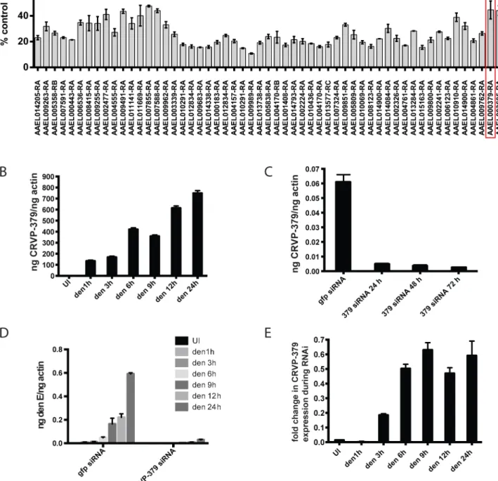
DENV infection, as expected (S2 Fig). The silencing of 9 genes caused cytotoxicity beyond our ability to accurately measure infection levels. Silencing approximately 55 individual genes decreased DENV infection of the cells to below 60% of control infection (Fig 1A), which is greater than 40% inhibition of infection. A number of these genes encode hypothetical proteins for which the function is not known. Several of our target genes do have putative known func-tions, including a cytochrome P450 (AAEL009762), histone H3 (AAEL003685) and a cysteine-rich venom protein (AAEL000379).
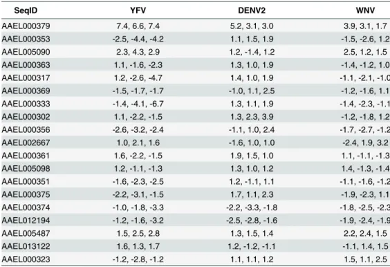
The cysteine-rich venom protein (CRVP), which we will call CRVP379, was a particularly interesting target. CRVP proteins are known to contain a trypsin inhibitor-like (TIL) domain, which indicates that the CRVP379 protein could be a serine protease inhibitor. Both serine proteases and their inhibitors are known to be involved in DENV infection and pathogenesis in the mosquito vector as well as in mammals [50–55]. The expression levels of 19 mosquito CRVP genes were examined during flavivirus infection with the previous microarray analysis. CRVP379 was the only CRVP significantly up-regulated inAe.aegyptiduring infection with any of the 3 prototypic flavivirus infections, including DENV, West Nile virus (WNV) and Yel-low Fever virus (YFV), at all timepoints tested (Table 1) [35]. In addition, the 19 CRVP genes that we looked at in our microarray actually have very low sequence identify at the amino acid level (S3 Fig). This indicates that they may not have identical or even similar functions, though they are grouped in a protein family due to the presence of multiple cysteines and a TIL domain. Therefore, we decided to assess the role of CRVP379 in DENV infection of the mos-quito vector in greater detail.
DENV infection requires CRVP379 in mosquito cells
Looking at gene expression over time in DENV-infected Aag2 cells, we found that CRVP379 expression increased more than 800-fold when compared to mock-infected cells (Fig 1B, P<0.01). Since the gene was upregulated in mosquito cells during DENV infection, we wanted to examine the phenotype during DENV infection with loss of function experiments. We used RNA interference (RNAi) with siRNA to reduce CRVP379 gene expression. To confirm gene knockdown, levels of CRVP379 were measured after siRNA transfection over time (Fig 1C), and the expression levels remained below 10% at 72h post-transfection. To determine how the reduction of CRVP379 altered DENV infection, we examined infection levels in Aag2 cells at various timepoints from 1 to 24h post-infection during siRNA knockdown. Silencing CRVP379 reduced DENV infection at all timepoints measured, as compared to infection in control cells transfected with siRNA against GFP (Fig 1D). Interestingly, we noticed that levels of CRVP379 were slightly elevated during DENV infection even during siRNA knockdown, when compared to uninfected cells. This can be seen by looking at the fold change in CRVP379 expression in GFP siRNA-transfected cells as compared to CRVP379 siRNA-transfected cells during DENV infection over time (Fig 1E). Together, these results indicated that the silencing effects of RNAi on CRVP379 are slightly overcome by the gene upregulation during DENV infection, but that infection levels still remained quite low when compared to cells with no CRVP379 silencing.
Endogenous CRVP379 is sufficient for optimal DENV infection
Since we found that the presence of CRVP379 is required for DENV infection, we wanted to test whether increasing CRVP379 levels would enhance infection levels. To do this, we cloned the CRVP379 coding region into the insect expression vector pAc5.1/V5-His (Life Technolo-gies, CA), resulting in pAcCRVP379 vector. This expression vector was transfected into anAe.
Fig 1. Silencing select virally-up-regulated genes reduces DENV infection in mosquito cells.A. The mosquito genes listed inS1 Figwere knocked down in Aag2 cells using RNAi and the effects on DENV infection were analyzed. The genes that reduced infection below 60% of control are shown. Aag2 cells were infected with DENV (MOI of 1.0) 72 h post-knockdown and analyzed for infection by qRT-PCR 24h post-infection. Data is displayed as percent control infection (using scrambled siRNA). Both DENV infection and qRT-PCR analysis were done in triplicate, data is pooled and error bars indicate standard deviation. B. DENV infection increases CRVP379 in Aag2 cells over time. Aag2 cells were infected with DENV (MOI of 1.0) and infection was measured using qRT-PCR analysis at the timepoints indicated. P<0.01. C. Expression of CRVP379 during RNAi knockdown. CRVP379 siRNA was
expressing GFP was transfected as a control into a separate group of DENV-infected cells. Transfection levels as measured by GFP transfection are over 50%, which will give meaningful results when looking at gene expression and effects on DENV infection (S4 Fig). At 48h post-infection, levels of CRVP379 expression were measured by qRT-PCR. The expression levels of CRVP379 were over 1000 times higher in the cells that were transfected with the CRVP379 plasmid (Fig 2A). We next infected the transfected cells with DENV at 48h post-transfection. At 24 hpi, RNA was isolated and qRT-PCR done to measure DENV infection in the cells. We found that the overexpression of CRVP379 did not increase DENV infection levels in the cells (Fig 2B). This indicated that the endogenous levels of CRVP379 protein are sufficient and that the virus has likely evolved to require only those amounts for optimum infection.
Acquisition of DENV requires CRVP379 in live mosquitoes
Since we found that CRVP379 was required for DENV infection of mosquito cells, we decided to next look at the requirements for infection in liveAe.aegypti. To do this, we designed dsRNA against the CRVP379 coding region and inoculated mosquitoes via intra-thoracic injection. At days 2, 4 and 8 post-injection, we dissected out midgut tissues and measured levels of CRVP379 expression by qRT-PCR analysis. Although knockdown was not achieved in all transfection of GFP control siRNA is also indicated. D. Reduction of CRVP379 reduces DENV infection over time. Either siRNA against CRVP379 or GFP was transfected into Aag2 cells and cells were infected with DENV (MOI of 1.0) at 72 h post-knockdown. Cells were analyzed for infection by qRT-PCR at the timepoints indicated. E. DENV infection increases CRVP379 expression during siRNA knockdown. Either siRNA against CRVP379 or GFP was transfected into Aag2 cells and cells were infected with DENV at 72 h post-knockdown. Gene expression was analyzed by qRT-PCR at the timepoints indicated. Data is expressed as the fold change in CRVP379 expression in cells with GFP siRNA versus cells with CRVP379 siRNA during DENV infection. B-E. Data is pooled from 6 separate experiments, error bars indicate standard deviation.
doi:10.1371/journal.ppat.1005202.g001
Table 1. Expression of CRVPs inAe.aegyptiduring flavivirus infection.
SeqID YFV DENV2 WNV
AAEL000379 7.4, 6.6, 7.4 5.2, 3.1, 3.0 3.9, 3.1, 1.7
AAEL000353 -2.5, -4.4, -4.2 1.1, 1.5, 1.9 -1.5, -2.6, 1.2
AAEL005090 2.3, 4.3, 2.9 1.2, -1.4, 1.2 2.5, 1.2, 1.5
AAEL000363 1.1, -1.6, -2.3 1.3, 1.0, 1.9 -1.4, -1.2, 1.0
AAEL000317 1.2, -2.6, -4.7 1.4, 1.0, 1.9 -1.1, -2.1, -1.0
AAEL000369 -1.5, -1.7, -1.7 -1.0, 1.1, 2.5 -1.2, -1.6, 1.1
AAEL000333 -1.4, -4.1, -6.7 1.3, 1.1, 1.9 -1.4, -2.3, -1.1
AAEL000302 1.1, -2.2, -1.5 1.3, 2.3, 3.9 -1.2, -1.8, 1.2
AAEL000356 -2.6, -3.2, -2.4 -1.1, 1.0, 2.4 -1.7, -2.7, -1.2
AAEL002667 1.0, 2.1, 1.6 -1.6, 1.0, 1.0 -2.4, 1.9, 3.2
AAEL000361 1.6, -2.2, -1.5 1.9, 1.5, 1.0 1.1, -1.1, -1.3
AAEL005098 1.2, -1.1, -1.3 1.3, 1.0, 1.2 1.4, -1.3, -1.4
AAEL000351 -1.6, -2.3, -2.5 1.2, -1.1, 1.1 -1.1, -1.6, -1.2
AAEL000375 -2.2, -3.1, -1.5 1.7, 1.1, 2.3 -1.9, -2.3, 1.1
AAEL000374 -1.0, -1.8, -3.3 -2.2, -3.3, -1.8 -1.8, -2.5, -2.3 AAEL012194 -1.2, -1.6, -3.2 -2.5, -2.8, -1.6 -1.9, -2.4, -1.9
AAEL005487 1.5, 2.5, 2.8 1.3, 1.5, 1.4 2.2, 2.4, 1.5
AAEL013122 1.6, 1.3, 1.7 1.2, -1.2, -1.1 -1.1, 1.4, 1.5
AAEL000323 -1.2, -2.8, -1.2 1.1, 1.1, 1.2 1.5, 1.1, 2.5
Values are expressed as fold-change over mock infection at time points Day 1, 2, 7.
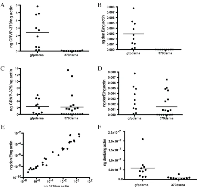
tissues tested, a near-complete reduction of CRVP379 expression (over 95%) in midguts was seen 70% of the time by day 8 (Fig 3-all panels). We next examined the effects of silencing CRVP379 on DENV acquisition in the mosquito midgut. Mosquitoes were again injected with dsRNA against CRVP379 or a control dsRNA against GFP protein. At day 4 post-injection, mosquitoes were infected with DENV by blood feeding using a hemotek apparatus. At day 4 post-infection, midgut tissues were dissected out and analyzed for both CRVP379 expression and DENV infection by qRT-PCR. Since not all midgut tissues had reduction in CRVP379 expression, we analyzed each midgut individually in order to examine the effects on DENV infection in midguts that did have reduced CRVP379. The levels of CRVP379 in the selected midguts are shown inFig 3A. In the midguts that had reduced CRVP379 expression, DENV infection was almost completely inhibited, as compared to infection in control mosquito mid-guts (Fig 3B, P<0.01). We also analyzed the data after adding back in the midguts that did not have sufficient gene knock down and looked at levels of DENV infection.Fig 3Cshows the lev-els of CRVP379 in these midguts. Interestingly, in midgut tissues where CRVP379 was not knocked down, DENV infection was comparable to levels in the GFP dsRNA-injected mosqui-toes (Fig 3D-squares). Plotting the data points as level of DENV versus level of CRVP379, there is a correlation between expression of CRVP379 in the mosquito midgut and level of DENV infection in that same midgut (Fig 3E, r = 0.6442, P<0.0001). This indicates that CRVP379 levels are directly related to levels of DENV infection in the mosquito midgut. Finally, we repeated the RNAi experiment and allowed the DENV infection to disseminate for 7 days. At day 7 post-infection, whole mosquitoes were homogenized and analyzed for both CRVP379 expression and DENV infection by qRT-PCR (Fig 3F). The mosquitoes that received the dsRNA against CRVP379 had a significant reduction in DENV infection levels as com-pared to the control mosquitoes. This indicates that the reduction of CRVP379 blocks DENV infection in the whole mosquito.
During DENV infection, CRVP379 interacts with mosquito prohibitin, a
putative DENV receptor
After establishing that DENV requires CRVP379 in both mosquito cells and liveAe.aegypti, we next wanted to investigate the mechanistic role that CRVP379 plays during infection. To do this, we used the tandem affinity purification (TAP) assay to identify putative mosquito pro-teins that bind CRVP379 during DENV infection. We cloned the coding region of CRVP379 into the NTAP vector (Stratagene, CA), which fuses the gene to purification tags, and
Fig 2. DENV infection optimally enhances CRVP379 expression.Aag2 cells were transfected with an insect expression vector encoding CRVP379 (AcCRVP379) or GFP (AcGFP) and A. CRVP379 expression was measured by qRT-PCR at 48 h post-transfection. B. Cells were infected with DENV (MOI of 1.0) at 48 h post-transfection and infection levels were measure by qRT-PCR at 24 hpi.
transfected this plasmid into Aag2 mosquito cells. Cells were infected with DENV 24h post-transfection and lysed 24h post-infection. The cell lysate was processed and CRVP379 was purified using the expressed tags, along with interacting mosquito proteins. The resulting solu-tion was sent for LC/MS-MS analysis to determine which proteins were pulled out of the
Fig 3. Silencing CRVP379 inhibits DENV acquisition in live mosquitoes.A-E. Mosquitoes were intra-thoracically injected with either dsRNA against the coding region of CRVP379 or dsRNA against GFP as control. At 4 dpmi, mosquitoes were infected with DENV through blood feeding. At 4 dpi, midgut tissues were dissected and individually analyzed for gene expression with qRT-PCR analysis. Each data point represents one mosquito midgut. A. Levels of CRVP379 in select midguts where RNAi was successful, as compared to levels in control mosquitoes. P<0.01. B. Levels of DENV infection in select midguts
where RNAi was successful, as compared to levels in control mosquitoes. P<0.01. Infection rates in midguts where CRVP379 RNAi was successful ranged from to .000000765–.0000315 ng DENV E/ng actin. C. Levels of CRVP379 in midguts where RNAi was both successful and unsuccessful, as compared to
levels in control mosquitoes. D. Levels of DENV infection in midguts where RNAi was both successful and unsuccessful, as compared to levels in control mosquitoes. C-D. Squares represent midguts where RNAi did not knock down CRVP379 successfully, cirlces represent midguts where RNAi did knock down CRVP379 successfully. E. Levels of DENV infection correspond to levels of CRVP379 expression. Both midguts where RNAi did and did not knock down CRVP379 were analyzed for both CRVP379 expression and DENV infection by qRT-PCR. Data is plotted as ngs of DENV E versus levels of CRVP379, normalized to mosquito actin. Data correlated with Pearson, r = 0.6442, P<0.0001 F. Silencing CRVP379 reduces DENV infection in whole mosquitoes.
Mosquitoes were intra-thoracically injected with either dsRNA against the coding region of CRVP379 or dsRNA against GFP as control. At 4 dpmi, mosquitoes were infected with DENV through blood feeding. At 7 dpi, homogenized whole mosquitoes were individually analyzed for gene expression with qRT-PCR analysis.
mosquito cell lysate by CRVP379 during DENV infection. A separate set of cells was trans-fected with the NTAP vector expressing GFP as control.Table 2lists the mosquito proteins that putatively bound CRVP379 and were not identified during control experiments. One of the proteins identified, prohibitin, was previously characterized as binding to DENV inAedes
A7 cells [56] and has also been suggested as a putative DENV receptor in mosquito cells [57]. Since we found that prohibitin binds CRVP379 during DENV infection using the TAP assay, this may indicate that the proteins act together to facilitate DENV entry into mosquito cells.
In establishing prohibitin as a putative receptor in mosquito cells, Kuadkitkan et al used siRNA against the mosquito prohibitin gene to inhibit protein production and saw a significant decrease in DENV infection in mosquito cells [57]. To confirm that prohibitin is required for DENV infection in mosquitoes, we designed dsRNA against the mosquito prohibitin gene and examined the impact of silencing prohibitin on DENV infection.Ae.aegyptiwere intra-thorac-ically inoculated with the dsRNA and at 4 days post micro-injection (dpmi), mosquitoes were infected with DENV via blood feeding. At 4 days post-infection (dpi), mosquito midguts (MG) were dissected and analyzed for infection by qRT-PCR analysis. Our results show that DENV infections levels were greatly inhibited by prohibitin silencing as compared to the control
Table 2. CRVP379 tandem affinity purification assay results.
SeqID Gene Score Expectation
AAEL012282 prohibitin 839 2.10E-80
AAEL003303 hypothetical 212 9.70E-18
AAEL008103 40S ribosomal 450 1.80E-41
AAEL007771 60S ribosomal 353 7.90E-32
AAEL011089 ribonucleoprotein 980 1.80E-94
AAEL001471 hypothetical 357 3.60E-32
AAEL006158 histone H3 152 9.80E-12
AAEL003415 lamin 611 1.30E-57
AAEL011197 actin 1458 0.00E+00
AAEL009883 26S proteasome subunit 276 4.00E-24
AAEL000482 histone H3 214 6.40E-18
AAEL006642 tubulin alpha chain 1350 0
AAEL007609 histone H2A 242 9.60E-21
AAEL008517 elongation factor tu 922 1.10E-88
AAEL015674 histone H2B 913 8.90E-88
AAEL010668 quinone oxioreductase 1276 4.10E-124
AAEL013739 electron transport oxioreductase 372 1.00E-33
AAEL010146 3_H-Coa dehydrogenase 608 2.50E-57
AAEL003530 ribosomal protein P1 288 2.80E-25
AAEL011180 hypothetical 250 1.90E-21
AAEL012736 ribosomal protein L15 344 6.70E-31
AAEL004317 hypothetical 212 1.10E-17
AAEL006511 ubiquitin 213 8.10E-18
AAEL008103 40S ribosomal 450 1.80E-41
AAEL015464 histone H1 339 2.10E-30
AAEL011584 chaperonin 60kD 743 8.60E-71
AAEL010608 succinate dehydrogenase 173 9.10E-14
AAEL004325 ribosomal L5 362 1.10E-32
AAEL008188 60S ribosomal 189 2.00E-15
mosquitoes (Fig 4A), confirming a requirement for prohibitin in DENV mosquito infection. We next performed co-immunoprecipitation to confirm the protein interaction between CRVP379 and prohibitin. Aag2 cells were transfected with an expression vector coding for His-tagged CRVP379 protein and/or infected with DENV. Cells were then lysed and antibody against the His tag was used to pull down His-CRVP379 from the cell lysate. Western blot anal-ysis identified prohibitin protein in the immunoprecipitate (Fig 4B), demonstrating that His-tagged CRVP379 bound prohibitin during DENV infection in the mosquito cells. We then used immunoflourescent imaging to visualize the putative prohibitin-CRVP379 protein inter-action. Aag2 cells were transfected with the His-tagged CRVP379 expression construct and infected with DENV 48 hours post-transfection. At 24 hours post-infection, cells were fixed and stained with antibodies against the His tag and prohibitin protein.Fig 4Cshows that the two proteins were highly colocalized during DENV infection. We next wondered whether pro-hibitin overexpression could rescue the mosquito cells that were resistant to DENV infection due to reduced CRVP379 expression. To investigate this, we transfected CRVP379 siRNA into
Fig 4. CRVP379 interacts with mosquito prohibitin during DENV infection.A. Prohibitin is required for DENV infection ofAedes aegypti. Mosquitoes were intra-thoracically injected with either dsRNA against the coding region of prohibitin (Prohdsrna) or dsRNA against GFP (gfpdsrna) as control. At 4 dpmi, mosquitoes were infected with DENV through blood feeding. At 4 dpi, midgut tissues were dissected and individually analyzed for gene expression with qRT-PCR analysis. Each data point represents one mosquito midgut. P<0.01. B. Aag2 cells were transfected with His-tagged CRVP379 and/or infected with
DENV. At 48h post-infection, antibodies against the His tag were used to pull the His-tagged CRVP379 protein out of cell lysates. The precipitated solution was run on SDS-PAGE gel and Western blot analysis was performed using antibody against prohibitin. C. CRVP379 and prohibitin co-localize in mosquito cells during DENV infection. Aag2 cells were transfected with His-tagged CRVP379 and infected with DENV 48h post-transfection. Cells were fixed in 4% paraformaldehyde 24h post-infection and stained with antibodies against the His-tag (green) and against prohibitin (red). DAPI was used to localize the nucleus. Representative images are shown.
Aag2 cells and then overexpressed mosquito prohitibin before infecting the cells with DENV. We found that the overexpression of prohibitin did not significantly increase DENV infection in cells with reduced CRVP379, though there was a slight enhancement (S5 Fig). This indicates that, though the proteins may act together to facilitate DENV infection in mosquito cells, pro-hibitin cannot replace the function of CRVP379 protein. Interestingly, the overexpression of prohibition in control Aag2 cells (with siRNA against GFP) did increase DENV infection (S5 Fig).
Antisera against CRVP379 inhibits DENV infection in mosquitoes
Since we found that inhibiting CRVP379 gene expression using RNA interference reduced DENV inAe.aegypti, we next wanted to try and inhibit protein function with antibody and examine the effects on DENV infection. To do this, recombinant protein consisting of residues 21–128 of CRVP379 was expressed inE.colialong with a GST tag for purification. To generate polyclonal antiserum, rabbits were immunized with the recombinant CRVP379 (rCRVP379). We used the antisera in Western blot analysis to confirm that antibodies would bind the recombinant protein (Fig 5A). We next ensured that the polyclonal antisera contained antibod-ies that recognized endogenous CRVP379 protein in the mosquito. To test this, we used the antisera to stain Aag2 cells and found that there was a strong reaction between the CRVP379 antisera and protein in the cells (Fig 5B). We then used the antisera to probe mosquito midgut tissue for endogenous protein.Fig 5Cdemonstrates that the CRVP379 antisera, but not the pre-immune control sera, recognized protein in the dissected mosquito MG tissue. We also used the antisera to probe MG tissue with reduced CRVP379 expression due to RNAi (S6 Fig). To confirm that the antisera did recognize the CRVP379 protein in the mosquito, we ectopi-cally expressed a His-tagged CRVP379 protein in Aag2 cells and used antibody against the His tag along with the CRVP379 antisera. Staining with the CRVP379 antisera colocalized with the anti-His staining, indicating that the antisera recognized the CRVP379 protein (Fig 5D). We then looked at tissue-specific expression of CRVP379 and found that levels are increased in both the salivary glands (SG) and midguts (MG) of DENV-infected mosquitoes, as compared to uninfected mosquito tissues, at all timepoints examined (Fig 5E). We also used ELISA analy-sis with the CRVP379 antisera using both Aag2 cell lysate,Ae.aegyptisalivary gland tissue andcompared to mosquitoes which fed on the preimmune sera (Fig 6C). The antisera against the control GST-tagged proteins did not reduce DENV acquisition in the mosquito MG tested (Fig 6D).
Discussion
Flaviviruses are known to modify gene expression in their mosquito transmission vectors dur-ing infection. Our previous results showed that infection ofAe.aegyptiwith either DENV, WNV or YFV, modifies expression levels of at least 405 genes [35]. The study of mosquito genes modified during flavivirus infection may lead to the identification of key vector antiviral mechanisms as well as key factors for interruption of the viral life cycle. One of the genes that we identified as being significantly upregulated during DENV infection was the CRVP379 gene. Cysteine-rich venom protein (CRVPs) are members of a large family of cysteine-rich
Fig 5. Antibodies against CRVP379 recognize native protein inAe.aegypti.A. CRVP379 antisera binds recombinant protein. An SDS-PAGE gel was run using rCRVP379 and Western blot analysis was done using the CRVP379 antisera. B. Antibodies were made against mosquito CRVP379 and used to bind endogenous protein in the Aag2 cell line. Aag2 cells were infected with DENV and cell fixed 24 hpi for staining. A representative image is shown at 20X. C. CRVP379 antibodies bind endogenous CRVP379 in mosquito midguts.Ae.aegyptiMG were dissected and fixed in 4% paraformaldehyde before staining. Both preimmune sera and sera from CRVP-379 injected mice were used for staining (green). Phalloidin594 was used to highlight midgut structure (red). D. Antisera against CRVP379 recognizes the CRVP379 protein in mosquito cells. Aag2 cells were transfected with an expression construct encoding His-tagged CRVP379 protein. Cells were fixed 48h post-transfection and stained with both CRVP379 antisera and antibody against the His tag. Secondary antibodies were used as indicated. E. CRVP379 is increased in both MG and SG of DENV-infected mosquitoes. Mosquitoes were either infected with DENV or mock solution and organs dissected at 1, 2 and 7 dpi. CRVP379 gene expression was analyzed by qRT-PCR analysis and is shown as ng CRVP379 normalized to actin. DENV was used at 105PFU/mL for infection in mosquitoes. F. CRVP379 antisera binds mosquito cells and tissues. ELISA analysis was
done with Aag2 cell lysate,Aedes aegyptisalivary gland extract (SGE) andAedes aegyptiextracted saliva. O.D. values at 450nm are presented on the graph.
secretory proteins (CRISPs), predominantly found in mammalian males and reptile venom [58]. CRISPs contain characteristic cysteine rich C-terminal domains thought to act as ion channel regulators [58] and are also characterized by their role in proteolytic and defense mechanisms [59]. CRISPs and CRVPs have been described in a broad spectrum of insects and higher vertebrates [59–62].
Recently, Bonizzoni et al found several CRVP genes to have differentially regulated expres-sion during DENV infection ofAe.aegypti, including CRVP379, which was shown to be
Fig 6. CRVP379 antisera blocks infection of DENV in mosquitoes.A/B. CRVP379 antisera inhibits DENV infection in mosquito cells. Aag2 (A) or Huh7 (B) cells were either incubated with antisera against CRVP379 or control preimmune sera for 2h at RT and then infected with DENV (pretreatment group) or antisera against CRVP379 or control preimmune sera was incubated with DENV for 1h at RT and then added to cells (simultaneous group). Infection was analyzed by qRT-PCR at 24 hpi. C/D. CRVP379 antisera inhibits DENV infection in mosquitoes.Ae.aegyptiwere fed a mixture of blood, DENV and either CRVP379 antisera or preimmune sera as indicated. Antisera were used at dilutions of 1/10 or 1/100 (C). Infection rates in midguts with CRVP379 antisera ranged from to .00008785–.07833 ng DENV E/ng actin. A separate group of mosquitoes was fed antisera against control mosquito proteins, MMP and PC
(D). At 3 dpi, mosquito MG were dissected and qRT-PCR analysis done to quantify DENV infection. Results are shown as ng DENV E normalized to mosquito actin. Each data point represents one MG.
upregulated on day 1 and day 14 in the mosquito MG during infection [42]. Here, we found that CRVP379 was the only CRVP significantly upregulated in mosquitoes after infection with DENV. Furthermore, knockdown of CRVP379 protein bothin vivoandin vitrowas able to reduce viral infection, and we found a significant positive association between the level of CRVP and DENV infection. This indicates that CRVP379 is specifically required for DENV infection ofAe.aegypti, at least in our current studies. The genetic variations between both DENV and mosquito strains likely contributes to differences among various studies, and con-sistent reporting of these discrepancies warrants further research into these variations along with additional transcriptomic analysis of the impact of DENV infection on mosquitoes.
Recent work has shown that another mosquito venom protein, a member of the antigen-5 family (Ag5), is upregulated in the salivary glands ofAe.aegyptiduring Chikungunya virus infection [63]. This protein is present in the saliva of several insects and is associated with platelet aggregation inhibition in blood sucking arthropods [46,64]. More investigation is needed to determine whether mosquito CRVP proteins and the Ag5 proteins are related or have similar functions during virus infection. Another group of CRVP proteins, the Cysteine-rich secretory proteins, Antigen 5, and Pathogenesis-related 1 proteins (CAP) superfamily, has been descried in the sialotranscriptome ofAe.Aegyptias well as inCulex[49,65,66]. Previous reports indicate that these genes could be preferentially expressed in the salivary glands of female mosquitos, perhaps suggesting an important role during blood feeding. Our study showed that up-regulation of CRVP379 occurs in both midgut and salivary glands of mosqui-toes, and expression in salivary glands increased from day 1 to day 7 during DENV infection. This suggests that CRVP379 may be found in the saliva of DENV-infected mosquitoes. We have previously found thatAe.aegyptiand certainAnophelessaliva have the ability to induce an antibody response in humans that can be correlated with the level of exposure to mosquito bites and disease status [67,68]. Several insect proteins from the CAP superfamily have also been reported to stimulate mammalian immune responses [69]. Given that CRVP379 has no homologous proteins in humans, we suspect that it will be a potent immunogen if used as a TBV.
Many CRVP proteins contain trypsin inhibitor-like (TIL) domains found in members of the serine protease inhibitor family [70] and functional sequence analysis confirmed that CRVP379 does contain a TIL domain from amino acids 23–79. Serine proteases and their inhibitors are known to have very specific interactions, and they play central roles in many cel-lular processes [71–73]. In addition, both serine proteases and their inhibitors have been shown to have an impact on DENV infectivity in both mammals and mosquitoes [50,55,74]. As such, we sought to identify the serine protease that CRVP379 potentially bound by using the TAP assay to investigate which mosquito proteins CRVP379 bound during DENV tion. We discovered that CRVP379 interacted with a number of mosquito genes during infec-tion, including histones, ubiquitin and prohibitin.
Previous research has suggested that prohibitin may be a receptor for DENV in mosquitoes, as expression levels of this protein correlate with the susceptibility of DENV infection in both
phagocytosis [78] as well as cell surface expressed binding protein [79]. In DENV infection, it has been suggested that prohibitin interacts directly at the cell surface with the viral envelope protein. Our current work shows that CRVP379 is able to interact with several other mosquito proteins including prohibitin, suggesting that CRVP379 may be involved in virus cell entry along with other putative roles.
In spite of decades of effort, there are currently no approved DENV vaccines available. A recent study with a live-attentuated tetravalent DENV vaccine developed by Sanofi Pasteur has demonstrated partial protection against DENV [22] and shows promising results, though efforts continue to develop vaccines that will confer full protection. A vector-based vaccine would nicely complement these efforts at traditional vaccine development and could contribute as an additional strategy to combat the increasing global spread of DENV. The development of vaccines targeting either a pathogen or vector protein to prevent transmission to human hosts is considered essential to the eradication of many emerging tropical diseases, including malaria. Transmission-blocking vaccines (TBVs) are currently being developed and have been shown to be successful at preventing malaria infection ofAnophelesmosquitoes (12–15). One of these, a TBV developed against thePlasmodiumprotein Pfs25, was able to prevent the transmission of malaria from infected mice to naïve mosquitoes (12,13). Another group found that vaccinating mice with the mosquito protein serpin-2 prevented the transmission ofPlasmodium bergheito a naïve group of mosquitoes (16). In addition, an arthropod-specific TBV based on the outer surface protein A (OspA) of Borrelia burgdorferi, the causative agent of Lyme disease, has been shown to protect mice from spirochete infection (17). Proteins of the sand fly have also been used successfully as TBVs to prevent the transmission ofLeishmania(18).
Dengue virus is transmitted to humans in saliva during mosquito probing and blood feed-ing. During this process, mosquitoes take in host factors contained in the blood including host antibodies, complement proteins and immune cells that remain active for several hours post-feeding [80,81]. Previous studies have shown that the presence of antibodies against mosquito proteins are able to disrupt mosquito infection and transmission of pathogens [82,83]. This type of TBV has several advantages over a TBV targeting pathogen antigens, including the abil-ity to target a conserved molecule among vector genera and that the targeted genes may also affect mosquito survival in nature. Recently, Cheng et al demonstrated that antibodies against mosquito C-type lectin proteins were able to block DENV infection inAedesmosquitoes [34]. Their data strongly suggested that a TBV targeting DENV acquisition in mosquitoes is possible and may be close at hand. Our results inhibiting DENV inAedesusing antisera targeting CRVP379 protein showed a similar reduction in viral infection and suggests that CRVP379 may also be a viable target for the development of a TBV. Importantly, CRVP379 has no homolog in humans and extremely low sequence identity to any protein in the human prote-ome. This means there should not be any off-target immune reaction targeting self if CRVP379 were to be used to stimulate antibody production in humans.
to correlate levels of these antibodies with putative protection against dengue virus infection and disease severity upon infection.
Materials and Methods
Cell culture and virus growth
The Aag2Ae.aegypticell line (ATCC, VA) was used for transfection and infection studies. The cells were grown at 30°C and 5% CO2 in DMEM supplemented with 10% heat-inactivated fetal bovine serum (Gemini, CA), 1% penicillin-streptomycin and 1% tryptose phosphate broth (Sigma, MO). Dengue virus stock was grown in C6/36Ae.albopictuscell line using the same media. The dengue strain used was DENV-2 New Guinea C. Cells were infected at an m.o.i. of 1.0, virus was allowed to propagate for 6–8 days, supernatant was removed, spun down and virus stock was stored at -80°C until use.
RNAi
S1 Figprovides a complete list of the siRNA molecules used in ourin vitroknock-down studies. Dharmafect4 reagent (Dharmacon,) was used to transfect the siRNA into the Aag2 cells according to manufacturer’s instrucitons. For gene knockdown in live mosquitoes, dsRNA was produced from 500bp coding regions of eitherAe.aegyptiCRVP379,Ae.aegyptiprohibitin or GFP. Briefly, PCR was used to produce a DNA template with T7 overhangs that was then used with the Ambion Megascript kit to produce the dsRNA molecules. The dsRNA was transfected into mosquitoes as described.
Mosquito infections
The Rockefeller strain ofAe.aegyptiwere infected by blood-feeding, using 400μL of DENV-infected C6/36 cell supernatant added to 1 mL serum-inactivated human donor blood (The Blood Center, New Orleans, LA). Mosquitoes were fed for 20 minutes at room temperature using a hemotek feeder and maintained in groups of 10 at 30°C, 80% humidity. Mosquitoes were supplied sucrose water as a source of dietary sugar. At the conclusion of experiments, mosquitoes were briefly washed in 70% ethanol and then rinsed in sterile PBS. Organs were dissected in sterile PBS and transferred to Eppendorf tubes separately. Mosquito organs were stored in PBS with protease inhibitors for protein assays and homogenized in RLT buffer (Qia-gen, CA) for gene expression assays.
qRT-PCR analysis
RNA was isolated from infectedAe.aegyptimosquitoes on Days 1, 2, 7 and purified using RNeasy kit (Qiagen, CA) according to manufacturer’s instructions. The quantitative RT-PCR (qRT-PCR) analysis was done using the QuantiFast kit according to manufacturer’s instruc-tions (Qiagen, CA). Oligos for the qRT-PCR reacinstruc-tions were:
DENV envelope: F: 5’-CATTCCAAGTGAGAATCTCTTTGTCA-3’
R: 5’-CAGATCTCTGATGAATAACCAACG-3’;Ae.aegyptiActin: F: 5’-GAACACCCAG TCCTGCTGACA-3’, R: 5’-TGCGTCATCTTCTCACGGTTAG-3’
Mosquito transfection
with 500ng plasmid/300 nL solution. The dsRNA was injected as previously described and as indicated in the figure legends.
Immunofluorescence analysis
Aag2Ae.aegypticells were infected with DENV at an MOI of 0.1. At 24 hours post-infection, infected cells and control cells were fixed in 4% paraformaldehyde for 20 min at RT, washed with PBS(-) and then stained for infection using antibodies against CRVP379 (L2 Diagnostics, CT), DENV envelope gene (Millipore, MA) and/or prohibitin (Abcam, MA). The antibodies were diluted in 1% BSA at 1/250 and cells were incubated for 20 minutes at RT. Any secondary antibodies used were standard (anti-mouse or anti-rabbit TRITC and FITC, DAPI and phalloi-din), and were diluted according to manufacturer’s instructions. Infection was visualized using fluorescent microscopy, equipment and specifics can be found in figure legends.
TAP expression plasmid constructs
All plasmids were prepared using Qiagen miniprep kits (Valencia, CA) after standard transfor-mation into DH5αcompetent bacterial cells. The tagged virus protein nTAP expression
plas-mids were made by cloning the coding regions for each viral protein into the N-terminal TAP plasmid (Stratagene, CA).
Western blots
Solutions were run on a 4–12% SDS-PAGE gel for 1.5 h at 15 milliamps per gel (unless figure legend indicates otherwise). The proteins were then transferred to PVDF membrane. The membrane was blocked with 5% milk in 1% TBST for 1h at RT and then incubated with the appropriate primary antibody overnight at 4°C. The membrane was washed and then incu-bated with the appropriate horseradish peroxidase secondary antibody for 1h at RT. The pro-tein blots were incubated with ECL substrates (Amersham, NJ) for 5 min at RT and then detected on Kodak film. Antisera production is described below and prohibitin antibody was purchased from Santa Cruz.
Transfection of plasmids (cells)
The expression plasmids were made from pAc5.1/V5-His A vector (Invitrogen, CA) and clon-ing was done usclon-ing PCR along with gene-specific primers as previously described [85]. We used the Qiagen mini-prep kit to isolate DNA from bacterial cultures after transforming DH5-alpha cells. Plasmids were transfected into cells using Effectene (Qiagen, CA) according to manufacturer’s instructions. Briefly, for a 10 cm2plate, 10μg of DNA was mixed with 500μL buffer EC and 32μL enhancer was added. This was allowed to incubate for 5 min on the benchtop. Then, 30μL Effectene reagent was added and the solution vortexed briefly. After 10 min incubation, the solution was added to the cells.
TAP assay
recently completedAedes aegyptimosquito genome [47]. Putative mosquito proteins were identified via amino acid sequence identity to both known mosquito proteins and their mam-malian counterparts using the BLAST software on the NCBI website. Mosquito proteins found to bind the tags alone as well as proteins found to bind tagged green fluorescent protein were eliminated as putative interacting partners.
Protein and antisera production
A recombinant protein consisting of residues 21–128 of CRVP379 was synthetically cloned into the pGEX-6p-1 expression vector (GE Life Sciences) into the BamH1 and Xho1 sites. The recombinant plasmid was transformed into Rosetta DE3 pLys2E.colicells. GST-CRVP379 protein was purifed from the bacteria cells as inclusion bodies by passing the E coli cells through a cell disruptor at 20 psi of pressure. Inclusion bodies were used to immunize rabbits to generate polycolonal antisera (CoCalico Biologicals, Reamstown, PA). Prior to immuniza-tion, rabbits were bled to obtain pre-immune control sera.
ELISA analysis
Serum samples were coated onto a 96-well ELISA plate (Thermo Fisher Sci, MA) and incu-bated overnight at 4°C. The plate was blocked with 1% BSA in PBS(-) and incuincu-bated with recombinant CRVP for an hour at RT. The proteins were washed off, antibodies were added for 30 min at RT, washed off and secondary-HRP was added for 30 min at RT, washed off and TMB substrate was added for 20 min at RT. Stop solution was added and the O.D. of the wells was read at 450 nm.
Supporting Information
S1 Fig. List of mosquito genes and siRNA sequences used in RNAi experiments.
(PDF)
S2 Fig. Full results of siRNA screen on DENV infection in mosquito cells.
(PDF)
S3 Fig. Alignment ofAe.aegyptiAAEL000374 & AAEL000379.
(PDF)
S4 Fig. Transfection of GFP control in Aag2 cells.
(PDF)
S5 Fig. Effects of Prohibitin overexpression on DENV infection in Aag2 cells with reduced CRVP379 expression.
(PDF)
S6 Fig. CRVP379 antibody staining in Aedes aegypti midguts with and without CRVP379 RNA intereference.
(PDF)
Acknowledgments
Author Contributions
Conceived and designed the experiments: BLR MJC TMC. Performed the experiments: BLR AT MJC DVe ML CMR EC SJ EF TMC. Analyzed the data: BLR AT DVe TMC. Contributed reagents/materials/analysis tools: DVa SH. Wrote the paper: BLR AT TMC.
References
1. Kao CL, King CC, Chao DY, Wu HL, Chang GJ (2005) Laboratory diagnosis of dengue virus infection: current and future perspectives in clinical diagnosis and public health. J Microbiol Immunol Infect 38: 5–16. PMID:15692621
2. Overgaard HJ, Alexander N, Matiz MI, Jaramillo JF, Olano VA, et al. (2012) Diarrhea and dengue con-trol in rural primary schools in Colombia: study protocol for a randomized concon-trolled trial. Trials 13: 182. doi:10.1186/1745-6215-13-182PMID:23034084
3. Gubler DJ (2002) Epidemic dengue/dengue hemorrhagic fever as a public health, social and economic problem in the 21st century. Trends Microbiol 10: 100–103. PMID:11827812
4. Guzman MG, Halstead SB, Artsob H, Buchy P, Farrar J, et al. (2010) Dengue: a continuing global threat. Nat Rev Microbiol 8: S7–16. doi:10.1038/nrmicro2460PMID:21079655
5. Hales S, de Wet N, Maindonald J, Woodward A (2002) Potential effect of population and climate changes on global distribution of dengue fever: an empirical model. Lancet 360: 830–834. PMID:
12243917
6. Kyle JL, Harris E (2008) Global spread and persistence of dengue. Annu Rev Microbiol 62: 71–92. doi:
10.1146/annurev.micro.62.081307.163005PMID:18429680
7. Gubler DJ (1998) Dengue and dengue hemorrhagic fever. Clin Microbiol Rev 11: 480–496. PMID:
9665979
8. Gubler DJ, Clark GG (1995) Dengue/dengue hemorrhagic fever: the emergence of a global health prob-lem. Emerg Infect Dis 1: 55–57. PMID:8903160
9. Guzman MG, Alvarez M, Halstead SB (2013) Secondary infection as a risk factor for dengue hemor-rhagic fever/dengue shock syndrome: an historical perspective and role of antibody-dependent enhancement of infection. Arch Virol 158: 1445–1459. doi:10.1007/s00705-013-1645-3PMID:
23471635
10. van den Berg H, Velayudhan R, Ebol A, Catbagan BH Jr., Turingan R, et al. (2012) Operational effi-ciency and sustainability of vector control of malaria and dengue: descriptive case studies from the Phil-ippines. Malar J 11: 269. doi:10.1186/1475-2875-11-269PMID:22873707
11. Jansen CC, Beebe NW (2010) The dengue vector Aedes aegypti: what comes next. Microbes Infect 12: 272–279. doi:10.1016/j.micinf.2009.12.011PMID:20096802
12. Miller M BS, Henderson DA. Control and Eradication. (2006) Disease Control Priorities in Developing Countries. In: Jamison DT BJ, Measham AR, et al., editors., editor. 2nd edition ed. Washington (DC): World Bank; 2006. Chapter 62.: Washington (DC): World Bank; 2006. Chapter 62.
13. Pinheiro FP, Corber SJ (1997) Global situation of dengue and dengue haemorrhagic fever, and its emergence in the Americas. World Health Stat Q 50: 161–169. PMID:9477544
14. Wilder-Smith A, Macary P (2014) Dengue: challenges for policy makers and vaccine developers. Curr Infect Dis Rep 16: 404. doi:10.1007/s11908-014-0404-2PMID:24781826
15. Scott RM, Nisalak A, Eckels KH, Tingpalapong M, Harrison VR, et al. (1980) Dengue-2 vaccine: viremia and immune responses in rhesus monkeys. Infect Immun 27: 181–186. PMID:6766903
16. Miller BR, Beaty BJ, Aitken TH, Eckels KH, Russell PK (1982) Dengue-2 vaccine: oral infection, trans-mission, and lack of evidence for reversion in the mosquito, Aedes aegypti. Am J Trop Med Hyg 31: 1232–1237. PMID:7149108
17. Kochel TJ, Raviprakash K, Hayes CG, Watts DM, Russell KL, et al. (2000) A dengue virus serotype-1 DNA vaccine induces virus neutralizing antibodies and provides protection from viral challenge in Aotus monkeys. Vaccine 18: 3166–3173. PMID:10856796
18. Hoffmann AA, Montgomery BL, Popovici J, Iturbe-Ormaetxe I, Johnson PH, et al. (1038) Successful establishment of Wolbachia in Aedes populations to suppress dengue transmission. Nature 476: 454–
457. doi:10.1038/nature10356PMID:21866160
19. Johnson AJ, Roehrig JT (1999) New mouse model for dengue virus vaccine testing. J Virol 73: 783–
20. Guirakhoo F, Weltzin R, Chambers TJ, Zhang ZX, Soike K, et al. (2000) Recombinant chimeric yellow fever-dengue type 2 virus is immunogenic and protective in nonhuman primates. J Virol 74: 5477–
5485. PMID:10823852
21. Fink K, Shi PY (2014) Live attenuated vaccine: the first clinically approved dengue vaccine? Expert Rev Vaccines 13: 185–188. doi:10.1586/14760584.2014.870888PMID:24350687
22. Sabchareon A, Wallace D, Sirivichayakul C, Limkittikul K, Chanthavanich P, et al. (2012) Protective effi-cacy of the recombinant, live-attenuated, CYD tetravalent dengue vaccine in Thai schoolchildren: a ran-domised, controlled phase 2b trial. Lancet 380: 1559–1567. doi:10.1016/S0140-6736(12)61428-7
PMID:22975340
23. Guzman MG, Hermida L, Bernardo L, Ramirez R, Guillen G Domain III of the envelope protein as a den-gue vaccine target. Expert Rev Vaccines 9: 137–147.
24. Kliks SC, Nisalak A, Brandt WE, Wahl L, Burke DS (1989) Antibody-dependent enhancement of den-gue virus growth in human monocytes as a risk factor for denden-gue hemorrhagic fever. Am J Trop Med Hyg 40: 444–451. PMID:2712199
25. Nikin-Beers R, Ciupe SM (2015) The role of antibody in enhancing dengue virus infection. Math Biosci 263: 83–92. doi:10.1016/j.mbs.2015.02.004PMID:25707916
26. Flipse J, Wilschut J, Smit JM (2013) Molecular mechanisms involved in antibody-dependent enhance-ment of dengue virus infection in humans. Traffic 14: 25–35. doi:10.1111/tra.12012PMID:22998156
27. Burke DS, Kliks S (2006) Antibody-dependent enhancement in dengue virus infections. J Infect Dis 193: 601–603; author reply 603–604.
28. Halstead SB (2003) Neutralization and antibody-dependent enhancement of dengue viruses. Adv Virus Res 60: 421–467. PMID:14689700
29. Villar L, Dayan GH, Arredondo-Garcia JL, Rivera DM, Cunha R, et al. (2015) Efficacy of a tetravalent dengue vaccine in children in Latin America. N Engl J Med 372: 113–123. doi:10.1056/
NEJMoa1411037PMID:25365753
30. Kay BH, Kemp DH (1994) Vaccines against arthropods. Am J Trop Med Hyg 50: 87–96. PMID:
8024089
31. Carter R (2001) Transmission blocking malaria vaccines. Vaccine 19: 2309–2314. PMID:11257353
32. Coutinho-Abreu IV, Ramalho-Ortigao M (2010) Transmission blocking vaccines to control insect-borne diseases: a review. Mem Inst Oswaldo Cruz 105: 1–12. PMID:20209323
33. Wang P, Conrad JT, Shahabuddin M (2001) Localization of midgut-specific protein antigens from Aedes aegypti (Diptera: Culicidae) using monoclonal antibodies. J Med Entomol 38: 223–230. PMID:
11296827
34. Liu Y, Zhang F, Liu J, Xiao X, Zhang S, et al. (2014) Transmission-blocking antibodies against mosquito C-type lectins for dengue prevention. PLoS Pathog 10: e1003931. doi:10.1371/journal.ppat.1003931 PMID:24550728
35. Colpitts TM, Cox J, Vanlandingham DL, Feitosa FM, Cheng G, et al. (2011) Alterations in the Aedes aegypti transcriptome during infection with West Nile, dengue and yellow fever viruses. PLoS Pathog 7: e1002189. doi:10.1371/journal.ppat.1002189PMID:21909258
36. Chisenhall DM, Londono BL, Christofferson RC, McCracken MK, Mores CN (2014) Effect of dengue-2 virus infection on protein expression in the salivary glands of Aedes aegypti mosquitoes. Am J Trop Med Hyg 90: 431–437. doi:10.4269/ajtmh.13-0412PMID:24445208
37. Chisenhall DM, Christofferson RC, McCracken MK, Johnson AM, Londono-Renteria B, et al. (2014) Infection with dengue-2 virus alters proteins in naturally expectorated saliva of Aedes aegypti mosqui-toes. Parasit Vectors 7: 252. doi:10.1186/1756-3305-7-252PMID:24886023
38. Baron OL, Ursic-Bedoya RJ, Lowenberger CA, Ocampo CB (2010) Differential gene expression from midguts of refractory and susceptible lines of the mosquito, Aedes aegypti, infected with Dengue-2 virus. J Insect Sci 10: 41. doi:10.1673/031.010.4101PMID:20572793
39. Behura SK, Gomez-Machorro C, deBruyn B, Lovin DD, Harker BW, et al. (2014) Influence of mosquito genotype on transcriptional response to dengue virus infection. Funct Integr Genomics 14: 581–589.
doi:10.1007/s10142-014-0376-1PMID:24798794
40. Campbell CL, Harrison T, Hess AM, Ebel GD (2014) MicroRNA levels are modulated in Aedes aegypti after exposure to Dengue-2. Insect Mol Biol 23: 132–139. doi:10.1111/imb.12070PMID:24237456
41. Zhang M, Zheng X, Wu Y, Gan M, He A, et al. (2013) Differential proteomics of Aedes albopictus sali-vary gland, midgut and C6/36 cell induced by dengue virus infection. Virology 444: 109–118. doi:10.
42. Bonizzoni M, Dunn WA, Campbell CL, Olson KE, Marinotti O, et al. (2012) Complex modulation of the Aedes aegypti transcriptome in response to dengue virus infection. PLoS One 7: e50512. doi:10. 1371/journal.pone.0050512PMID:23209765
43. Behura SK, Severson DW (2012) Intrinsic features of Aedes aegypti genes affect transcriptional responsiveness of mosquito genes to dengue virus infection. Infect Genet Evol 12: 1413–1418. doi:
10.1016/j.meegid.2012.04.027PMID:22579482
44. Sim S, Ramirez JL, Dimopoulos G (2012) Dengue virus infection of the Aedes aegypti salivary gland and chemosensory apparatus induces genes that modulate infection and blood-feeding behavior. PLoS Pathog 8: e1002631. doi:10.1371/journal.ppat.1002631PMID:22479185
45. Dai J, Narasimhan S, Zhang L, Liu L, Wang P, et al. (2010) Tick histamine release factor is critical for Ixodes scapularis engorgement and transmission of the lyme disease agent. PLoS Pathog 6: e1001205. doi:10.1371/journal.ppat.1001205PMID:21124826
46. Arca B, Lombardo F, Francischetti IM, Pham VM, Mestres-Simon M, et al. (2007) An insight into the sia-lome of the adult female mosquito Aedes albopictus. Insect Biochem Mol Biol 37: 107–127. PMID:
17244540
47. Nene V, Wortman JR, Lawson D, Haas B, Kodira C, et al. (2007) Genome sequence of Aedes aegypti, a major arbovirus vector. Science 316: 1718–1723. PMID:17510324
48. Calvo E, Pham VM, Lombardo F, Arca B, Ribeiro JM (2006) The sialotranscriptome of adult male Anopheles gambiae mosquitoes. Insect Biochem Mol Biol 36: 570–575. PMID:16835022
49. Ribeiro JM, Charlab R, Pham VM, Garfield M, Valenzuela JG (2004) An insight into the salivary tran-scriptome and proteome of the adult female mosquito Culex pipiens quinquefasciatus. Insect Biochem Mol Biol 34: 543–563. PMID:15147756
50. Conway MJ, Watson AM, Colpitts TM, Dragovic SM, Li Z, et al. (2014) Mosquito saliva serine protease enhances dissemination of dengue virus into the mammalian host. J Virol 88: 164–175. doi:10.1128/
JVI.02235-13PMID:24131723
51. Furuta T, Murao LA, Lan NT, Huy NT, Huong VT, et al. (2012) Association of mast cell-derived VEGF and proteases in Dengue shock syndrome. PLoS Negl Trop Dis 6: e1505. doi:10.1371/journal.pntd. 0001505PMID:22363824
52. Bonizzoni M, Britton M, Marinotti O, Dunn WA, Fass J, et al. (2013) Probing functional polymorphisms in the dengue vector, Aedes aegypti. BMC Genomics 14: 739. doi:10.1186/1471-2164-14-739PMID: 24168143
53. Gulley MM, Zhang X, Michel K (2013) The roles of serpins in mosquito immunology and physiology. J Insect Physiol 59: 138–147. doi:10.1016/j.jinsphys.2012.08.015PMID:22960307
54. Xi Z, Ramirez JL, Dimopoulos G (2008) The Aedes aegypti toll pathway controls dengue virus infection. PLoS Pathog 4: e1000098. doi:10.1371/journal.ppat.1000098PMID:18604274
55. Brackney DE, Foy BD, Olson KE (2008) The effects of midgut serine proteases on dengue virus type 2 infectivity of Aedes aegypti. Am J Trop Med Hyg 79: 267–274. PMID:18689635
56. Paingankar MS, Gokhale MD, Deobagkar DN (2010) Dengue-2-virus-interacting polypeptides involved in mosquito cell infection. Arch Virol 155: 1453–1461. doi:10.1007/s00705-010-0728-7PMID:
20571839
57. Kuadkitkan A, Wikan N, Fongsaran C, Smith DR (2010) Identification and characterization of prohibitin as a receptor protein mediating DENV-2 entry into insect cells. Virology 406: 149–161. doi:10.1016/j.
virol.2010.07.015PMID:20674955
58. Gibbs GM, O'Bryan MK (2007) Cysteine rich secretory proteins in reproduction and venom. Soc Reprod Fertil Suppl 65: 261–267. PMID:17644967
59. Udby L, Calafat J, Sorensen OE, Borregaard N, Kjeldsen L (2002) Identification of human cysteine-rich secretory protein 3 (CRISP-3) as a matrix protein in a subset of peroxidase-negative granules of neutro-phils and in the granules of eosinoneutro-phils. J Leukoc Biol 72: 462–469. PMID:12223513
60. Parkinson NM, Conyers C, Keen J, MacNicoll A, Smith I, et al. (2004) Towards a comprehensive view of the primary structure of venom proteins from the parasitoid wasp Pimpla hypochondriaca. Insect Bio-chem Mol Biol 34: 565–571. PMID:15147757
61. Arensburger P, Megy K, Waterhouse RM, Abrudan J, Amedeo P, et al. (2010) Sequencing of Culex quinquefasciatus establishes a platform for mosquito comparative genomics. Science 330: 86–88. doi:
10.1126/science.1191864PMID:20929810
63. Tchankouo-Nguetcheu S, Bourguet E, Lenormand P, Rousselle JC, Namane A, et al. (2012) Infection by chikungunya virus modulates the expression of several proteins in Aedes aegypti salivary glands. Parasit Vectors 5: 264. doi:10.1186/1756-3305-5-264PMID:23153178
64. Ribeiro JM, Arca B, Lombardo F, Calvo E, Phan VM, et al. (2007) An annotated catalogue of salivary gland transcripts in the adult female mosquito, Aedes aegypti. BMC Genomics 8: 6. PMID:17204158 65. Gibbs GM, Roelants K, O'Bryan MK (2008) The CAP superfamily: cysteine-rich secretory proteins,
anti-gen 5, and pathoanti-genesis-related 1 proteins—roles in reproduction, cancer, and immune defense.
Endocr Rev 29: 865–897. doi:10.1210/er.2008-0032PMID:18824526
66. Ribeiro JM, Arca B, Lombardo F, Calvo E, Phan VM, et al. (2007) An annotated catalogue of salivary gland transcripts in the adult female mosquito, Aedes aegypti. BMC Genomics 8: 6. PMID:17204158 67. Londono-Renteria B, Cardenas JC, Cardenas LD, Christofferson RC, Chisenhall DM, et al. (2013) Use
of anti-Aedes aegypti salivary extract antibody concentration to correlate risk of vector exposure and dengue transmission risk in Colombia. PLoS One 8: e81211. doi:10.1371/journal.pone.0081211 PMID:24312537
68. Londono-Renteria BL, Eisele TP, Keating J, James MA, Wesson DM (2010) Antibody response against Anopheles albimanus (Diptera: Culicidae) salivary protein as a measure of mosquito bite exposure in Haiti. J Med Entomol 47: 1156–1163. PMID:21175067
69. Ameri M, Wang X, Wilkerson MJ, Kanost MR, Broce AB (2008) An immunoglobulin binding protein (antigen 5) of the stable fly (Diptera: Muscidae) salivary gland stimulates bovine immune responses. J Med Entomol 45: 94–101. PMID:18283948
70. Suzuki M, Itoh T, Anuruddhe BM, Bandaranayake IK, Shirani Ranasinghe JG, et al. (2010) Molecular diversity in venom proteins of the Russell's viper (Daboia russellii russellii) and the Indian cobra (Naja naja) in Sri Lanka. Biomed Res 31: 71–81. PMID:20203422
71. Plotnick MI, Mayne L, Schechter NM, Rubin H (1996) Distortion of the active site of chymotrypsin com-plexed with a serpin. Biochemistry 35: 7586–7590. PMID:8652540
72. Wei A, Rubin H, Cooperman BS, Christianson DW (1994) Crystal structure of an uncleaved serpin reveals the conformation of an inhibitory reactive loop. Nat Struct Biol 1: 251–258. PMID:7656054
73. Han J, Zhang H, Min G, Kemler D, Hashimoto C (2000) A novel Drosophila serpin that inhibits serine proteases. FEBS Lett 468: 194–198. PMID:10692585
74. Rizvi N, Chaturvedi UC, Mathur A (1991) Inhibition of the presentation of dengue virus antigen by mac-rophages to B cells by serine-protease inhibitors. Int J Exp Pathol 72: 23–29. PMID:1888663
75. Morrow IC, Parton RG (2005) Flotillins and the PHB domain protein family: rafts, worms and anaes-thetics. Traffic 6: 725–740. PMID:16101677
76. McClung JK, Danner DB, Stewart DA, Smith JR, Schneider EL, et al. (1989) Isolation of a cDNA that hybrid selects antiproliferative mRNA from rat liver. Biochem Biophys Res Commun 164: 1316–1322.
PMID:2480116
77. Kuadkitkan A, Smith DR, Berry C (2012) Investigation of the Cry4B-prohibitin interaction in Aedes aegypti cells. Curr Microbiol 65: 446–454. doi:10.1007/s00284-012-0178-4PMID:22767320
78. Sharma A, Qadri A (2004) Vi polysaccharide of Salmonella typhi targets the prohibitin family of mole-cules in intestinal epithelial cells and suppresses early inflammatory responses. Proc Natl Acad Sci U S A 101: 17492–17497. PMID:15576509
79. Kolonin MG, Saha PK, Chan L, Pasqualini R, Arap W (2004) Reversal of obesity by targeted ablation of adipose tissue. Nat Med 10: 625–632. PMID:15133506
80. Grotendorst CA, Carter R (1987) Complement effects of the infectivity of Plasmodium gallinaceum to Aedes aegypti mosquitoes. II. Changes in sensitivity to complement-like factors during zygote develop-ment. J Parasitol 73: 980–984. PMID:3116195
81. Margos G, Navarette S, Butcher G, Davies A, Willers C, et al. (2001) Interaction between host comple-ment and mosquito-midgut-stage Plasmodium berghei. Infect Immun 69: 5064–5071. PMID:
11447187
82. Chugh M, Adak T, Sehrawat N, Gakhar SK (2011) Effect of anti-mosquito midgut antibodies on devel-opment of malaria parasite, Plasmodium vivax and fecundity in vector mosquito Anopheles culicifacies (Diptera: culicidae). Indian J Exp Biol 49: 245–253. PMID:21614887
83. Ramasamy MS, Sands M, Kay BH, Fanning ID, Lawrence GW, et al. (1990) Anti-mosquito antibodies reduce the susceptibility of Aedes aegypti to arbovirus infection. Med Vet Entomol 4: 49–55. PMID:
1966777