online | memorias.ioc.fiocruz.br
Over the past several decades, a new set of viruses that are non-pathogenic to humans have been isolated from mosquito vectors, such as Aedes sp. (Kittayapong et al. 1999, Jousset et al. 2000). Arbovirology researchers are particularly interested in these viruses because of their potential use as biological controls and expression vec-tors (Afanasiev et al. 1994, Carlson et al. 2006, Wei et al. 2006). In this study, we identified and molecularly charac-terised a mosquito densovirus (MDV) that belongs to the
Brevidensovirus genus of the Densovirinae sub-family, which belongs to the Parvoviridae family (Bergoin & Tijs-sen 2000). MDVs are non-enveloped viruses that have a single-stranded DNA genome of approximately 4 kb and predominantly negative polarity (85%), packaged within the capsid (Afanasiev et al. 1994). Viruses belonging to this genus have three major open reading frames in the posi-tive polarity strand, which encode for non-structural (NS1 and NS2) and structural proteins (VP1 and VP2). These ge-nomic sequences possess self-complementary palindromic sequences at their 5’ and 3’ ends, which are essential for the replication and encapsidation of their genetic material (Afanasiev et al. 1994). These sequences differ between the various brevidensoviruses in terms of their length and se-quence pairing generates a Y or T shaped structure (Bergoin & Tijssen 2000).These viruses are thought to be non-patho-genic to humans, but they show varying degrees of pathoge-nicity in mosquitoes (O’Neill et al. 1995, Kittayapong et al. 1999, Carlson et al. 2006, Suchman et al. 2006).
In this paper, we isolated and characterised a MDV that was a contaminant in C6/36 cell cultures infected with yellow fever virus. Our results from in vitro co-infection assays with this MDV and a recent clinical isolate of dengue virus (DENV) indicate that the MDV interfered with dengue viral morphogenesis.
MATERIALS AND METHODS
Cells and viruses - The Aedes albopictus cell line (Igarashi 1978) (C6/36, ATCC, CRL-1660) was cultured in Leibovitz’s medium (L-15, Gibco/Invitrogen, Grand Is-land, NY, USA) supplemented with 5% fetal bovine serum (FBS) (Gibco/Invitrogen, Grand Island, NY, USA), 0.26% tryptose (Sigma-Aldrich, St. Louis, MO, USA) and 25 µg/ mL gentamicin (Gibco/Invitrogen, Grand Island, NY, USA) at 28ºC. Kidney epithelial cells from Cercopithecus aethiops (Vero E6, ATCC, CRL-1586) were maintained at 37ºC with 5% CO2 in Iscove’s Modified Dulbecco’s me-dium (Gibco/Invitrogen, Grand Island, NY, EUA) supple-mented with 5% FBS and 25 µg/mL gentamicin.
The MDV stocks, which are referred to as BR/07 MDV, were prepared by infecting C6/36 cell cultures.
BR DEN3 290-02, which is a recent clinical isolate of DENV serotype 3 (DENV-3), was used for the co-infection experiments (GenBank accession EF629369) (Nogueira et al. 2008). The viral stock that was used for the experiments was the supernatant from the fifth pas-sage of the C6/36 cell cultures.
The YF17DD virus was kindly provided by Dr Ri-cardo Galler [Biomanguinhos, Oswaldo Cruz Founda-tion (Fiocruz), Rio de Janeiro]; the viral supernatants used in the experiments were prepared through serial passage in Vero cells.
Total nucleic acid extraction - The total cellular nu-cleic acid content was extracted from the cells by phenol extraction as follows. The cells were lysed by three cycles of freeze/thaw. The cell debris was removed by
centrifu-Financial support:FIOCRUZ, CNPq, CNPq/PROSUL, Fundação Araucária ALPM is supported by a CNPq fellowship. CNDS is a CNPq fellowship recipient.
+ Corresponding author: [email protected] Received 29 September 2010
Accepted 19 January 2011
Genetic and biological characterization of a densovirus isolate
that affects dengue virus infection
Ana Luiza Pamplona Mosimann1, Juliano Bordignon1, Giovanny Camacho Antevêre Mazzarotto1, Maria Cristina M Motta2, Federico Hoffmann3, Claudia Nunes Duarte dos Santos1/+
1Instituto Carlos Chagas-Fiocruz, Rua Prof. Algacyr Munhoz Mader 3775, Curitiba, PR, Brasil 2Instituto de Biofísica Carlos Chagas Filho, Universidade Federal do Rio de Janeiro, Rio de Janeiro, RJ, Brasil
3Department of Biochenisty and Molecular Biology, Mississippi State University, MS, USA
Brevidensoviruses have an encapsidated, single-stranded DNA genome that predominantly has a negative po-larity. In recent years, they have received particular attention due to their potential role in the biological control of pathogenic arboviruses and to their unnoticed presence in cell cultures as contaminants. In addition, brevidensovi-ruses may also be useful as viral vectors. This study describes the first genetic and biological characterization of a mosquito densovirus that was isolated in Brazil; moreover, we examined the phylogenetic relationship between this isolate and the other brevidensoviruses. We further demonstrate that this densovirus has the potential to be used to biologically control dengue virus (DENV) infection with in vitro co-infection experiments. The present study pro-vides evidence that this densovirus isolate is a fast-spreading virus that affects cell growth and DENV infection.
gation (10 min at 400 g) and sodium dodecyl sulfate and
β-mercaptoethanol were added to the supernatant at a fi -nal concentration of 1%. Following incubation for 3 min at 72ºC, an equal volume of phenol:chlorophorm:isoamyl-alcohol (25:24:1) was added. The aqueous phase was removed and re-extracted with an equal volume of chlorophorm:isoamyl-alcohol (24:1). The nucleic acids were precipitated with 10% 3 M sodium acetate, pH 5.3, and 2.5 volumes of ethanol; the extracted nucleic acids were finally resuspended in nuclease-free water.
Panchip - The nucleic acids that were extracted from the C6/36 cell line were randomly amplified and hy-bridized to a pan-viral DNA microarray, as described by Wang et al. (2003).
Transmission electron microscopy - For routine trans-mission electron microscopy, the cells were fixed for 1 h in 2.5% glutaraldehyde and diluted in 0.1 M cacodylate buffer, pH 7.2. The samples were washed twice with the same buffer and subsequently fixed in 1% OsO4, 0.8% KFe(CN)6 and 5 mM CaCl2diluted in 0.1 M cacodylate buffer. After fixation, the cells were washed, dehydrated in a graded series of acetone solutions and embedded in Epoxy resin. Ultrathin sections were stained with uranyl acetate and lead citrate before analysis with a Zeiss 900 transmission electron microscope.
MDV isolation - BR/07 MDV was initially identi-fied in a cell culture supernatant sample that was also infected with yellow fever virus (BR/01). A neutraliza-tion assay with yellow fever virus polyclonal antibod-ies was performed to isolate the MDV. The sample was serially diluted (10-1-10-7) in L-15 medium supplemented
with 0.26% tryptose and 25 µg/mL gentamicin to a final volume of 500 µL. A yellow fever polyclonal antibody
(50 µL) was then added to each dilution and the mixture was incubated for 2 h at 37ºC. Following the incubation period, each mixture was used to infect 3.5 x 10⁵ C6/36
cells. After incubation for 1 h at 28ºC, the inoculum was discarded and replaced with fresh medium. Five days af-ter infection, the total cellular nucleic acid content was extracted from the cells.
Yellow fever reverse transcription-polymerase chain reaction (RT-PCR) and MDV PCR - Yellow fever virus (BR/01) infection of the samples was assessed with re-verse transcription of the total nucleic acid extract. An ImProm-IITM Reverse Transcription System (Promega,
Madison, WI, USA) with random hexamers (Invitrogen, Carlsbad, CA, USA) was used according to manufac-turer’s instructions. The synthesised cDNA and the total nucleic acid were tested with specific yellow fever and MDV primers, respectively, in a PCR reaction with re-combinant Taq DNA polymerase (Invitrogen, Carlsbad, CA, USA) according to the manufacturer’s instructions. The primers used for the PCR amplification were YF21, YF34, DNV3F and DNV3R (Table I). Positive and nega-tive samples were included for each reaction.
MDV genome amplification and sequencing - Whole genome PCR amplification was used to geneti-cally characterise BR/07 MDV. We used the following high-fidelity enzymes according to the manufacturer’s instructions: Platinum® Taq DNA Polymerase High
Fidelity (Invitrogen, Carlsbad, CA, USA), QIAGEN®
LongRange PCR System (QIAGEN, Valencia, CA, USA) and TripleMaster® PCR System (Eppendorf, Westbury,
NY, USA). The primers were initially designed based on the Ae. albopictus densovirus sequence (GenBank ac-cession NC_004285) and subsequently from the BR/07
TABLE I
Primers used in reverse transcription-polymerase chain reaction (PCR), PCR and sequencing
Primer Sequence 5’ → 3’ Position
DNV1F TGTTGGGAGCATGACGCACAGT 375-396a
DNV1R GGTTCTCTGTCTGCGTCTGCGATGAACA 3072-3045a
DNV2F TCGTGATACGGATACTCCAAGATACAG 43-69a
DNV3F AATCGAGAAACAGCATACTACACATTCGT 1134-1162a
DNV3R TTTATTTCCATAGATATTGACTGTTTCGAT 1457-1428a
DNV4F TATAAGTCCATATTCCATATAAGAAATATTATT 10-42a
DNV4R ACGGTTGATATACACGTTCCATCA 913-890a
DNV5F GGAGAAGACAACAGCAAAACAGCAA 1860-1884a
DNV5R CTGGTGTTATCATTGGTTCTTCAAATAGGA 2163-2134a
DNV6F TACTTGGACTTCAACTACGTATCAAATCA 2783-2811a
DNV6R TTCTGAGGTGGAATACGGAGGTGGA 3791-3767a
DNV7F CACAACCACAAATTCCAGACGAAACC 3438-3463a
DNV7R CAACGCAGACTGAATTCATCACTCGAT 327-301a
DNV8R GGCTCAATGAGAGTTGGAGGACGACT 2437-2412a
YF21 CACATATCCTAGGTATTGTAAGCC 9193-9170b
YF34 GCCAATGAGGCTGTCCAAGACC 8903-8924b
MDV nucleotide sequence as it became available. The primer sequences are listed in Table I. To amplify the 5’ and 3’ ends of BR/07 MDV, the total nucleic acid extract from the supernatant of infected cell cultures was dena-tured at 95ºC for 5 min, ligated with T4 RNA ligase (New England Biolabs, Ipswich, MA, USA) at 37ºC for 30 min and incubated at 16ºC for 16 h. The ligated product was purified by phenol extraction as previously described. The purified DNA was used as the template for a PCR reaction with the DNV7F and DNV7R primers. The PCR product was purified with a High Pure PCR Product Puri-fication Kit (Roche Diagnostics, Mannheim, Alemanha) and was inserted into a pGEM®-T-Easy vector (Promega,
Madison, WI, USA) for nucleotide sequencing.
The nucleotide sequencing was performed with a BigDye® Terminator v3.1 Cycle Sequencing Kit
(Ap-plied Biosystems, Foster City, IA, USA) in an ABI PRISM® 3100 Genetic Analyzer (Applied Biosystems,
Foster City, IA, USA). Alternatively, the PCR products were sequenced by Macrogen Inc (Seoul, Korea). The sequences were assembled with the phred/Phrap/consed software package (www.phrap.org) (Ewing et al. 1998, Ewing & Green 1998, Gordon et al. 1998, 2001). The final consensus sequence was deposited in GenBank un-der the accession GU452720.
Secondary structure prediction for the 5’ and 3’ ends
- The DNA mfold server (Zuker 2003) (www.bioinfo.rpi. edu/applications/mfold/cgi-bin/dna-form1.cgi) was used to predict the secondary structure of the BR/07 MDV 5’ and 3’ ends. For comparison, the same procedure was used to predict the secondary structure of Aedes denso-nucleosis virus (GenBank accession M37899), which has previously been described (Afanasiev et al. 1991). The default settings were used for all of the analyses.
Phylogenetic analysis - Bioinformatic searches for similar sequences were performed online with the National Center for Biotechnology Information Blast server. Based upon the results, we assembled a reference panel of densovirus sequences from GenBank (Table II). Sequences ranging from the start codon of the open reading frame (ORF) on the 5’ end to the stop codon of the ORF on the 3’ end were aligned using the ClustalW algorithm (Thompson et al. 1994); distance calculations and phylogenetic analysis were performed using Molec-ular Evolutionary Genetics Analysis version 4 (Tamura et al. 2007). The pairwise distance comparisons were based on the proportion of nucleotide differences be-tween the sequences and phylogenies were estimated by neighbour-joining (Saitou & Nei 1987) using the maxi-mum composite likelihood distance (Tamura & Kumar 2002). Support for the nodes was evaluated with 1,000 bootstrap pseudoreplicates (Felsenstein 1985).
Polyclonal and monoclonal antibody production - Adult BALB/c mice were immunised five times with viable BR/07 MDV virus particles obtained from the supernatant of C6/36 infected cell cultures. Prior to immunisation, a pre-immune serum sample was col-lected. On day zero, the mice were immunised intra-peritoneally with a 1:1 mixture (200 µL) of virus and Freund’s complete adjuvant (Sigma-Aldrich, St. Louis,
MO, USA). On days seven, 14 and 21, the mice were immunised intraperitoneally with a 1:1 mixture (200 µL) of virus and Alu-Gel-S (SERVA Electrophoresis, Heidelberg, Germany). Finally, on day 28, the mice were immunised intravenously and intraperitoneally with virus only (100 µL/route). Three days after the last immunisation, a post-immune serum sample and the spleen were collected. The serum was evaluated with an indirect immunofluorescence assay (IFA) with in-fected cell cultures to validate its use as a source of a polyclonal antibody (Supplementary data). The spleen was processed for monoclonal antibody production as described by Mazzarotto et al. (2009). Our protocol was approved by the Ethical Committee on Animal Experi-ments of Fiocruz (LW-26/09).
MDV concentration - Viral stock titration was done by quantitative PCR as described by Ledermann et al. (2004). DNA from the viral supernatants was purified with an AxyPrep Body Fluid Viral DNA/RNA Miniprep Kit (Axygen Biosciences, Union City, CA, USA) and quantified in an ABI 7500 thermocycler/fluorescence reader with a TaqMan Universal PCR Master Mix (Ap-plied Biosystems, Foster City, IA, USA). Standard curves were constructed using serial 10-fold dilutions (1010-101
genome copies) of the plasmid T&A cloning vector (Real
TABLE II
Densovirus sequences used in phylogenetic analysis
GenBank
accession Identification
EF579760 Culex pipiens pallens densovirus strain XJ057 EF579763 Cux. pipiens pallens densovirus strain XJ0511 EF579762 Cux. pipiens pallens densovirus strain XJ059 EF579764 Cux. pipiens pallens densovirus strain XJ0545 EF579770 Cux. pipiens pallens densovirus strain YN05169 EF579758 Cux. pipiens pallens densovirus strain GZWN2 EF579759 Cux. pipiens pallens densovirus strain GZWN5 EF579761 Cux. pipiens pallens densovirus strain XJ058 EF579769 Cux. pipiens pallens densovirus strain YN05159 EF579765 Cux. pipiens pallens densovirus strain YN0569 EF579768 Cux. pipiens pallens densovirus strain YN05152 EF579767 Cux. pipiens pallens densovirus strain YN05150 EF579757 Cux. pipiens pallens densovirus strain GZWN1 EF579766 Cux. pipiens pallens densovirus strain YN05145 EF579771 Cux. pipiens pallens densovirus strain YN05217 EF579756 Cux. pipiens pallens densovirus strain JZ-16 EU233812 Anopheles gambiae densonucleosis virus NC_011317 An. gambiae densonucleosis virus
M37899 Aedes densonucleosis virus
AY095351 Aedes albopictus C6/36 cell densovirus AY310877 Ae. albopictus C6/36 cell densovirus
AY605055 Haemagogus equinus densovirus
NC_004285 Ae. albopictus densovirus
Biotech Corporation, Banqiao, Taiwan) with the PCR in-sert, which was cloned according to the manufacturer’s instructions. The plasmid molecular weight was calcu-lated with the sequence manipulation software available at http://www.bioinformatics.org/sms2/dna_mw.html. The plasmid was purified with a Wizard Plus SV Mini-prep kit (Promega, Madison, WI, USA), the concentra-tion was estimated with a Nano Drop ND-1,000 Spec-trophotometer (Nano Drop technologies, Wilmington, DE, USA) and the number of copies was calculated ac-cording to Ke et al. (2006). The number of copies was calculated by taking into account that our target consists of single-stranded DNA, although double-stranded DNA was used for the standard curve.
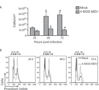
Cell growth and cell-cycle kinetics - Six-well plates were seeded with 4 x 10⁴ C6/36 cells/cm2 and incubated
for 24 h; the cells were subsequently infected with a mul-tiplicity of four genomes/cell [4 mulmul-tiplicity of genome (MOG)] of BR/07 MDV or mock infected. At 24, 48 and 72 h post-infection, the cells were detached, stained with trypan blue and the number of viable cells was counted with a Neubauer chamber. The same conditions were used to evaluate the cell cycle stage via flow cytometry, as described by Fried et al. (1978).
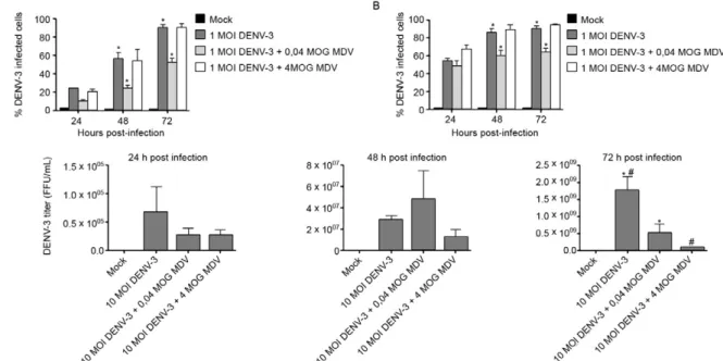
Co-infection experiments - C6/36 cell cultures were infected with a multiplicity of infection (MOI) of one or 10 of DENV-3 (BR DEN3 290-02), four or 0.04 MOG of BR/07 MDV or a mixture of these two viruses. Non-in-fected cultures were used as the negative control (mock). The extent of infection was evaluated at 24, 48 and 72 h with flow cytometry in a BD FACSCalibur™ Flow Cy-tometer (BD Biosciences, San Jose, CA, USA), as well as by titration of the DENV particles from the cell cul-ture supernatants. The titration was performed using the focus immunodetection technique (Desprès et al. 1993); flow cytometry was performed as previously described by Bordignon et al. (2002). The results were analysed with the FlowJo flow cytometry analysis software (Tree-Star, Inc, Ashland, OR, USA).
Replicon experiments - C6/36 cell cultures prepared and infected with BR/07 MDV (3 MOG or 0.03 MOG), as described above, were subsequently transfected with the BR DEN3 290-02 replicon (Mosimann et al. 2010). Five days post-infection/transfection, the monolayer was fixed and stained for immunofluorescence analysis with either anti-MDV or anti-DENV-3 polyclonal antibodies and the percentage of positive cells was estimated.
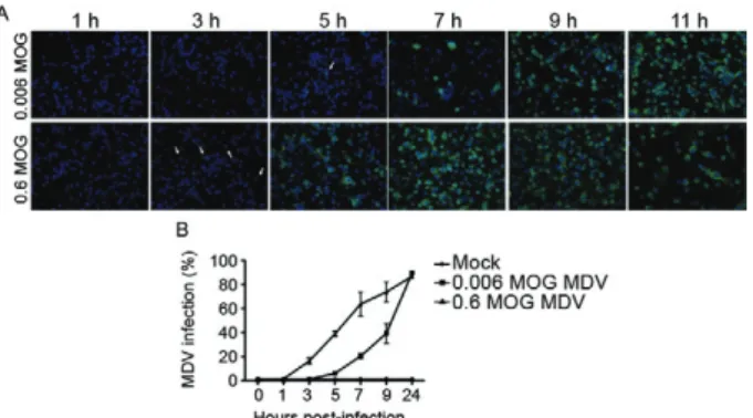
Virus spread experiments - The C6/36 cells were seeded at a density of 5 x 10⁴ cells/cm2 and incubated
for 48 h; the cells were subsequently infected with 0.6 or 0.006 MOG of BR/07 MDV (or a mock infection for the control). After incubation for 1 h at 28ºC, the inoculum was removed and cells were washed with phosphate buff-ered saline (PBS) buffer before replenishment with cell medium. At various time points (0, 1, 3, 5, 7, 9, 11 h and 24 h), the cells were either detached and immunolabeled for flow cytometry analysis or fixed and permeabilized with a 1:1 solution of methanol:acetone at -20ºC for at least 2 h for an indirect immunofluorescence assay.
IFA - The C6/36 cell monolayers that were previ-ously fixed and permeabilised were incubated with mouse anti-MDV polyclonal antibodies at 37ºC for 45 min, washed three times with PBS and incubated for 45 min at 37ºC with goat anti-mouse conjugated to fluo- fluo-rescein isothiocyanate diluted 1:100 in PBS containing 0.3% (v/v) of 10 µg/mL Evan’s blue. The cells were sub-sequently incubated at room temperature for 5 min with 1 mg/mL 4’-6-diamidine-2-phenyl indole, washed five times with PBS buffer and, once dry, were overlaid with a solution of PBS and 10% (v/v) glycerol.
The immunofluorescence images were captured with a Nikon Eclipse TE300 microscope (Nikon, Tokyo, Ja-pan) attached to a CoolSNAPTM-Pro
cf camera (Media
Cy-bernetics, Bethesda, MD, EUA). The images were visu-alised and edited using the Image-Pro® PLUS v.4.5.1.29
software (Media Cybernetics, Bethesda, MD, EUA).
Statistics - The results are expressed as the mean ±
standard deviation of the data. Differences between the groups were analysed with a one-way or two-way analy-sis of variance, followed by a Bonferroni post-test. A p value < 0.05 was considered significant.
RESULTS
The BR/07 MDV was initially detected in the super-natant of C6/36 cells infected with YFV (BR/01) that had been sent to our laboratory for research purposes (Supplementary data). Because of the atypical cytopath-ic effect that was observed in the C6/36 cell cultures, we suspected that there was additional microorganism con-tamination. A putative contaminating virus was identi-fied by electron microscopyt, which showed viral parti-cles of various sizes within the same cell. Hybridization of randomly amplified nucleic acids from the culture to a pan-viral DNA microarray yielded strong hybridisation for multiple oligonucleotide probes from known denso-viruses. Viral identification was later confirmed by PCR using primers for conserved regions of the Brevidenso-virus genome, which were retrieved from the public data banks (Supplementary data).
The BR/07 MDV was subsequently isolated by elimi-nating the yellow fever virus, which was also present in the sample, by neutralization. BR/07 MDV isolation was con-firmed by RT-PCR and PCR with yellow fever and MDV-specific primers, respectively (Supplementary data).
Due to the limited amount of sequence data available for the 5’ and 3’ ends of these viruses, it was difficult to predict the first and last nt of the BR/07 MDV genome. However, the BR/07 MDV end sequences that were defined by the genome assembly display the predicted “Y”-shaped structure, similar to the predicted structure for Ae. densonucleosis virus, although with longer arms (Supplementary data). The arms formed by the 5’ and 3’ end of the BR/07 MDV isolate were 108 and 164 nt long, respectively, whereas the analogous structures for Ae. densonucleosis virus are 102 and 146 nt long.
Our initial biological characterization of the BR/07 MDV isolate involved assessment of the cell growth and cell cycle kinetics. Preliminary observations indicated that infection with this virus affected cell growth. In-fected C6/36 cell growth was significantly inhibited relative to the non-infected cell growth (Fig. 2A). Cell cycle kinetic experiments showed that the infected cells were arrested in the G2 phase (Fig. 2B).
Our next step was to quantify the viral inoculum. We tried several titration protocols for the BR/07 MDV isolate, including the TCID50 protocol described by Jous-set et al. (1993), or the focus immunodetection technique
(Desprès et al. 1993), which were both unsuccessful. The number of genome copies was quantified through quan-titative PCR as described by Ledermann et al. (2004).
The observations made while attempting to titrate the virus suggest that the BR/07 MDV infection spread in the culture within a few hours. To evaluate the rate of viral spread, an experiment was performed with two different virus concentrations (0.6 MOG and 0.006 MOG). The re-sults of these experiments (Fig. 3A) clearly show that the BR/07 MDV infection spread rapidly. At an MOG of 0.6, we were able to detect protein synthesis in the infected cells as early as 3 h infection (white arrows) and by 7 h post-infection, the whole monolayer was infected. This process was delayed with 0.006 MOG, and the whole monolayer was infected by 9 h post-infection. These IFA results are consistent with the flow cytometry data (Fig. 3B).
Several previous studies (Jousset et al. 1993, Carlson et al. 2006, Suchman et al. 2006, Wei et al. 2006) have shown that MDV can be used as a biological control for arboviral infection. Based on these observations, we eval-uated the capacity of BR/07 MDV to affect the in vitro in-fection time course with a clinical isolate of dengue virus. Co-infection experiments were performed with either 4 MOG or 0.04 MOG of BR/07 MDV in combination with DENV-3 at a MOI of one or 10. We found that there was significant inhibition of DENV infection, which was mea-sured by the percentage of infected cells, irrespective of the MOI of DENV used when the cells were co-infected at 0.04 MOG of BR/07 MDV (Fig. 4A, B). In contrast, there was no difference in the percentage of DENV infection in cells co-infected at 4 MOG of BR/07 MDV and cells infected exclusively with DENV-3. Titration of DENV in the supernatants of the cultures infected with 4 MOG of
Fig. 1A: schematic representation of the BR/07 mosquito densovi-rus (MDV) viral isolate genome (above) and its open reading frames (below) (drawn with Vector NTI software from Vector NTI Advance 10.3.1 Software package, Invitrogen, Carlsbad, CA, USA); B: phylo-genetic tree of viruses belonging to the Brevidensovirus genus. The tree was generated based on the region contained within the dashed box in A. Aa: Aedes albopictus; Aae: Aedes aegypti; Ag: Anopheles gambiae; Cp: Culex pipiens; He: Haemagogus equinus; NS: non-structural; VP: structural proteins.
BR/07 MDV (Fig. 4C) typically had lower values at 24 h and 48 h post-infection, although these observations were not statistically significant. At 72 h post-infection, a sig-nificant decrease in the DENV-3 viral titre was observed when the cells were co-infected with BR/07 MDV at both MOIs tested (4 MOG and 0.04 MOG).
To determine whether BR/07 MDV infection inter-fered with DENV-3 replication, we transfected MDV-infected C6/36 cells with a DENV-3 replicon (BR DEN3 290-02 replicon) and estimated the number of anti-DENV-3 reactive cells at five days post-infection/trans-fection. The results suggest that (Supplementary data) there was no significant difference in the percentage of anti-DENV-3 reactive cells for the MDV infected and non-infected cells that were subsequently transfected with the DENV-3 replicon.
DISCUSSION
The main purpose of this work was to genetically and biologically characterise a new Brevidensovirus isolate (BR/07 MDV), as well as to assess its ability to affect DENV morphogenesis. BR/07 MDV was first detected in a C6/36 cell culture infected with an YFV sample (BR/01) that was sent to our laboratory for research purposes. The atypical YFV phenotype in the mosquito cell culture suggested the presence of a contaminating co-infecting microorganism. Electron microscopy, mi-croarray hybridisation, PCR and genome sequencing identified this microorganism as a MDV.
Based on the previously published data (Gorziglia et al. 1980, Jousset et al. 1993, O’Neill et al. 1995, Chen et al. 2004), we hypothesize that the cell line initially used in the YFV isolation, most likely a mosquito cell line, may have been contaminated. Unfortunately, because the cell culture supernatant sample was sent seven years ago for research purposes, there is no way to track the infection origin.
Thus, we suggest that researchers who work with ar-bovirus isolation from mosquitoes should check their cell lines periodically for MDV infection. The potential of these viruses to pass unnoticed in cell cultures is due to their ability to establish persistent infections that are not associated with any obvious cytopathic effect (Gorziglia et al. 1980, Jousset et al. 1993, O’Neill et al. 1995).
Phylogenetic analysis grouped BR/07 MDV with Ae. aegypti densovirus, which was supported by high bootstrap values (Fig. 1). This could be due to the fact that the viral isolate reported in this article was clearly present as a cell culture contaminant, without means of tracing its real ori-gin. Similar results were described by Chen et al. (2004).
Previous findings by Paterson et al. (2005) suggest that the expression of Ae. aegypti densonucleosis vi-rus NS1 in C6/36 cells causes the cells to halt in the G2 phase. Consistent with this, we saw that BR/07 MDV af-fected Ae. albopictus cell growth (Fig. 2A) and appeared to cause cell cycle arrest in G2 (Fig. 2B). Cell cycle ar-rest has also been previously described by Op De Beeck and Caillet-Fauquet (1997) in a study with another par-vovirus, the Minute virus of mice. This study showed that NS1 interferes with cellular DNA replication and induces chromatin damage. Similarly, Oleksiewicz and Alexandersen (1997) saw that infection of Crandell fe-line kidney cells with Aleutian mink disease parvovirus induced cell-cycle arrest.
Previous studies have also suggested that MDV re-duces the severity of DENV infection (Burivong et al. 2004). Thus, we tested the ability of MDV to act as a biological control for DENV infection in vitro with co-infection experiments. We observed significantly lower levels of cell infection and a lower virus titre in C6/36 cells that were co-infected with BR/07 MDV and DENV-3 compared to cells infected with DENV-3 alone. However, the discrepancy observed between the DENV titre results (Fig. 4C) and the percentage of in-fected cells determined with flow cytometry (Fig. 4A, B) remains unclear. Although co-infection at 4 MOG of BR/07 MDV did not significantly inhibit dengue infec-tion (Fig. 4A, B), DENV-3 viral progeny generainfec-tion was significantly inhibited by BR/07 MDV at both MOG tested 72 h post co-infection (Fig. 4C). Therefore, the effect of BR/07 MDV on DENV infection does not ap-pear to be related to viral entry into the cells or genome replication/translation but may be due to changes in vi-ral assembly or budding. Replicons are especially well suited to study viral replication once they have all the elements needed for the replication of the viral genetic material in cells, but they do not encode the functional structural proteins and consequently are not able to gen-erate new viral particles (Jones et al. 2005). We there-fore used a DENV-3 replicon (Mosimann et al. 2010) in co-infection experiments with BR/07 MDV as described above to confirm our findings. DENV-3 replication was not significantly affected by MDV infection (3 MOG or 0.03 MOG) (Supplementary data). However, because the percentage of transfected cells was low, the possibility that MDV infection interferes with DENV-3 replication cannot be completely ruled out.
Wei et al. (2006) obtained promising results for the use of MDV as a biological control. Specifically, they observed that DENV-2 infection was 100-fold decreased in MDV-co-infected Ae. albopictus mosquitoes, com-pared to mosquitoes infected with DENV-2 alone. Us-ing a different approach, Burivong et al. (2004) showed that C6/36 cells that were persistently infected with
Ae. albopictus densovirus had a less severe infection, although they were not protected from infection itself. Significantly decreased DENV-2 viral particle produc-tion was observed in cells that were persistently infected with MDV at 72 h and 96 h but was not observed at 120 h post-infection. However, the use of different co-infec-tion protocols makes it difficult to compare the previous findings with our own.
Viruses that belong to the Densovirus genus display certain features that make them suitable for use as bio-logical controls. They are non-enveloped viruses that can survive for long periods of time in nature. They also act as larvicides and can be vertically transmitted to mosqui-to offspring. Additionally, infection in adult mosquimosqui-toes has been associated with a decreased lifespan (Jousset et al. 1993, Carlson et al. 2006, Suchman et al. 2006). Be-cause these viruses possess a small genome, they may also prove useful as expression vectors, although this remains to be confirmed (Afanasiev et al. 1994). They may also be useful for immunisation strategies, where se-quences that are able to interfere with either the replica-tion of clinically important arboviruses or the reproduc-tion of mosquito vectors could be inserted into the MDV genome (Carlson et al. 2006). However, such possibilities must be addressed carefully, because previous findings have suggested that arthropod cells can have balanced, persistent infections with two or three heterologous vi-ruses (Burivong et al. 2004, Kanthong et al. 2008, 2010).
ACKNOWLEDGEMENTS
To Paulo Arauco and Vanessa Stella, for their technical help with sequencing and cell culture procedures, respective-ly, to Dr Ricardo Galler, for providing the YF17DD strain, to Dr David Wang, for assistance with the microarray analysis, to Dr Eleonora Campos, for helpful discussion, and to Dr André Báfica, for critical reading.
REFERENCES
Afanasiev BN, Galyov EE, Buchatsky LP, Kozlov YV 1991. Nucle-otide sequence and genomic organization of Aedes densonucleo-sis virus. Virology 185: 323-336.
Afanasiev BN, Kozlov YV, Carlson JO, Beaty BJ 1994. Densovirus of Aedes aegypti as an expression vector in mosquito cells. Exp Parasitol 79: 322-339.
Bergoin M, Tijssen P 2000. Molecular biology of Densovirinae. In S Faisst, J Rommelaere (eds.), Parvoviruses from molecular biol-ogy to patholbiol-ogy and therapeutic uses, Karger, Basel, p. 12-32.
Bordignon J, Pires Ferreira SC, Medeiros Caporale GM, Carrieri ML, Kotait I, Lima HC, Zanetti CR 2002. Flow cytometry assay for in-Flow cytometry assay for in-tracellular rabies virus detection. J Virol Methods 105: 181-186.
Burivong P, Pattanakitsakul SN, Thongrungkiat S, Malasit P, Flegel TW 2004. Markedly reduced severity of dengue virus infection in mosquito cell cultures persistently infected with Aedes al-bopictus densovirus (AalDNV). Virology 329: 261-269.
Carlson J, Suchman E, Buchatsky L 2006. Densoviruses for control and genetic manipulation of mosquitoes. Adv Virus Res 68: 361-392.
Chen S, Cheng L, Zhang Q, Lin W, Lu X, Brannan J, Zhou ZH, Zhang J 2004. Genetic, biochemical and structural characterization of a new densovirus isolated from chronically infected Aedes al-bopictus C6/36 cell line. Virology 318: 123-133.
Ewing B, Green P 1998. Base-calling of automated sequencer traces using phred. II. Error probabilities. Genome Res 8: 186-194.
Ewing B, Hillier L, Wendl MC, Green P 1998. Base-calling of au-tomated sequencer traces using phred. I. Accuracy assessment.
Genome Res 8: 175-185.
Felsenstein J 1985. Confidence limits on phylogenies: an approach using the bootstrap. Evolution 39: 783-791.
Fried J, Perez AG, Clarkson BD1978. Rapid hypotonic method for flow cytofluorometry of monolayer cell cultures. Some pitfalls in staining and data analysis. J Histochem Cytochem 26: 921-933.
Gordon D, Abajian C, Green P 1998. Consed: a graphical tool for se-quence finishing. Genome Res 8: 195-202.
Gordon D, Desmarais C, Green P 2001. Automated finishing with autofinish. GenomeRes11: 614-625.
Gorziglia M, Botero L, Gil F, Esparza J 1980. Preliminary character-ization of virus-like particles in a mosquito (Aedes pseudoscutel-laris) cell line (Mos. 61). Intervirology 13: 232-240.
Igarashi A 1978. Isolation of a Singh’s Aedes albopictus cell clone sensi-tive to dengue and Chikungunya viruses. J Gen Virol40: 531-544.
Jones CT, Patkar CG, Kuhn RJ 2005. Construction and applications of yellow fever virus replicons. Virology331: 247-259.
Jousset FX, Baquerizo E, Bergoin M 2000. A new densovirus iso-lated from the mosquito Culex pipiens (Diptera: Culicidae).
Virus Res 67: 11-16.
Jousset FX, Barreau C, Boublik Y, Cornet M 1993. A parvo-like virus persistently infecting a C6/36 clone of Aedes albopictus
mosquito cell line and pathogenic for Aedes aegypti larvae.
Virus Res 29: 99-114.
Kanthong N, Khemnu N, Pattanakitsakul SN, Malasit P, Flegel TW 2010. Persistent, triple-virus co-infections in mosquito cells. BMC Microbiol 10: 14.
Kanthong N, Khemnu N, Sriurairatana S, Pattanakitsakul S, Mala-sit P, Flegel TW 2008. Mosquito cells accommodate balanced, persistent co-infections with a densovirus and dengue virus. Dev Comp Immunol32: 1063-1075.
Ke GM, Cheng HL, Ke LY, Ji WT, Chulu JL, Liao MH, Chang TJ, Liu HJ 2006. Development of a quantitative Light Cycler real-time RT-PCR for detection of avian reovirus. J Virol Methods 133: 6-13.
Kittayapong P, Baisley KJ, O’Neill SL 1999. A mosquito densovi-rus infecting Aedes aegypti and Aedes albopictus from Thailand.
Am J Trop Med Hyg 61: 612-617.
Ledermann JP, Suchman EL, Black WC 4th, Carlson JO 2004. In-fection and pathogenicity of the mosquito densoviruses AeDNV, HeDNV and APeDNV in Aedes aegypti mosquitoes (Diptera: Culicidae). J Econ Entomol 97: 1828-1835.
Mazzarotto GA, Raboni SM, Stella V, Carstensen S, de Noronha L, Levis S, Zanluca C, Zanetti CR, Bordignon J, Duarte dos Santos CN 2009. Production and characterization of monoclonal
anti-bodies against the recombinant nucleoprotein of Araucaria hanta-virus. J Virol Methods 162: 96-100.
Mosimann AL, de Borba L, Bordignon J, Mason PW, dos Santos CN 2010. Construction and characterization of a stable subgenomic replicon system of a Brazilian dengue virus type 3 strain (BR DEN3 290-02). J Virol Methods 163: 147-152.
Nogueira MB, Stella V, Bordignon J, Batista WC, de Borba L, Silva LHP, Hoffmann FG, Probst CM, Santos CND 2008. Evidence for the co-circulation of dengue virus type 3 genotypes III and V in the Northern Region of Brazil during the 2002-2004 epidemics.
Mem Inst Oswaldo Cruz 103: 483-488.
Oleksiewicz MB, Alexandersen S 1997. S-phase-dependent cell cycle disturbances caused by Aleutian mink disease parvovirus. J Virol 71: 1386-1396.
O’Neill SL, Kittayapong P, Braig HR, Andreadis TG, Gonzalez JP, Tesh RB 1995. Insect densoviruses may be widespread in mos-quito cell lines. J Gen Virol 76: 2067-2074.
Op De Beeck A, Caillet-Fauquet P 1997. The NS1 protein of the au-tonomous parvovirus Minute virus of mice blocks cellular DNA replication: a consequence of lesions to the chromatin? J Virol 71: 5323-5329.
Paterson A, Robinson E, Suchman E, Afanasiev B, Carlson J 2005. Mosquito densonucleosis viruses cause dramatically different infection phenotypes in the C6/36 Aedes albopictus cell line.
Virology 337: 253-261.
Saitou N, Nei M 1987. The neighbor-joining method: a new method for reconstructing phylogenetic trees. Mol Biol Evol 4: 406-425.
Suchman EL, Kononko A, Plake E, Doehling M, Kleker B, Black WC 4th, Buchatsky L, Carlson J 2006. Effects of AeDNV infection on
Aedes aegypti lifespan and reproduction. Biol Control 39: 465-473.
Tamura K, Dudley J, Nei M, Kumar S 2007. MEGA4: Molecular Evo-lutionary Genetics Analysis (MEGA) software version 4.0. Mol Biol Evol 24: 1596-1599.
Tamura K, Kumar S 2002. Evolutionary distance estimation under heterogeneous substitution pattern among lineages. Mol Biol Evol 19: 1727-1736.
Thompson JD, Higgins DG, Gibson TJ 1994. CLUSTALW: improv-ing the sensitivity of progressive multiple sequence alignment through sequence weighting, position-specific gap penalties and weight matrix choice. Nucleic Acids Res 22: 4673-4680.
Wang D, Urisman A, Liu YT, Springer M, Ksiazek TG, Erdman DD, Mardis ER, Hickenbotham M, Magrini V, Eldred J, Latreille JP, Wilson RK, Ganem D, DeRisi JL 2003. Viral discovery and se-quence recovery using DNA microarrays. PLoS Biol 1: E2.
Wei W, Shao D, Huang X, Li J, Chen H, Zhang Q, Zhang J 2006. The pathogenicity of mosquito densovirus (C6/36DNV) and its inter-action with dengue virus type II in Aedes albopictus. Am J Trop Med Hyg 75: 1118-1126.
Characterization of anti-mosquitodensovirus (MDV) polyclonal an-tibodies. Indirect immunofluorescence assay (IFA) of C6/36 cultures infected with MDV or mock infected. For IFA, the cells were fixed and incubated with pre or post-immune serum diluted 1:200 followed by anti-mouse conjugated to fluorescein isothiocyanate. Cell nuclei were stained with 4’-6-diamidine-2-phenyl indole (DAPI). Pictures were taken with 400x magnification factor. The scale bar in the bright field images corresponds to 50 µm.
YF BR/01 sample characterization. A: indirect immunofluorescence assay of C6/36 and Vero cells infected with the YF BR/01 sample, a yel-low fever positive control (YF17DD P2) or a negative control (mock). Cells were fixed at six (C6/36) or seven (Vero) days post-infection and labelled with yellow fever polyclonal antibody followed by anti-mouse conjugated to fluorescein isothiocyanate; B: bright field micros-copy of C6/36 cells infected or not (mock) with the samples described
for A. For all experiments 4 x 10⁴ cells/cm2 were seeded and were
in-fected 24 h later with the same amount of virus. In A C6/36 and Vero images were taken with magnification factors of 400x and 200x, re-spectively, in B a magnification factor of 100x was used.
Transmission electron microscopy of C6/36 cells infected with the sample YF BR/01 at 48 h post-infection. Arrows point to the large particles and arrowheads to an aggregate of smaller particles associ-ated to the endoplasmic reticulum. Bar = 0.5 µm.
Mosquito densovirus polymerase chain reaction (PCR) product
Isolation of mosquitodensovirus (MDV). Polymerase chain reaction (PCR) from C6/36 cells infected with different dilutions (10-1-10-6) of the YF
BR/01 sample that had been pre-incubated with anti-yellow fever polyclonal antibody; products were visualized by electrophoresis on 0.8% agarose gels. MDV PCR was performed with primers DNV-3F and DNV-3R, giving an expected size of 324 bp. Yellow fever (YF) PCR was carried out using primers YF34 and YF21, giving an expected size of 291 bp. Gels were loaded with 250 ng of 1 Kb Plus DNA Ladder and 25% (v/v) of PCR reactions. Bl: blank; Co+: positive control.
Predicted secondary structures for 5’ and 3’ ends of the viral genome. 5’ ends of (A) BR/07 mosquitodensovirus (MDV) and (B) Aedes
Replicon experiments. A: indirect immunofluorescence assay of C6/36 cultures transfected with 1 µg of RNA of the Replicon BR DEN3 290-02
previously infected or not at 0.03 or 3 multiplicity of genomes (MOG) of BR/07 mosquitodensovirus (MDV). Cells were fixed and incubated with
anti-dengue virus-3 (DENV-3) polyclonal antibody followed by anti-mouse conjugated to fluorescein isothiocyanate. Cell nuclei were stained with 4’-6-diamidine-2-phenyl indole. Images were obtained with a 400x magnification factor. Pictures representative of three experiments; B: estima-tion of the percentage of anti-DENV-3 reactive cells in C6/36 cultures transfected with 1 µg of RNA of the Replicon BR DEN3 290-02 previously