Vaccinia Infection
Martha A. Hutchens1, Kathryn E. Luker2, Joanne Sonstein3, Gabriel Nu´n˜ez1,4,5, Jeffrey L. Curtis1,3,6, Gary D. Luker1,2,7*
1Graduate Program in Immunology, University of Michigan Medical School, Ann Arbor, Michigan, United States of America,2Department of Radiology, University of Michigan Medical School, Ann Arbor, Michigan, United States of America,3Division of Pulmonary and Critical Care Medicine, Department of Internal Medicine, University of Michigan Medical School, Ann Arbor, Michigan, United States of America,4Department of Pathology, University of Michigan Medical School, Ann Arbor, Michigan, United States of America,5Comprehensive Cancer Center, University of Michigan Medical School, Ann Arbor, Michigan, United States of America,6Department of Veterans Affairs Health System, University of Michigan Medical School, Ann Arbor, Michigan, United States of America,7Department of Microbiology and Immunology, University of Michigan Medical School, Ann Arbor, Michigan, United States of America
Abstract
Innate immune responses are essential for controlling poxvirus infection. The threat of a bioterrorist attack usingVariola major, the smallpox virus, or zoonotic transmission of other poxviruses has renewed interest in understanding interactions between these viruses and their hosts. We recently determined that TLR3 regulates a detrimental innate immune response that enhances replication, morbidity, and mortality in mice in response to vaccinia virus, a model pathogen for studies of poxviruses. To further investigate Toll-like receptor signaling in vaccinia infection, we first focused on TRIF, the only known adapter protein for TLR3. Unexpectedly, bioluminescence imaging showed that mice lacking TRIF are more susceptible to vaccinia infection than wild-type mice. We then focused on TLR4, the other Toll-like receptor that signals through TRIF. Following respiratory infection with vaccinia, mice lacking TLR4 signaling had greater viral replication, hypothermia, and mortality than control animals. The mechanism of TLR4-mediated protection was not due to increased release of proinflammatory cytokines or changes in total numbers of immune cells recruited to the lung. Challenge of primary bone marrow macrophages isolated from TLR4 mutant and control mice suggested that TLR4 recognizes a viral ligand rather than an endogenous ligand. These data establish that TLR4 mediates a protective innate immune response against vaccinia virus, which informs development of new vaccines and therapeutic agents targeted against poxviruses.
Citation:Hutchens MA, Luker KE, Sonstein J, Nu´n˜ez G, Curtis JL, et al. (2008) Protective Effect of Toll-like Receptor 4 in Pulmonary Vaccinia Infection. PLoS Pathog 4(9): e1000153. doi:10.1371/journal.ppat.1000153
Editor:Mark L. Buller, Saint Louis University, United States of America
ReceivedMay 5, 2008;AcceptedAugust 13, 2008;PublishedSeptember 19, 2008
Copyright:ß2008 Hutchens et al. This is an open-access article distributed under the terms of the Creative Commons Attribution License, which permits unrestricted use, distribution, and reproduction in any medium, provided the original author and source are credited.
Funding:This research was supported by R21AI066192 and RO1 HL082480 from the National Institutes of Health (NIH) and Merit Review funds from the Department of Veterans Affairs. Support for imaging experiments was provided by NIH grant R24CA083099 for the University of Michigan Small Animal Imaging Resource.
Competing Interests:The authors have declared that no competing interests exist. * E-mail: [email protected]
Introduction
In 1980, the World Health Organization declared that smallpox had been eliminated as a human disease [1]. Nevertheless, potential bioterrorist release of Variola major, the causative agent for smallpox, and human infection with monkeypox or other zoonotic orthopoxviruses has heightened interest in this family of viruses [2]. Variola major is particularly feared as a bioterrorism agent because of the high rate of transmission and up to 30% mortality caused by smallpox [3]. Fatal cases of smallpox were characterized by clinical findings similar to septic shock, likely mediated by the host inflammatory response to infection. However, molecules and signaling pathways that initiate and control protective and detrimental immune responses to Variola majorremain poorly defined. Identifying molecular determinants of the innate immune response to poxviruses is critical to under-standing pathogenesis of poxvirus infections and developing better therapies to prevent or ameliorate the sepsis-like disease manifestations. This knowledge also may lead to development of a safer smallpox vaccine that eliminates the high risk of severe, life-threatening complications associated with the current live,
attenuated vaccinia virus vaccine. Improved understanding of the innate immune response to poxviruses will have benefits beyond advancing new vaccines and therapies to prevent and treat infection. Vaccinia virus is being investigated as a gene delivery, oncolytic, or immunizing vector for a wide variety of diseases, including cancer, HIV and malaria [4–8]. Greater knowledge of normal host-pathogen interactions will enable more efficient targeting and efficacy of these vectors in patients. Finally, insights gained from studying pulmonary infection with poxviruses are expected to inform research on protective and harmful aspects of host immunity to other respiratory pathogens.
TLR4-dependent recognition of LPS is well-established as a central regulator of effective host immunity to bacterial pathogens, TLR4 also may signal in response to a wide variety of endogenous ligands, such as heat shock proteins [9,10]. TLR4 also may respond to some viral proteins, and TLR4-dependent signaling may be necessary to limit viral replication and disease morbidity in vivo [11,12]. These studies emphasize that functions of TLRs in host immunity may extend to pathogens that do not carry known ligands for specific receptors, particularly as TLRs respond to infections in living animals.
We recently established that TLR3 controls a detrimental innate immune response to pulmonary infection with vaccinia virus, a model virus for studies of orthopoxviruses [13]. Compared with wild-type mice, mice lacking TLR3 (TLR32/2) had reduced viral replication and were protected against disease morbidity and mortality. Adverse effects of TLR3 signaling were caused in part by an excessive inflammatory response to infection. To further investigate TLR3 in poxvirus infection, we initially focused on functions of TIR domain-containing adapter inducing interferon-b (TRIF), the only known downstream adapter molecule for TLR3. Unexpectedly, mice lacking TRIF (TRIF2/2) did not reproduce protective effects of deleting TLR3, but TRIF2/2 was more susceptible to vaccinia infection. These data prompted us to analyze functions of TLR4, the only other TLR known to signal through TRIF, in response to respiratory infection with vaccinia virus. We determined that TLR4 signaling protects mice against vaccinia infection, limiting viral replication and local inflamma-tion.
Results
TRIF2/2mice have a distinct phenotype from TLR32/2 mice
We recently reported that TLR32/2 mice are protected from pulmonary vaccinia infection compared to wild type C57BL/6 controls [13]. The only known adaptor molecule for TLR3 is TRIF, so TLR3 is thought to signal exclusively through TRIF to control secretion of type I interferons and pro-inflammatory cytokines [14]. Because of this direct TLR3 to TRIF signaling
Unexpectedly, susceptibility of TRIF mice to vaccinia infection was distinct from that of the TLR32/2mice. TRIF2/2 mice had less weight loss than wild-type mice on days 1–4 post-infection (p,0.05), which is similar to our published results for TLR32/2versus wild-type mice, (Figure 1A). However, TRIF2/2 mice differed from TLR32/2 animals in that replication of Vac-GFL was greater in mice lacking TRIF, as quantified by region of interest analysis of head, chest, and abdomen sites on biolumines-cence images. By area under the curve (AUC) analysis, TRIF2/2 mice had significantly greater luminescence in their chests (from lung infection) than wild-type mice (Figure 1B;p,0.01). These data indicate that a different and/or additional host molecule(s) controls responses to vaccinia in TRIF2/2mice relative to those mediated solely by TLR3. This experiment continued until day 7 post-infection, when the animals were euthanized to obtain plasma and bronchoalveolar (BAL) fluid for quantification of cytokines. Levels of IL-6, IL-4, IFN-c, MCP-1, TNF-a, and TGF-bwere measured in these samples, but no significant differences were seen between the TRIF2/2and WT mice (data not shown).
TLR4 confers protection in pulmonary vaccinia infection
We hypothesized that TLR4, the only other Toll-like receptor known to signal through TRIF, may control differing host responses to vaccinia in TLR32/2versus TRIF2/2mice. TLR4 is reported to limit replication of a limited number of viruses [11,16], although functions of this receptor in vaccinia infection have not been established. To investigate TLR4 in host defense against vaccinia virus, we used C3H/HeJ mice, which have a point mutation in the cytoplasmic region of TLR4 that renders them unresponsive to LPS [17]. As controls, we used C3HeB/FeJ mice, which have normal, functional TLR4. C3HeB/FeJ mice are genetically similar to C3H/HeJ mice and are well-established as a control strain for experiments using C3H/HeJ animals [18–20].
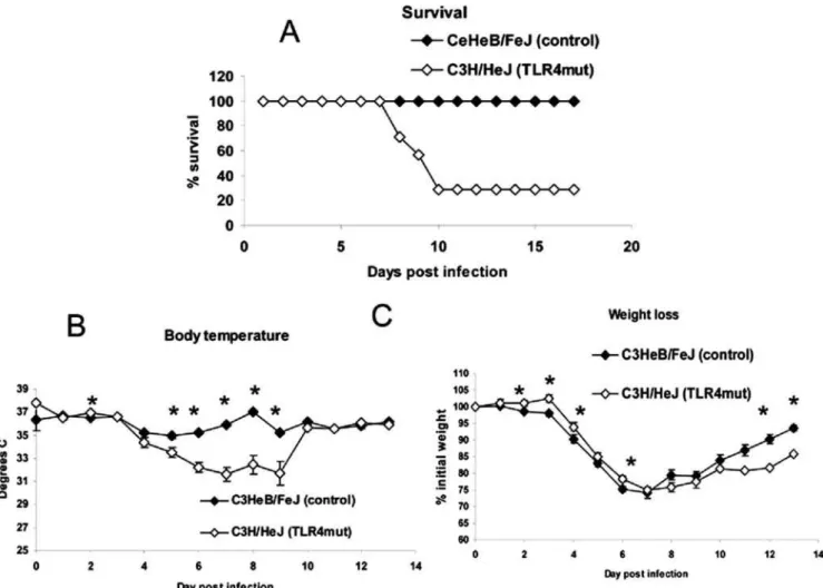
We infected C3H/HeJ and C3HeB/FeJ mice with 16104pfu Vac-GFL. Systemic effects of disease were monitored by weight loss and rectal temperature, while viral replication and spread were assessed with bioluminescence imaging. While both strains of mice lost body temperature in response to vaccinia infection [21], temperatures decreased to a greater extent over the course of the infection in C3H/HeJ (TLR4 mutant) mice relative to C3HeB/ FeJ (Figure 2A). C3H/HeJ mice had a slightly lower temperature than the controls prior to infection, but there were no differences between strains on days 1, 2, and 3 post-infection. Beginning on day 4, however, rectal temperatures were significantly lower in C3H/HeJ mice, reaching a mean temperature of 33uC on day 6 post-infection (p,0.05). In contrast, the lowest mean temperature recorded in C3HeB/FeJ mice was 34.7uC on day 7. C3H/HeJ mice had significantly lower temperatures than control C3HeB/ FeJ mice on days 4–7 (p,0.05). As a second marker of disease severity, weight loss was monitored over the course of the disease
(Figure 2B). Surprisingly, the TLR4 mutant mice lost slightly less weight than the controls with significant differences on days 1–3 and 7–8 post-infection (p,0.05). Although the pattern of the weight loss difference is opposite that of the body temperature, it is consistent with the reduced weight loss observed in TLR32/2and TRIF2/2mice compared with wild-type controls.
Bioluminescence imaging showed that C3H/HeJ (TLR4 mutant) mice had significantly greater viral replication in the chest region on days 1, 4, and 5 post-infection (p,0.05; Figure 2C). Light measured in the chest region of interest predominantly represents viral replication in the lung. C3H/HeJ mice also had significantly more Vac-GFL bioluminescence in the chest over the full course of the experiment as determined by AUC analysis. AUC values for bioluminescence were 1.616108vs. 3.186107for C3H/HeJ and C3HeB/FeJ mice, respectively (p,0.01). C3H/ HeJ mice also had increased abdominal luminescence compared to control animals on day 4 (p,0.05) and over the course of the experiment by AUC analysis (Figure 2D). AUC values for photons produced in abdominal regions were 1.686107and 5.216106for C3H/HeJ and control C3HeB/FeJ mice, respectively (p,0.05). Bioluminescence in the head region did not differ between groups, and all mice recovered from infection (data not shown). Collectively, these data suggest that TLR4 limits respiratory infection and systemic spread of vaccinia virus.
To establish effects of TLR4 on survival, we infected C3H/HeJ and control C3HeB/FeJ mice with 56105pfu Vac-GFL, a dose 1.5 logs higher than used previously. Using this inoculum, TLR4 mutant mice were clearly more susceptible to vaccinia infection.
By day 10 post-infection, 70% of C3H/HeJ mice had died, while all control mice recovered from infection (Figure 3A). As in the previous experiment, loss of body temperature was measured as a sign of morbidity. C3H/HeJ mice were significantly more hypothermic than control C3HeB/FeJ mice on days 2 and 5–9 post-infection (p,0.05; Figure 3B). The rapid recovery of mean temperature in C3H/HeJ mice between days 9 and 10 is caused by death of the most hypothermic mice, while the surviving animals recovered temperature comparable to control C3HeB/ FeJ mice. These data were consistent over 5 independent experiments. Weight loss also was monitored over the course of the disease. C3H/HeJ mice exhibited less weight loss than control C3HeB/FeJ animals over the first 7 d post-infection, and these differences were significant on days 2–4 and 6 (p,0.05; Figure 3C). The same trend was observed in two subsequent experiments. However, C3H/HeJ mice recovered weight more quickly than control C3HeB/FeJ animals on days 8–13, with significant differences observed on days 12 and 13 (p,0.05). Both body temperature and weight loss are reported to be regulated by cytokines, including IL-1, IL-6, and TNF-a, as part of the ‘‘sickness response’’ [22]. The discrepancy between these param-eters during vaccinia infection suggests underlying differences in mechanisms and pathways that regulate these two global measures of disease. These data highlight limitations of using weight loss alone as a measure of disease severity in vaccinia infection.
With an inoculum of 56105pfu Vac-GFL, differences in viral replication between genotypes were even more pronounced than in the earlier experiment. Bioluminescence from Vac-GFL was greater in the head region of C3H/HeJ mice compared with controls. Differences between strains were statistically significant over the latter part of infection on days 5 and 7–10 (p,0.05; Figure 4A). Over the course of the experiment, there was a trend for higher head bioluminescence in C3H/HeJ mice as determined by AUC analysis, although this difference was not statistically significant. Similarly, bioluminescence in the chests of TLR4 mutant mice was significantly increased over the controls (p,0.05) on days 3–7 and 10 post-infection (Figure 4B,C). At the peak of infection on day 6, the chest luminescence of the TLR4 mutant mice was 8-fold higher than that of the control mice. Moreover, the AUC for biolumines-cence in C3H/HeJ TLR4 mutant mice was significantly greater than that for controls (7.226108 vs. 8.206107, respectively;
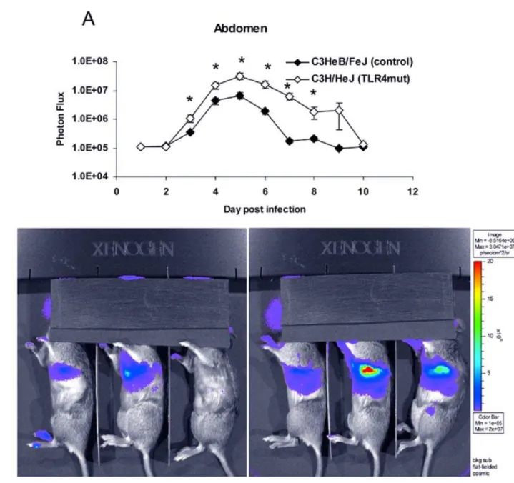
p,0.05). Increased viral replication in lungs of C3H/HeJ was also confirmed by plaque assay (Figure 5). Finally, C3H/HeJ mice had greater systemic spread of the virus to the abdomen (Figure 6A and 6B). At the peak on day 5, the TLR4 mutant mice had 4.7-fold higher luminescence in the abdomen than wild-type controls. Differences between the two genotypes were significant on days 3–8 post-infection. The AUC of the abdominal luminescence in the C3H/HeJ mice was 4.396107 compared with 9.396106 in the control C3HeB/FeJ mice, respectively (p,0.05). These data extend our initial observations of increased viral replication and dissem-ination in mice lacking functional TLR4. Taken together, loss of functional TLR4 renders C3H mice more susceptible to pulmonary vaccinia infection, as measured by multiple parameters. Therefore, TLR4 must recognize some exogenous or endogenous ligand present in vaccinia infection.
To exclude the possibility of our results being affected by endotoxin contamination of our viral preparation, we infected RAW cells with Vac-GFL in the presence or absence of 10mg/mL polymyxin B [23]. Levels of IL-6, TNF-a, and MCP-1 in the cell culture supernatants were assayed by ELISA. Adding polymyxin B did not affect levels of IL-6, MCP-1, or TNF-ain the supernatants of infected cells (data not shown), establishing that contaminating endotoxin did not affect our in vivo studies.
Figure 1. TRIF2/2 mice are more susceptible to vaccinia infection than WT. TRIF2/2and WT BL/6 mice were infected with 16104 pfu Vac-FL. (A) Weight loss, expressed as percent of initial
weight. *p,0.05. (B) Chest luminescence, expressed as photon flux. Error bars denote SEM.
Loss of TLR4 signaling does not abolish IFN-bproduction
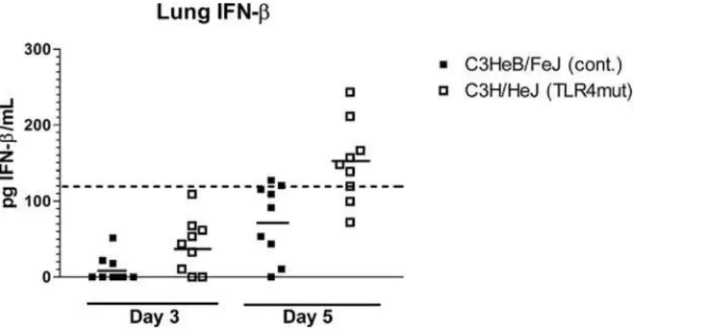
Type I interferons are essential to effective host defense against vaccinia infection [24]. TLR4 signaling results in production of Type I interferons through activation of transcription factors interferon regulatory factor 3 (IRF3) and NF-kB. To determine to what extent TLR4 regulates secretion of type I interferons during vaccinia infection, we measured concentrations of interferon-b (IFN-b) in lung tissue of C3H/HeJ and control mice. Mice were infected i.n. with 56105pfu Vac-GFL, and lungs were harvested on days 3 and 5 post-infection. The day 3 time point is early in the course of infection, at the beginning, or just before, differences in luminescence and body temperature appear. Day 5 is near the peak of the infection where differences between TLR4 mutant and control mice are most pronounced. Lungs were homogenized and concentrations of IFN-b in supernatants were measured by ELISA. Levels of IFN-bwere below the limit of reliable detection on day 3 in both groups of mice. On day 5 post-infection, IFN-b levels in 8 of 9 control mice remained below the limit of detection. On the other hand, six of 9 C3H/HeJ mice had IFN-babove the limit of reliable detection on day 5 (Figure 7). Therefore, C3H/ HeJ mice are capable of producing IFN-b despite the lack of TLR4 signaling, showing redundancy in signaling pathways that activate a type I interferon response to vaccinia.
Loss of TLR4 does not eliminate the inflammatory response to vaccinia in the lung
We hypothesized that protective effects of TLR4 may be mediated by pro-inflammatory cytokine responses, limiting viral replication and spread directly, or indirectly through recruitment of immune cells. To test this hypothesis, we infected C3H/HeJ and control mice with 56105 pfu Vac-GFL i.n. and harvested lungs on days 3 and 5 post-infection. Supernatants from homogenized lungs were analyzed by ELISA for IL-6, TNF-a, and MCP-1. There were no significant differences in levels of any of these cytokines between groups of mice on day 3. On day 5, TNF-a and MCP-1 levels were the same in TLR4 mutant and control mice, but IL-6 levels were significantly higher in C3H/HeJ lungs (Figure 8). No significant differences were detected in the plasma at either time (data not shown) (p.0.4). As with type I interferon, redundant signaling pathways are able to elicit NF-kB-dependent cytokine production in response to vaccinia infection.
To analyze the degree and composition of leukocyte infiltrates in the lung, immune cells were isolated from uninfected mice and mice on days 3 and 5 post-infection. Numbers and types of cells were analyzed by flow cytometry. At day 3, there was a trend towards higher total CD45+cells in the C3H/HeJ cell, but these differences were not significant. We also measured subsets of immune cells in the Figure 2. TLR4 mutant mice are more susceptible to vaccinia than controls.C3HeB/FeJ and C3H/HeJ mice were infected with 16104pfu
Vac-GFL. (A) Body temperature. (B) Weight loss, expressed as percent of initial weight; (C) Chest luminescence. (D) Abdominal luminescence, expressed as photon flux. *p,0.05. Error bars denote SEM.
lung, including B lymphocytes, CD4 and CD8 lymphocytes, macrophages, dendritic cells, and neutrophils. However, there were no consistent differences in cell types recruited to lungs of infected C3H/HeJ and control C3HeB/FeJ mice (data not shown).
To further assess the pattern of inflammation and tissue damage in TLR4 mutant lungs, we examined the lungs of vaccinia-infected mice by histology. Hematoxylin and eosin–stained sections showed foci of mixed and lymphocytic peribronchial and perivascular infiltrate (Figure 9A and 9B). Occasionally, infiltrating cells could be seen in alveoli separate from any peribronchial or perivascular focus. In foci of severe inflammation, epithelial cell necrosis was observed, and some inflammatory cells had apoptotic morphology. As a quantitative measure of inflammation, numbers of foci in each section were counted. On day 3 post-infection, the beginning of the interval when increased levels of virus could be discerned in lungs of TLR4 mutant mice, C3H/HeJ TLR4 mutant mice had significantly more foci of inflammation than controls (p,0.05; Figure 10). No consistent differences were detected on day 5. These data indicate that TLR4 is not required for producing an early local inflammatory response to vaccinia infection.
Vaccinia predominantly infects epithelial cells in the lung
To investigate the cell type(s) involved in the propagation of infection in the lungs, we performed immunohistochemical
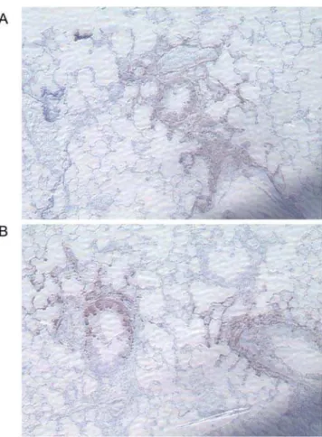
staining on paraffin-embedded lung sections with anti-GFP. Mice were infected with 56105pfu Vac-GFL, and lungs were harvested on days 3 and 5 post-infection. In all samples, intense anti-GFP staining was localized to bronchial epithelial cells with less extensive infection detected in alveolar epithelial cells (Figure 11A and 11B). Samples of both genotypes also showed some staining of cells among the inflammatory infiltrate, possibly macrophages, although firm identification could not be made. In all samples, the regions of positive anti-GFP antibody staining were associated with foci of inflammation, but many foci of inflammation had no regions of anti-GFP antibody staining. The distribution and types of infected cells did not differ between strains of mice. These findings suggest that in the lungs, vaccinia primarily replicates and spreads through epithelial cells.
TLR4 recognizes a viral particle ligand
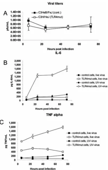
In order to determine whether TLR4 was signaling in response to an endogenous or a viral ligand, we treated bone marrow macrophages isolated from C3H/HeJ and control mice with live or UV-inactivated virus (MOI = 5), and measured levels of TNF-a and IL-6 in the supernatant. The undiluted stock of UV-inactivated Vac-GFL (9.06107pfu/mL) produced no plaques or cytopathic effect in cultured Vero cells (data not shown). TLR4 mutant and control macrophages were equally resistant to viral Figure 3. Increased susceptibility of TLR4 mutant mice is more pronounced at higher viral dose.C3HeB/FeJ and C3H/HeJ mice were infected with 56105pfu Vac-GFL intranasally. (A) Survival curve, expressed as percentage of mice surviving. (B) Body temperature. (C) Weight loss,
replication, even when challenged with live virus at a high MOI (Figure 12A). TLR4 mutant and control macrophages treated with UV-inactivated virus produced significantly (p,0.05) higher levels of IL-6 and TNF-athan cells of the same genotype treated with live virus (Figure 12B and 12C). This is likely due to the absence vaccinia-encoded inhibitors of TLR and other signaling pathways, such as N1L, A46R, and A52R [25–27], produced by replicating vaccinia virus. TLR4 mutant macrophages produced significantly higher levels of both IL-6 and TNF-athan control cells (p,0.05; both live and UV-inactivated virus). This shows that TLR4 not only is unnecessary for the cytokine response of bone marrow macrophages to vaccinia virus, but it actually dampens that response. C3H/HeJ (TLR4 mutant) cells treated with UV-inactivated virus produced by far the highest levels of any condition, rising above IL-6 levels in C3H/HeJ-with-live virus cultures by 6- to 7-fold and 2- to 3-fold for TNF-a in the same cells. Cytokine levels in UV-inactivated C3H/HeJ cultures were approximately 6- to 10-fold higher than those in UV-inactivated Figure 4. Increased viral replication in TLR4 mutant mice is more pronounced at higher viral dose.C3HeB/FeJ and C3H/HeJ mice were infected with 56105pfu Vac-GFL intranasally. (A) Head luminescence. (B) Chest luminescence. (C) Representative chest images. C3HeB/FeJ (left) and
C3H/HeJ (right), 30 s exposure, f-stop 1. Purple denotes lower luminescence intensity; red, higher luminescence intensity.*p,0.05. Error bars denote SEM.
doi:10.1371/journal.ppat.1000153.g004
Figure 5. Increased viral titers in TLR4 mutant lungs.C3HeB/FeJ and C3H/HeJ mice were infected with 56105pfu Vac-GFL intranasally.
Lung viral titer expressed as pfu/mL; lungs harvested day 6 post-infection. *p,0.05. Error bars denote SEM.
C3HeB/FeJ cultures. The fact that macrophages were able to produce TNF-aand IL-6 in response to UV-inactivated virus, and that TLR4-intact macrophages produce significantly less of these cytokines, indicates that neither viral replication nor cell death is necessary for TLR4 recognition of vaccinia virus. This suggests that TLR4 recognizes a component of the viral particle rather than an endogenous ligand released from infected host cells.
Discussion
The innate immune system is vital for host defense against poxviruses, but molecular mechanisms of virus recognition and host defense are incompletely understood. While a robust Th1 immune response is necessary to eliminate vaccinia and other poxviruses, an exaggerated innate immune response also may
threaten the life of the host. In septic shock, a systemic ‘‘cytokine storm’’ causes blood vessel dilation and activation of the clotting cascade, leading to hypotension, hemolysis, and multi-organ failure. Severe and fatal cases of smallpox are characterized by fever, hypotension, coagulopathy, blood vessel dilatation, and leukocyte extravasation, all of which are resemble the pathophys-iology of septic shock [3]. These observations, coupled with the absence of lesions from any location except the skin, suggest that an uncontrolled systemic immune response is the most dangerous aspect of poxvirus infection.
As one mechanism through which immunity contributes to disease manifestations of poxviruses, we recently reported that TLR3 has a detrimental effect in vaccinia infection [13]. Specifically, mice lacking TLR3 had decreased viral replication, morbidity, and mortality following infection with vaccinia virus. Figure 6. Increased viral replication in TLR4 mutant abdomens is more pronounced at higher viral dose.C3HeB/FeJ and C3H/HeJ mice were infected with 56105pfu Vac-GFL intranasally. (A) Abdominal luminescence. (B) Representative images of splenic luminescence C3HeB/FeJ (left)
and C3H/HeJ (right) mice, 30 s exposure, f-stop 1. Purple denotes lower luminescence intensity; red, higher luminescence intensity.*p,0.05. Error bars denote SEM.
These data established TLR3 as a key determinant of poxvirus pathogenesis and highlight the critical balance between effective and excessive innate immune responses during poxvirus infection. To further investigate signaling pathways by which TLR3 exacerbates poxvirus disease, we first analyzed vaccinia infection in mice lacking TRIF, the only known adapter protein for TLR3. Unlike TLR32/2mice, viral replication was significantly greater in TRIF2/2 mice relative to wild-type animals. These data suggested the possibility that protective effects of TRIF against vaccinia infection were mediated through TLR4. Besides TLR3, TLR4 is the only other TLR known to use TRIF as a signaling adaptor. Although TLR4 canonically recognizes bacterial LPS, this receptor also has been implicated in host defense against some viruses. For example, TLR4 is reported to recognize respiratory syncytial virus (RSV) protein F or vesicular stomatitis virus (VSV) glycoprotein G, thereby initiating protective innate immune responses [11,16].
Unlike TLR3, TLR4 does not rely on TRIF exclusively, but also can signal through the adaptor myeloid differentiation factor 88 (MyD88). We hypothesized that in TLR32/2 mice, the normal inflammatory response was attenuated sufficiently to minimize injury to the host while still eliminating vaccinia virus. In TRIF2/2
mice, we reasoned that signaling inputs from TLR3 and TLR4 were both blocked, thus decreasing the inflammatory response to such a degree that the host was not able to make an effective defense against the virus. Consistent with this hypothesis, we demonstrated a protective effect for TLR4 in pulmonary vaccinia infection. Mice with an inactivating mutation in TLR4 suffered increased mortality, more severe hypothermia, and increased viral replication in the head, chest, and abdomen. Further investigation into the mecha-nism of this protection, however, revealed a more complicated picture.
The TLR4 signaling pathway results in activation of NF-kB and interferon regulatory factor 3, suggesting that TLR4-deficient mice would have increased viral replication because of impaired cytokine production and recruitment of immune cells to the lung and other sites of infection. However, levels of TNF-a, MCP-1, IL-6, and IFN-bin TLR4 mutant mice were equal to or even greater than those of the controls. Moreover, histological examination of infected lungs showed that TLR4 mutant mice had significantly more foci of inflammation in their lungs than did controls as early as day 3 post-infection. The results suggest either that TLR4 does not function in these aspects of host immunity to vaccinia virus or that other pattern recognition receptors compensate for loss of Figure 7. Lack of TLR4 does not impair IFN-bproduction.IFN-bconcentrations in lung homogenate supernatants were measured by ELISA. Points represent individual mice. Dashed line represents lower limit of reliable detection on standard curve. Solid lines represent mean IFN-b concentration.
doi:10.1371/journal.ppat.1000153.g007
Figure 8. Lack of TLR4 does not impair proinflammatory cytokine production. IL-6 levels in the lung homogenate supernatants measured by ELISA. Error bars denote SEM. *p,0.05.
TLR4. For example, recent studies suggest protective functions of TLR2 and TLR9 in poxvirus infection [28,29]. The fact that the TLR4 mutant mice are still more susceptible to disease indicates that other pattern recognition receptors are not fully redundant to TLR4 in poxvirus infection.
Immunohistochemical staining revealed vaccinia infection predominantly in bronchiolar epithelium with lesser amounts of viral GFP in alveolar epithelial cells. These data are consistent with previous studies showing that respiratory infection with poxviruses causes a necrotizing bronchopneumonia [30]. We also identified viral GFP antigen in immune cells in the lung, likely macrophages. Previous studies suggest that monocyte/ macro-phage cell types are responsible for systemic spread of poxviruses [30]. While we cannot exclude the possibility that GFP is present in these cells because of phagocytosis rather than infection, our data are compatible with a model in which cells in the monocyte lineage are responsible for systemic dissemination of virus. The observation that both genotypes exhibited a similar repertoire of infected cells suggests that a difference in susceptibility of specific cell types does not account for increased susceptibility of the TLR4 mutant mice.
Increased IL-6 and TNF-alevels in TLR4 mutant macrophages treated with UV-inactivated virus show that viral replication and
cell damage are dispensable for TLR4 recognition of vaccinia. This suggests that TLR4 recognizes a component of the viral particle rather than a host ligand. In our model, the ligand recognized by TLR4 likely would be located in/on the intracellular mature virion (IMV) particle, the predominant form of virus isolated by standard purification procedures such as those used in this research. TLR4 predominantly localizes to the cell
Figure 9. TLR4 alters the inflammatory response to vaccinia infection: representative photomicrographs.Mice were infected intranasally with 56105pfu Vac-GFL. Lungs were harvested on days 3
and 5 post-infection, preserved in 10% formalin, paraffin-embedded, and stained with H&E. (A, B) representative sections of C3H/HeJ (A) and C3HeB/FeJ (B) lung tissue obtained on day 3 post-infection. B = bron-chiole; V = blood vessel; arrows denote inflammatory foci.
doi:10.1371/journal.ppat.1000153.g009
Figure 10. TLR4 alters the inflammatory response to vaccinia infection: quantification of foci of inflammation. Data are expressed as number of foci per section. *p,0.01. Error bars denote SEM.
doi:10.1371/journal.ppat.1000153.g010
Figure 11. Vaccinia localizes to the bronchial and alveolar epithelium. Lungs harvested from Vac-GFL-infected mice (56105pfu)
on day 5 post-infection were stained with anti-GFP and counterstained with hematoxylin. (A) Representative section of C3H/HeJ lung, 1506. (B)
Representative section of C3HeB/FeJ lung, 1506.
membrane, so candidate TLR4 ligands likely would be on the surface of the intracellular mature virion. However, crosslinking DNA in the viral genome with UV/psoralen treatment does not prevent vaccinia from entering the cell and uncoating, so the TLR4 ligand also could be a capsid protein or another protein present in the viral particle.
Increased inflammation in TLR4 mutant mice may be secondary to increased viral burden or a primary effect of the loss of TLR4. Because TLR4 mutant macrophages secrete increased levels of IL-6 and TNF-a even when challenged with UV-inactivated virus, we propose that lack of TLR4 signaling causes increased inflammation. This interpretation also is supported by our data showing equal viral titers in TLR4 mutant and control macrophage cultures infected with live virus despite the higher cytokine levels in TLR4 mutant cell cultures. Consistent with our observations in TLR32/2mice, TLR4 may provide its protection by dampening the inflammatory response elicited in response to vaccinia infection.
However, TLR4 differentially activates an aspect(s) of antiviral defense that is essential for early control of vaccinia replication and spread. Understanding details of this differential regulation will reveal strategies to enhance beneficial immunity to poxviruses and suppress detrimental host responses.
Materials and Methods
Mice
TRIF2/2 mice backcrossed to a C57BL/6 background were originally developed by the S. Akira laboratory and were bred at the University of Michigan. Wild-type (WT) C57BL/6J control mice were obtained from The Jackson Laboratory. Adult male and female mice ages 7 to 9 wk old were used for experiments. C3HeB/FeJ and C3H/HeJ mice were obtained from The Jackson Laboratory. Adult male mice ages 6 to 10 wk old were used for experiments. 17-wk-old mice were used as uninfected controls for histological studies.
Vaccinia virus
We prepared stocks of Vac-GFL, a recombinant Western Reserve (WR) vaccinia virus that expresses firefly luciferase and GFP, and determined viral titers as described previously [13]. Viral titers in excised organs were analyzed by serial dilution on Vero cells [15].
Cells
Vero cells were maintained as we previously have described [15]. Primary bone marrow macrophages were obtained by flushing the femurs and tibiae of mice with cold PBS. This suspension was filtered through a 100mm filter and a 40mm filter. Macrophages were cultured in Dulbecco’s modified Eagle medium supplemented with 20% L929-cell-conditioned media, 10% heat-inactivated fetal bovine serum, 1% L-glutamine, and 0.1% penicillin-streptomycin. Macrophages were cultured 1 wk in this media before performing experiments with them.
Animal procedures
All animal procedures were approved by the University of Michigan Committee on the Use and Care of Animals. Mice were infected i.n. with vaccinia virus as described previously [15]. Weights and rectal temperatures (Physitemp Instruments) were recorded on conscious mice immediately before infection and on each day throughout experiments.
Bioluminescence imaging
Bioluminescence imaging was performed on each day after infection using an IVIS 200 system (Caliper). Imaging and data analysis were performed as described previously [15].
Figure 12. TLR4 recognizes an endogenous ligand and downregulates cytokine secretion. Control C3HeB/FeJ and TLR4 mutant C3H/HeJ bone marrow macrophages were treated with live or UV-inactivated Vac-GFL at MOI = 5 (56105 pfu). (A) Viral titers in
macrophage cultures treated with live virus. IL-6 (B) and TNF-a (C) concentrations in supernatants measured by ELISA. For all combina-tions of pairs of points, p,0.05 except the pair denoting TNF-ain control cells treated with live vs. UV-inactivated virus at 6 hours post-infection.
Histology
To prepare lungs for histology, mice were euthanized on days 3 and 5 post-infection, and lungs were inflated with 1 mL of 10% formalin in PBS. Lungs were excised, preserved in 10% formalin overnight or longer, and then transferred to 70% ethanol solution. Fixed tissues were paraffin embedded and sectioned by the Morphology Core Facility at the University of Michigan. Tissue sections were stained with Gill’s hematoxylin and counterstained with eosin. Sites of viral replication in the lungs were identified by immunohistochemistry, based on detection of GFP from Vac-GFL. Paraffin-embedded lung sections were stained using the Vector Laboratories ABC staining kit. Tissue sections were stained with rabbit polyclonal anti-GFP antibody (1/500 dilution) (Invitrogen) and goat anti-rabbit secondary antibody (1/200 dilution; Vector Laboratories). Blocking solution consisted a 1/ 67 dilution of goat serum in PBS with 250 mM total NaCl.
To quantify foci of inflammation in lung sections, we analyzed transverse lung sections through comparable portions of the upper and lower lobes of each lung. Sections were viewed under a 46 objective, and numbers of inflammatory foci were counted. Mean values for numbers of foci and SEM were calculated.
Serum and tissue cytokines
Blood was obtained from the abdominal aorta of euthanized mice and collected into heparinized tubes. Plasma was separated from cells by centrifugation. Bronchoalveolar lavage of TRIF2/2 mice was performed by intratracheal instillation and withdrawal of 1 mL PBS in lungs of euthanized mice. Plasma and bronchoal-veolar lavage fluid concentrations of TNF-a, IL-6, and MCP-1 were determined by ELISA performed by the University of Michigan Cancer Center Cellular Immunology Core Facility. Concentrations of IFN-b were measured by ELISA (PBL Biomedical Laboratories) according to the manufacturer’s instruc-tions.
Lungs were harvested on day 3 or 5 post-infection and homogenized in 5 mL PBS with a Polytron tissue homogenizer (Brinkmann). Lung homogenates were centrifuged at 21116g for 10 minutes at 4uC. Supernatants were removed and concentra-tions of TNF-a, IL-6, and MCP-1 measured by ELISA as described above.
Flow cytometry
Lungs were excised on day 3 or 5 post-infection and disaggregated by mechanical disruption in a blender (VWR). Cells were counted and analyzed by flow cytometry as described previously [31]. The following mAbs obtained from BD Pharmingen were used: RM4-4 (anti-murine CD4, rat IgG2b), 53-6.72 (anti-murine CD8, rat IgG2b), 1D3 (anti-murine CD19, rat IgG2a),M1/70 murine CD11b, rat IgG2b), HL3 (anti-murine CD11c, hamster IgG1), 2.4G2 (anti-(anti-murine CD16/CD32 Fc block, rat IgG2b), 30-F11 (anti-murine CD45, rat IgG2b), and RB6-8C5 (anti-murine Ly6G Gr-1, rat IgG2b). Monoclonal Abs were primarily conjugated with FITC, PE, APC and APC-Cy7; biotinylated Abs were visualized using streptavidin-PerCP-Cy5.5 (BD Pharmingen). Isotype matched control mAbs (BD Pharmin-gen or eBioscience) were tested simultaneously in all experiments. All samples were analyzed on the BD LSR II flow cytometer with 3 lasers (488 nm blue, 405 nm violet and 633 nm HeNe red). CD45 APC-Cy7 and Invitrogen LIVE/DEAD Fixable Violet Dead Cell Stain were added to all lung mince samples. Subset analysis was performed on gated CD45-positive live cells. A minimum of 10,000 cells were analyzed for each sample. For all analyses, percentages for matched isotype control Abs were subtracted from values obtained for staining with specific Abs for individual markers.
UV inactivation of virus
Vac-GFL was UV-inactivated by irradiation for 90 s on ‘‘sterilize’’ setting in a GS Genelinker UV-chamber (BioRad) following incubation in a Hank’s Balanced Salt Solution (HBSS) solution containing 1.0mg Psoralen according to the protocol of Puhlmann and colleagues [32].
Statistics
Data were analyzed byt test for pairwise comparisons, using Microsoft Excel or GraphPad Prism software. Differences with
p,0.05 were regarded as statistically significant. Author Contributions
Conceived and designed the experiments: MAH KEL JS JLC GDL. Performed the experiments: MAH JS GDL. Analyzed the data: MAH KEL JS GN JLC GDL. Contributed reagents/materials/analysis tools: MAH GDL. Wrote the paper: MAH JLC GDL.
References
1. Breman JG, Arita I (1980) The confirmation and maintenance of smallpox eradication. N Engl J Med 303: 1263–1273.
2. Reynolds MG, Davidson WB, Curns AT, Conover CS, Huhn G, et al. (2007) Spectrum of infection and risk factors for human monkeypox, United States, 2003. Emerg Infect Dis 13: 1332–1339.
3. Bray M, Buller M (2004) Looking back at smallpox. Clin Infect Dis 38: 882–889. 4. Kirn DH, Wang Y, Le Boeuf F, Bell J, Thorne SH (2007) Targeting of interferon-beta to produce a specific, multi-mechanistic oncolytic vaccinia virus. PLoS Med 4: e353. doi:10.1371/journal.ppat.1000353.
5. Erbs P, Findeli A, Kintz J, Cordier P, Hoffmann C, et al. (2008) Modified vaccinia virus Ankara as a vector for suicide gene therapy. Cancer Gene Ther 15: 18–28.
6. Santra S, Sun Y, Parvani JG, Philippon V, Wyand MS, et al. (2007) Heterologous prime/boost immunization of rhesus monkeys by using diverse poxvirus vectors. J Virol 81: 8563–8570.
7. Yang S, Guo ZS, O’Malley ME, Yin X, Zeh HJ, et al. (2007) A new recombinant vaccinia with targeted deletion of three viral genes: its safety and efficacy as an oncolytic virus. Gene Ther 14: 638–647.
8. Bejon P, Mwacharo J, Kai OK, Todryk S, Keating S, et al. (2006) Immunogenicity of the candidate malaria vaccines FP9 and modified vaccinia virus Ankara encoding the pre-erythrocytic antigen ME-TRAP in 1-6 year old children in a malaria endemic area. Vaccine 24: 4709–4715.
9. Chase MA, Wheeler DS, Lierl KM, Hughes VS, Wong HR, et al. (2007) Hsp72 induces inflammation and regulates cytokine production in airway epithelium
through a TLR4- and NF-kappaB-dependent mechanism. J Immunol 179: 6318–6324.
10. Aneja R, Odoms K, Dunsmore K, Shanley TP, Wong HR (2006) Extracellular heat shock protein-70 induces endotoxin tolerance in THP-1 cells. J Immunol 177: 7184–7192.
11. Georgel P, Jiang Z, Kunz S, Janssen E, Mols J, et al. (2007) Vesicular stomatitis virus glycoprotein G activates a specific antiviral Toll-like receptor 4-dependent pathway. Virology 362: 304–313.
12. Haeberle HATR, Casola A, Brasier AR, Dieterich HJ, Van Rooijen N, Gatalica Z, Garofalo RP (2002) Respiratory syncytial virus-induced activation of nuclear factor-kappaB in the lung involves alveolar macrophages and toll-like receptor 4-dependent pathways. J Infect Dis 186: 1199–1206.
13. Hutchens M, Luker K, Sottile P, Sonstein J, Lukacs N, et al. (2008) TLR3 increases disease morbidity and mortality from vaccinia infection. J Immunol 180: 483–491.
14. Kawai T, Akira S (2007) SnapShot: Pattern-recognition receptors. Cell 129: 1024.
15. Luker K, Hutchens M, Schultz T, Pekosz A, Luker G (2005) Bioluminescence imaging of vaccinia virus: effects of interferon on viral replication and spread. Virology 341: 284–300.
16. Kurt-Jones E, Popova L, Kwinn L, Haynes L, Jones L, et al. (2000) Pattern recognition receptors TLR4 and CD14 mediate response to respiratory syncytial virus. Nat Immunol 1: 398–401.
as inhibitor of LPS contamination ofSchistosoma mansonirecombinant proteins in human cytokine analysis. Microb Cell Fact 6: 1.
24. van den Broek M, Muller U, Huang S, Aguet M, Zinkernagel R (1995) Antiviral defense in mice lacking both alpha/beta and gamma interferon receptors. J Virol 69: 4792–4796.
25. DiPerna G, Stack J, Bowie A, Boyd A, Kotwal G, et al. (2004) Poxvirus protein N1L targets the I-kappa B kinase complex, inhibits signaling to NF-kappa B by
CCR6, but not endothelial selectins, mediate the accumulation of immature dendritic cells within the lungs of mice in response to particulate antigen. J Immunol 175: 874–883.