Submitted7 October 2015
Accepted 5 January 2016
Published28 January 2016
Corresponding author
Azlina Abdul Aziz, [email protected]
Academic editor
Alberto Davalos
Additional Information and Declarations can be found on page 16
DOI10.7717/peerj.1628
Copyright
2016 Kong et al.
Distributed under
Creative Commons CC-BY 4.0
Protective effects of the extracts of
Barringtonia racemosa
shoots against
oxidative damage in HepG2 cells
Kin Weng Kong1, Sarni Mat-Junit1, Norhaniza Aminudin2,
Fouad Abdulrahman Hassan3, Amin Ismail3and Azlina Abdul Aziz1
1Department of Molecular Medicine, Faculty of Medicine, University of Malaya, Kuala Lumpur, Malaysia 2Institute of Biological Sciences, Faculty of Science, University of Malaya, Kuala Lumpur, Malaysia
3Department of Nutrition and Dietetics, Faculty of Medicine and Health Sciences, Universiti Putra Malaysia,
Serdang, Selangor, Malaysia
ABSTRACT
Barringtonia racemosais a tropical plant with medicinal values. In this study, the ability of the water extracts of the leaf (BLE) and stem (BSE) from the shoots to protect HepG2 cells against oxidative damage was studied. Five major polyphenolic compounds consisting of gallic acid, ellagic acid, protocatechuic acid, quercetin and kaempferol were identified using HPLC-DAD and ESI-MS. Cell viability assay revealed that BLE and BSE were non-cytotoxic (cell viabilities >80%) at concentration less than 250µg/ml and 500µg/ml, respectively. BLE and BSE improved cellular antioxidant status measured by FRAP assay and protected HepG2 cells against H2O2-induced cytotoxicity. The extracts
also inhibited lipid peroxidation in HepG2 cells as well as the production of reactive oxygen species. BLE and BSE could also suppress the activities of superoxide dismutase and catalase during oxidative stress. The shoots ofB. racemosacan be an alternative bioactive ingredient in the prevention of oxidative damage.
SubjectsBiochemistry, Food Science and Technology
Keywords Polyphenols,Barringtonia racemosa, HPLC-ESI-MS, Antioxidant enzymes, Lipid peroxidation, Oxidative stress
INTRODUCTION
Oxidative stress is attributed to physiological imbalance between the production of reactive oxygen species (ROS) and antioxidant defense capability, in favour of the former (Choi et al., 2010). It is a crucial factor that contributes to aging and multiple degenerative diseases owing to the alteration of biological molecules such as DNA, proteins and lipids (Yoshihara, Fujiwara & Suzuki, 2010). Endogenous and exogenous antioxidants are the important candidates for maintaining the oxidative balance of human physiology and diminishing the impact of ROS.
Barringtonia racemosa(L.) Spreng is a tropical or subtropical plant belonging to the Lecythidaceae family. In Malaysia, the shoots of this wildly grown plant are usually consumed as salad, either fresh or blanched (Lim, 2012). Previous studies by our group using chemical and biological antioxidant assays demonstrated that the water extracts of B. racemosashoots had excellent antioxidant properties as a result of their high amounts of polyphenols (Kong et al., 2012). The prominent polyphenolic compounds identified in theB. racemosaextracts were gallic acid, ellagic acid and quercetin (Kong et al., 2014). Antioxidant analyses ofB. racemosausing cellular model has never been conducted and information obtained from such study can provide useful data particularly with regards to their ability to protect cells against oxidative damage.
Hepatocellular carcinoma cells, HepG2, are a well established cell line and a reliable model in studying the antioxidant effects of dietary compounds (Alía et al., 2006b). Phenolic acids and flavonoids from plants are metabolised by the liver after absorption, mainly, in the small intestine (Martín et al., 2008). In this study, HepG2 cells were used as a cellular model to further investigate the effects of the water extracts ofB. racemosaon the antioxidant defense systems as well as their ability to protect the cells against oxidative damage. Data obtained will provide further evidence to support the biological action of B. racemosaextracts, particularly as a potent source of antioxidative agents.
MATERIALS AND METHODS
Analytical reagents and chemicals
HPLC grade or analytical grade solvents and chemicals were purchased from the general suppliers. Polyphenolic standards used were of HPLC grade (purity >95%) including gal-lic acid, protocatechuic acid, ellagic acid, quercetin and kaempferol. These polyphenogal-lic standards were purchased from Sigma-Aldrich Chemical Co. (St. Louis, MO, USA).
Sample preparation and extraction
The shoots ofB. racemosawere obtained from the state of Kedah, located in northern Peninsular Malaysia. The voucher specimen (KLU48175) of the sample was deposited in the Herbarium of Rimba Ilmu, University of Malaya. The shoots were separated into two parts; the leaf and the stem portions. The lyophilised samples were ground and sieved via a 1 mm mesh. Plant extraction was performed following the method ofKong et al. (2012). Briefly, 2 g of dried sample was extracted with 40 ml of water at 30◦C for 24 h. Following centrifugation, the resulting supernatant was subjected to lyophilisation and re-dissolved in water to give theB. racemosaleaf (BLE) and stem (BSE) extracts. The extracts were passed through a sterilised 0.22µm syringe filter before the cell culture treatments. Gallic acid standard was used for comparison in the cell-based assays, as it is one of the major polyphenols found inB. racemosa.
Analysis of polyphenols inB. racemosausing HPLC-DAD and ESI-MS
PTFE membrane filters prior to chromatographic analysis. Hydrolysis was performed in order to release the free polyphenols (aglycone) from the conjugated forms, hence allowing easier identification of the polyphenols in the samples. High performance liquid chromatography-diode array detector (HPLC-DAD) (Agilent 1100, Santa Clara, USA) and electrospray ionisation-mass spectrometry (ESI-MS) analyses were conducted following the method ofHassan et al. (2011). For the HPLC analyses, the stationary phase comprised of a reversed-phased Lichrospher C18column (250 mm×4 mm, i.d. 5µm, Merck, Germany), at a temperature of 30◦C. Gradient elution system was applied using 0.2% acetic acid (solvent A) and methanol (solvent B) with a flow rate of 0.8 ml/min. A linear gradient system was employed for the separation: 5–90% B in 20 min, 90% B in 5 min, 90–5% B in 5 min. The polyphenolic compounds were detected by DAD at 280 nm. Identification of polyphenolic compounds was done by comparing the retention times with that of the authentic standards.
Polyphenolic compounds detected in the extracts were further confirmed using ESI-LC-MS using an Applied TSQ Quantum Ultra-LCMS system (Thermo Fisher, USA). Both negative and positive modes electrospray ionisation (ESI±) of the mass spectrometer was applied. The capillary temperature was set at 270◦C and the spray voltage was 3,500 V. The sheath/auxiliary/sweep gas was 99% pure nitrogen, and the sheath gas pressure was 30 psi with 5 psi for the auxiliary gas pressure. The injection volume was 10µl and flush speed was 100µl/s. The mass to charge ratio (m/z) was obtained through the full scan mass in the range ofm/z100–800. The identified polyphenolic compounds were confirmed by comparing them/zwith their molecular weight and them/z of the authentic standards.
Cell culture
Human hepatoma HepG2 cell line was obtained from the American Type Culture Collec-tion (ATCC) (Manassas, VA, USA). Cells were cultivated in DMEM with 2.0 g/l sodium bicarbonate, antibiotics (100 units of penicillin/ml and 100µg of streptomycin/ml) and 10% fetal bovine serum (FBS). Cells were maintained in a humidified atmosphere of 5% CO2at 37◦C.
Cytotoxicity effects
Cellular antioxidant status
Cellular antioxidant status was determined using the ferric reducing power (FRAP) assay (Benzie & Strain, 1996). HepG2 cells were plated at 5×10³cells per well in 100µl DMEM in a 96 well plate. Following stabilisation, the cells were treated with 200µl of BLE and BSE using two levels of concentrations prepared in DMEM: high concentrations (5, 10, 20µg/ml) and low concentrations (0.5, 1, 2µg/ml); or gallic acid: high concentrations (5, 10, 20µM) and low concentrations (0.5, 1, 2µM). The concentration of gallic acid was estimated based on the total polyphenolic content reported in our previous study (Kong et al., 2012). After 24 h of incubation, cells were washed 3 times with PBS and 100µl of 25 mM Tris–HCl (pH 7.4) was added to the medium. The plate was ultrasonicated for 5 min to induce cell rupture. Freshly prepared FRAP reagent (300 mM acetate buffer, 10 mM ferric-tripyridyl triazine, 20 mM iron (III) chloride, 10:1:1) was added and incubated at 37◦C for 30 min. The absorbance was read at 595 nm. Iron sulphate (FeSO
4) at a
concentration range of 0–1,000µM was used as standard and analysed as above. Results were expressed asµM of ferrous ion (Fe2+).
Cytoprotective effects
The cytoprotective effects of BLE, BSE and gallic acid were determined by a modified method ofKong et al. (2010). HepG2 cells were seeded in 96 well plates at 5×10³cells per well. The cells were supplemented with 100µl DMEM for 24 h at 37◦C with 5% CO2
in a humidified atmosphere. Then, cells were pre-incubated with BLE, BSE (0–20µg/ml) or gallic acid (0–20µM) for 24 h. After three washes with PBS, 200µl of H2O2(300µM) solution was added to induce cellular damage or cell death. After 24 h, cell viability was measured using MTT assay as previously described. Positive and negative controls included cells treated with H2O2or medium alone, respectively. The cytoprotective effect
was expressed as the percentage of viable cells following treatments.
Analysis of cellular reactive oxygen species (ROS)
The changes of intracellular ROS levels were measured accordingly based on a modified method ofChoi et al. (2010). HepG2 cells (5×10³cells per well) were plated into 96-well
plates and allowed to stabilise for 24 h before being pre-treated with BLE, BSE (0–20µg/ml) or gallic acid (0–20µM) for 24 h. After three washes with PBS, the cells were incubated in the dark with 100µM 2,7-dichlorodihydrofluorescein diacetate (DCFH-DA), prepared in serum-free media, for 30 min, at 37◦C. Subsequently, cells were washed twice with PBS and incubated with 1 mM H2O2for 1 h. Fluorescence reading was taken with the excitation
and emission wavelengths set at 485 nm and 530 nm (Varian Cary Eclipse Fluorescence Spectrophotometer, USA), respectively. Positive and negative controls consisted of cells treated with H2O2but without sample treatment and cells containing medium alone,
respectively. Results were expressed as relative fluorescence unit.
Analysis of lipid peroxidation
1 h (Puiggròs et al., 2005). Cells were then gently washed thrice with PBS and harvested in 1.5 ml of PBS by scraping. Following centrifugation, the pellet was re-suspended in 100µl of 25 mM Tris–HCl buffer (pH 7.4) and subjected to ultrasonication for 5 min. Protein content of the cell suspension was measured using bovine serum albumin as standard (Bradford, 1976).
The extent of lipid peroxidation was estimated by measuring levels of malondialdehyde (MDA) using the thiobarbituric acid reactive substances (TBARS) assay (Buege & Aust, 1978). Ninety microlitres of the reaction mixture was mixed with 180µl of thiobarbituric acid (0.37%), trichloroacetic acid (15%), and hydrochloric acid (0.25 N) at a ratio of 1:1:1. The mixture was heated in a 90◦C water bath for 20 min and cooled at room temperature for 10 min. Following centrifugation, absorbance of the supernatant was measured at 532 nm. Positive and negative controls consisted of cells treated with H2O2and medium alone,
respectively. A standard calibration curve was prepared from 1,1,3,3-tetraethoxypropane (TEP) (0–0.02µmol/ml), a commercial form of MDA. Results were expressed as nmol MDA equivalents/µg protein.
Analysis of cellular antioxidant enzyme activities
HepG2 cells (1.5×10⁵cells per well) were plated into 6-well plates and stabilised for 24 h prior to treatment with 2 ml of BLE, BSE (5–20µg/ml) or gallic acid (5–20µM). After the treatment, the cells were subjected to induction of oxidative stress by incubating the cells for 1 h with 2 ml of H2O2(1 mM). After the incubation, cells were washed three times with
PBS and harvested by scraping. The cells were ultrasonicated for 5 min in 0.2 ml of PBS containing 25 mM Tris–HCl (pH 7.4). Protein content of the cell suspension was measured (Bradford, 1976). The cells were subsequently centrifuged and the supernatant was kept at −20◦C until further analysis. Positive and negative controls consisted of cells treated with H2O2and medium alone, respectively. Superoxide dismutase (SOD) and catalase (CAT)
activities were determined using assay kits following the manufacturer’s instructions.
Superoxide dismutase activity
SOD activity was conducted according to the manufacturer’s instructions (Cayman, USA). The capability of SOD to cause dismutation of superoxide anion radicals (O−•2 ) generated from xanthine oxidase and hypoxanthine was measured. A diluted tetrazolium salt was used as radical detector. One unit (U) of SOD is defined as the amount of enzyme needed to produce 50% dismutation of O−•2 . The SOD activity was expressed as U/mg protein.
Catalase activity
CAT activity was assayed according to the manufacturer’s instructions (Cayman, USA). This assay is based on the peroxidatic activity caused by CAT on the reaction between methanol and H2O2that forms formaldehyde and water. Formaldehyde formed can be
Statistical analysis
All data were expressed as mean±standard error of means (SEM) of three independent experiments. Data were statistically analysed using the SPSS statistical software version 15 (SPSS Inc, Chicago, Illinois, USA). One-way analysis of variance (ANOVA) and Fisher’s least significant difference test were used to compare means among the groups. Independent t-test was used for comparison between groups. The level of significance was set atp<0.05.
RESULTS AND DISCUSSION
HPLC-DAD and ESI-MS analyses of polyphenols in B. racemosa
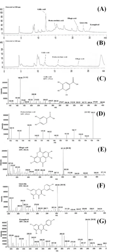
HPLC-ESI-MS is an effective tool for identification and characterisation of polyphenolic compounds (Hassan et al., 2011). MS ionises polyphenolic compounds to their charged forms, from which their mass to charge ratios (m/z) can be determined. HPLC analysis of the shoots ofB. racemosaidentified the presence of gallic acid, protocatechuic acid, ellagic acid, quercetin and kaempferol in BLE whereas only gallic acid, protocatechuic acid and ellagic acid were detected in BSE. The presence of these polyphenols in the plant extracts were further confirmed using ESI-MS (Figs. 1Aand1B).Figures 1C–1Gshows the mass to charge ratio (m/z) of the polyphenols detected using ESI-MS analyses. The polyphenolic compounds identified in BLE and BSE were in agreement with our previous study, analysed using ultra high performance liquid chromatography (Kong et al., 2014).
In mass spectrometry analyses, gallic acid and kaempferol were detected in ESI (−) modes, with [M−H]− peak observed atm/z 168.96 for gallic acid (Fig. 1C) andm/z 284.98 for kaempferol (Fig. 1G). Protocatechuic acid was monitored in ESI (+) mode, with [M+H]+peak observed atm/z155.21 (Fig. 1D). On the other hand, both ESI negative and positive modes were able to detect ellagic acid and quercetin in the samples, however ESI (−) mode was selected due to better sensitivity and lower background noise. The [M−H]− peak of ellagic acid and quercetin were observed at m/z 301.05 and 300.95, respectively (Figs. 1Eand1F). The polyphenols detected inB. racemosawere confirmed as they were in agreement with them/z of their standard and molecular weight.
Cytotoxicity effects
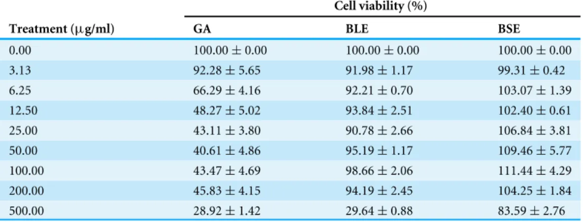
Toxicity study was conducted to ascertain that the extracts were safe for the proposed treatments on the HepG2 cells. High dosage of dietary compounds could be toxic or mutagenic in cell culture system, producing adverse metabolic reactions in mammals (Alía et al., 2006a). Hence, the direct effects of BLE, BSE and gallic acid on cell viability of HepG2 cells at different dosages were investigated (Table 1).
Table 1 The effects of gallic acid, BLE and BSE on cell viability of HepG2 cells. Cell viability (%)
Treatment (µg/ml) GA BLE BSE
0.00 100.00±0.00 100.00±0.00 100.00±0.00
3.13 92.28±5.65 91.98±1.17 99.31±0.42
6.25 66.29±4.16 92.21±0.70 103.07±1.39
12.50 48.27±5.02 93.84±2.51 102.40±0.61
25.00 43.11±3.80 90.78±2.66 106.84±3.81
50.00 40.61±4.86 95.19±1.17 109.46±5.77
100.00 43.47±4.69 98.66±2.06 111.44±4.29
200.00 45.83±4.15 94.19±2.45 104.25±1.84
500.00 28.92±1.42 29.64±0.88 83.59±2.76
Notes.
Cells (5×10³cells/well) were treated with gallic acid, BLE and BSE for 48 h before subjected to MTT assay. Results are
ex-pressed as means±SEM.
BLE, Leaf water extract ofB. racemosa; BSE, Stem water extract ofB. racemosa; GA, Gallic acid.
(Yeum et al., 2004). These preliminary analyses showed that theB. racemosaextracts have very low toxicity and are only cytotoxic at very high concentrations (>200µg/ml), which are not physiologically achievable.
In contrast, increasing concentrations of gallic acid was toxic to HepG2 cells whereby the concentration that inhibited 50% of cell proliferation (IC50) was calculated as 11.6µg/ml or 68µM. Pure gallic acid was cytotoxic at high concentrations and its reported pro-oxidant activities could have caused the cell death, possibly by activating the Fenton reactions, leading to generation of H2O2(Kobayashi et al., 2004). The pro-oxidant activity of gallic
acid was also reported in a study using Caco-2 human colon and F344 rat liver cells (Lee et al., 2005). However, pure gallic acid at concentration less than 4.3µg/ml or 25µM was non-cytotoxic, with cell viability more than 80%.
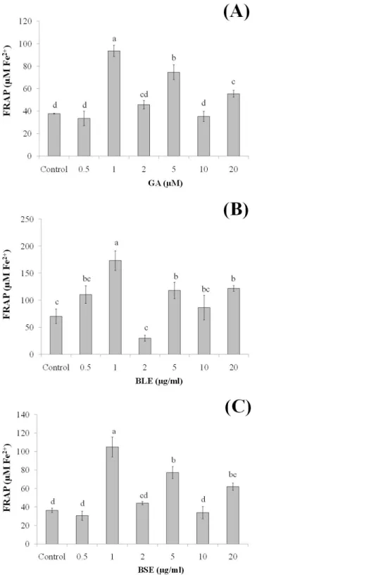
Since the plant extracts was found to be non-cytotoxic, determination of its cellular antioxidant effects was conducted using two levels of concentrations, i.e., low concentrations (0.5, 1, 2 µg/ml) and high concentrations (5, 10, 20 µg/ml). Low concentrations were used to ascertain if changes in antioxidant responses could be seen at these concentrations.
Cellular antioxidant status
to pure gallic acid alone could indicate potential synergistic effects of the polyphenols in conferring the antioxidant effects.
The plasma concentration of polyphenols is relatively low, about 0.001–6µM, due to their extensive metabolism (Boulton, Walle & Walle, 1998;Spencer et al., 2008). In a human bioavailability study, following the oral administration of gallic acid, its plasma level increased to 2µM (Shahrzad et al., 2001). This concentration is slightly higher than the concentrations of gallic acid (1µM) and the plant extracts (1µg/ml) in our study in which high antioxidant activity was observed. This indicates that physiological concentration of the plant extracts was adequate to induce antioxidant protection. Furthermore, higher concentration of plant extracts may introduce xenobiotic stress to the cells (D’Archivio et al., 2010). The improved antioxidant status of the treated-cells in this study indicated the ability of exogenous antioxidants fromB. racemosa to protect HepG2 cells against oxidative stress.
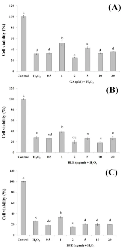
Cytoprotective effects
This assay was conducted to ascertain the ability of gallic acid and the plant extracts to protect the HepG2 cells against cell death following induction of oxidative damage. Treatment of HepG2 cells with gallic acid (1 and 5µM) and BLE and BSE (1µg/ml), significantly protected the cells against H2O2-induced oxidative damage (Figs. 3A–3C).
The increased antioxidant activities at this concentration, as measured by FRAP assay could have protected the cells against oxidative damage. However, increasing the concentrations of gallic acid, BLE and BSE did not further protect the cells from H2O2-induced oxidative
damage. HepG2 cells treated with antioxidant-rich extracts such as olive oil, cocoa and common sage also improved antioxidant status of the cells and protected the cells against oxidative damage, further supporting the results from this study (Goya, Mateos & Bravo, 2007;Lima et al., 2007;Martín et al., 2008).
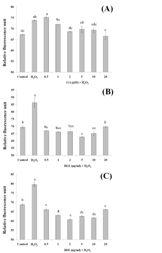
Reactive oxygen species production
Measurement of ROS would give an indication on levels of oxidative stress. H2O2was used
as the source of ROS whereby H2O2was converted to hydroxyl radicals and subsequently
caused oxidation of dichlorodihydrofluorescein (DCFH) to dichlorofluorescein (DCF) complex, a fluorescent compound. In addition to hydroxyl radicals, other ROS including peroxyl radicals and lipid hydroperoxides can also contribute to formation of this fluorescent complex.
Pre-treatment of HepG2 cells with BLE and BSE prior to H2O2-induced oxidative stress
gave lower fluorescent values compared to cells treated with H2O2alone (Figs. 4Band4C).
This study demonstrates the potential synergistic effect of polyphenols inB. racemosa extracts in reducing oxidative damage as opposed to using a single bioactive compound. The polyphenols in BLE and BSE comprise of a mixture of polar phenolic acids to semipolar flavonoids. Due to the nature of their varying polarity, these polyphenolic antioxidants are able to react at the hydrophilic and hydrophobic phases of the cells to eliminate ROS (Yeum et al., 2004). Additionally, mutual synergistic effects of different polyphenolic compounds can enhance the antioxidative effect (Dai & Mumper, 2010).
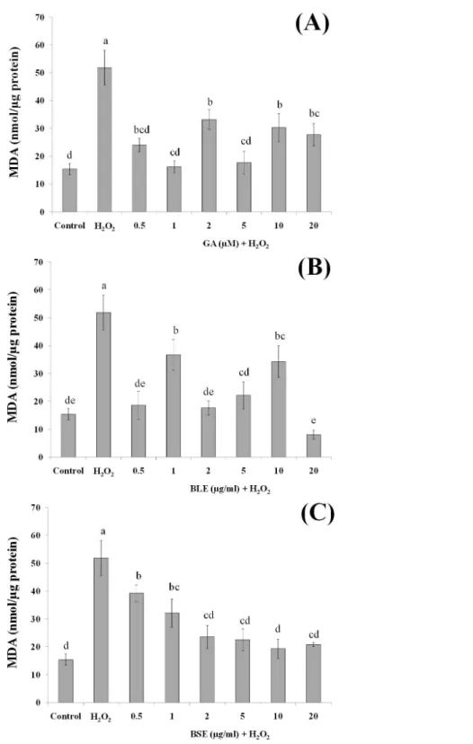
Analysis of lipid peroxidation
Since lipids in cell membrane are prone to oxidation, the effects of BLE and BSE in protecting against lipid peroxidation were also investigated. Lipids, especially polyunsaturated fatty acids (PUFA) at the membrane are susceptible to oxidative damage by ROS, forming lipid hydroperoxides and subsequently MDA (Martín et al., 2008), the latter being a widely used biomarker for oxidative stress (Martín et al., 2010).
Figures 5A–5Cshows the MDA levels of the different treatment groups. HepG2 cells treated with H2O2alone evoked a significant increase in the MDA levels, approximately
three folds higher than the negative control containing medium alone. HepG2 cells treated with gallic acid, BLE and BSE showed significant reduction (p<0.05) in MDA levels compared to positive control, indicating the ability of the samples to protect the cells against H2O2-induced lipid peroxidation. Results from this analysis also showed that
low concentration of gallic acid and the plant extracts were adequate to prevent lipid peroxidation and that increasing the concentration of the extracts did not necessarily lead to higher inhibition of lipid peroxidation.
Polyphenols including gallic acid, quercetin and kaempferol that were detected in the extracts are strong scavengers of hydroxyl radicals (Carocho & Ferreira, 2013). Gallic acid was also able to protect liver cells,in vitro, against oxidative damage (Senevirathne et al., 2012). Previous studies reported that pre-incubation of HepG2 cells with rutin and quercetin could reduce lipid peroxidation (Alía et al., 2006b). Indeed, studies utilising polyphenolic-rich extracts such as purple sweet potato and common sage reported reduced lipid peroxidation in HepG2 cells, indicating the important roles of antioxidant polyphenols in providing protection against oxidative damage (Hwang et al., 2011;Lima et al., 2007).
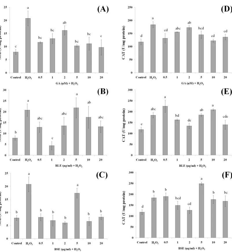
Activities of antioxidant enzymes
In addition to the direct effects of antioxidants inB. racemosain scavenging ROS, bioactive compounds in the plant could protect against oxidative damage by influencing activities of antioxidant enzymes. Antioxidant enzymes play a vital role in modulating the redox balance of cells especially during oxidative stress. Changes in antioxidant enzyme activities is a fairly sensitive indicator of oxidative stress and can also be used to predict responses of antioxidants in plants (Martín et al., 2008). In this study, the activities of two major antioxidant enzymes; SOD and CAT were measured. SOD catalyses the dismutation of superoxide anion radicals (O−•2 ) to produce O2 and H2O2(Pieme et al., 2010) whereas
CAT catalyses the transformation of H2O2to H2O (Alía et al., 2006b).
Figure 6 The effects of gallic acid, BLE and BSE on activities of SOD (A–C) and CAT (D–F) in HepG2 cells following H2O2-induced oxidative
damage.Cells (1.5×10⁵cells/well) were pre-treated with the plant extracts or gallic acid for 24 h prior to induction of oxidation with H
Pre-treatment of HepG2 cells with 1 µg/ml BLE significantly reduced SOD activity by 79%. Although the remaining concentrations showed a reduced trend in SOD activity, this was not significant. BSE on the other hand, caused significant decrease in SOD activity (60–71%) at all tested concentrations except 5µg/ml. Gallic acid reduced SOD activity significantly by 37–53% at all concentrations except 2µM.
Similar to SOD, positive control cells with H2O2-induced oxidation showed higher
activities of CAT than negative control cells without H2O2-induced oxidation.
Pre-treatment with gallic acid at 0.5µM and 5–20µM significantly suppressed the activities of CAT by 20–30% in cells subjected to H2O2-induced oxidation. BLE, at 2 and 20µg/ml significantly reduced 23–26% of CAT activity whereas a 31% decrease in CAT activity was observed in cells treated with 2µg/ml BSE.
Positive control or cells treated only with H2O2 showed elevation of SOD and CAT
activities, indicating a positive response of the cells in adapting towards increased production of ROS (Martín et al., 2010). The actions of SOD and CAT are closely related, whereby SOD reacts with O−•2 to produce H2O2that is subsequently reacted upon by CAT.
Pre-treatment of the cells with gallic acid, BLE and BSE prior to induction of oxidative stress, led to reduced activities of SOD and CAT. Although in some instances, these reductions were not statistically significant, a reduced trend was observed. Epicatechin, quercetin and phenolic-rich cranberry powders were reported to prevent the increment of antioxidant enzyme activities during oxidative stress (Alía et al., 2006b;Martín et al., 2010;Martín et al., 2015). The ability of theB. racemosaextracts to regulate the activities of SOD and CAT indicate the potential of these extracts to assist the cells defense mechanism in responding towards oxidative stress.
CONCLUSIONS
BLE and BSE at non-cytotoxic levels protected HepG2 cells against oxidative damage by acting as antioxidants, thus inhibiting ROS production and lipid peroxidation. In addition, the plant extracts also suppressed activities of the antioxidant enzymes SOD and CAT under conditions of oxidative stress. This current study indicates the potential use of the shoots ofB. racemosaand its bioactive ingredients for the development of functional foods. Its antioxidant properties could provide the added ability to increase the antioxidant defense mechanism and to provide protection against oxidative stress-related diseases.
ADDITIONAL INFORMATION AND DECLARATIONS
Funding
Grant Disclosures
The following grant information was disclosed by the authors: University of Malaya Research Grants: RG458/12HTM, FP015-2013B. High-Impact Research Grant: H-20001-00-E000009.
Competing Interests
The authors declare there are no competing interests.
Author Contributions
• Kin Weng Kong conceived and designed the experiments, performed the experiments, analyzed the data, wrote the paper, prepared figures and/or tables, reviewed drafts of the paper.
• Sarni Mat-Junit and Azlina Abdul Aziz conceived and designed the experiments, analyzed
the data, contributed reagents/materials/analysis tools, wrote the paper, reviewed drafts of the paper.
• Norhaniza Aminudin contributed reagents/materials/analysis tools, reviewed drafts of
the paper.
• Fouad Abdulrahman Hassan performed the experiments.
• Amin Ismail contributed reagents/materials/analysis tools.
Data Availability
The following information was supplied regarding data availability: The raw data is provided inSupplemental Information.
Supplemental Information
Supplemental information for this article can be found online athttp://dx.doi.org/10.7717/ peerj.1628#supplemental-information.
REFERENCES
Alía M, Mateos R, Ramos S, Lecumberri E, Bravo L, Goya L. 2006a.Influence of
quercetin and rutin on growth and antioxidant defense system of a human hepatoma cell line (HepG2).European Journal of Nutrition45:19–28
DOI 10.1007/s00394-005-0558-7.
Alía M, Ramos S, Mateos R, Granado-Serrano AB, Bravo L, Goya L. 2006b.Quercetin
protects human hepatoma HepG2 against oxidative stress induced by tert-butyl hydroperoxide.Toxicology and Applied Pharmacology212:110–118
DOI 10.1016/j.taap.2005.07.014.
Benzie IFF, Strain JJ. 1996.The ferric reducing ability of plasma (FRAP) as a measure
of ‘antioxidant power’: the FRAP assay.Analytical Biochemistry239:70–76
DOI 10.1006/abio.1996.0292.
Boulton DW, Walle UK, Walle T. 1998.Extensive binding of the bioflavonoid quercetin
Bradford MM. 1976.A rapid and sensitive method for the quantitation of microgram quantities of protein utilizing the principle of protein dye binding.Analytical Biochemistry72:248–254DOI 10.1016/0003-2697(76)90527-3.
Buege JA, Aust SD. 1978.Microsomal lipid peroxidation.Methods in Enzymology
52:302–310DOI 10.1016/S0076-6879(78)52032-6.
Carocho M, Ferreira ICFR. 2013.A review on antioxidants, prooxidants and related
controversy: natural and synthetic compounds, screening and analysis method-ologies and future perspectives.Food and Chemical Toxicology51:15–25
DOI 10.1016/j.fct.2012.09.021.
Choi JY, Kim H, Choi YJ, Ishihara A, Back K, Lee SG. 2010.Cytoprotective activities of
hydroxycinnamic acid amides of serotonin against oxidative stress-induced damage in HepG2 and HaCaT cells.Fitoterapia81:1134–1141
DOI 10.1016/j.fitote.2010.07.015.
Dai J, Mumper RJ. 2010.Plant phenolics: extraction, analysis and their antioxidant and
anticancer properties.Molecules15:7313–7352DOI 10.3390/molecules15107313.
D’Archivio M, Filesi C, Varì R, Scazzocchio B, Masella R. 2010.Bioavailability of the
polyphenols: status and controversies.International Journal of Molecular Sciences
11:1321–1342DOI 10.3390/ijms11041321.
Goya L, Mateos R, Bravo L. 2007.Effect of the olive oil phenol hydroxytyrosol
on human hepatoma HepG2 cells: protection against oxidative stress in-duced by tert-butylhydroperoxide.European Journal of Nutrition46:70–78
DOI 10.1007/s00394-006-0633-8.
Hassan FA, Ismail A, Abdulhamid A, Azlan A. 2011.Identification and quantification
of phenolic compounds in bambangan (Mangifera pajangKort.) peels and their free radical scavenging activity.Journal of Agricultural and Food Chemistry59:9102–9111
DOI 10.1021/jf201270n.
Hwang YP, Choi JH, Choi JM, Chung YC, Jeong HG. 2011.Protective mechanisms of
anthocyanins from purple sweet potato against tert-butyl hydroperoxide-induced hepatotoxicity.Food and Chemical Toxicology49:2081–2089
DOI 10.1016/j.fct.2011.05.021.
Kobayashi H, Oikawa S, Hirakawa K, Kawanishi S. 2004.Metal-mediated oxidative
damage to cellular and isolated DNA by gallic acid, a metabolite of antioxidant propyl gallate.Mutation Research—Genetic Toxicology and Environmental Mutage-nesis558:111–120DOI 10.1016/j.mrgentox.2003.11.002.
Kong KW, Mat-Junit S, Aminudin N, Ismail A, Abdul-Aziz A. 2012.Antioxidant
activities and polyphenolics from the shoots ofBarringtonia racemosa(L.) Spreng in a polar to apolar medium system.Food Chemistry134:324–332
DOI 10.1016/j.foodchem.2012.02.150.
Kong KW, Mat-Junit S, Ismail A, Aminudin N, Abdul-Aziz A. 2014.Polyphenols in
Barringtonia racemosaand their protection against oxidation of LDL, serum and haemoglobin.Food Chemistry 146:85–93DOI 10.1016/j.foodchem.2013.09.012.
Kong KW, Rajab NF, Nagendra Prasad K, Ismail A, Markom M, Tan CP. 2010.
activity towards hydrogen peroxide-induced cellular and DNA damage.Food Chemistry123:1142–1148DOI 10.1016/j.foodchem.2010.05.077.
Lee WK, Haeng JH, Hyong JL, Chang YL. 2005.Antiproliferative effects of dietary
phenolic substances and hydrogen peroxide.Journal of Agricultural and Food Chemistry53:1990–1995DOI 10.1021/jf0486040.
Lim TK. 2012. Barringtonia racemosa. In:Edible medicinal and non medicinal plants:
volume 3, fruits. Dordrecht: Springer, 114–121.
Lima CF, Valentao PCR, Andrade PB, Seabra RM, Fernandes-Ferreira M,
Pereira-Wilson C. 2007.Water and methanolic extracts of Salvia officinalis protect HepG2
cells from t-BHP induced oxidative damage.Chemico-Biological Interactions
167:107–115DOI 10.1016/j.cbi.2007.01.020.
Martín MA, Ramos S, Mateos R, Izquierdo-Pulido M, Bravo L, Goya L. 2010.Protection
of human HepG2 cells against oxidative stress by the flavonoid epicatechin. Phy-totherapy Research24:503–509 DOI 10.1002/ptr.2961.
Martín MA, Ramos S, Mateos R, Marais JPJ, Bravo-Clemente L, Khoo C, Goya L.
2015.Chemical characterization and chemo-protective activity of cranberry
phenolic powders in a model cell culture. Response of the antioxidant defenses and regulation of signaling pathways.Food Research International71:68–82
DOI 10.1016/j.foodres.2015.02.022.
Martín MA, Ramos S, Mateos R, Serrano ABG, Izquierdo-Pulido M, Bravo L, Goya
L. 2008.Protection of human HepG2 cells against oxidative stress by cocoa
phenolic extract.Journal of Agricultural and Food Chemistry56:7765–7772
DOI 10.1021/jf801744r.
Mosmann T. 1983.Rapid colorimetric assay for cellular growth and survival: application
to proliferation and cytotoxicity assays.Journal of Immunological Methods65:55–63
DOI 10.1016/0022-1759(83)90303-4.
Pieme CA, Penlap VN, Ngogang J, Costache M. 2010.In vitrocytotoxicity and
antioxi-dant activities of five medicinal plants ofMalvaceaefamily from Cameroon. Environ-mental Toxicology and Pharmacology29:223–228DOI 10.1016/j.etap.2010.01.003.
Puiggròs F, Llópiz N, Ardévol A, Bladé C, Arola L, Salvadó MJ. 2005.Grape seed
procyanidins prevent oxidative injury by modulating the expression of antioxi-dant enzyme systems.Journal of Agricultural and Food Chemistry53:6080–6086
DOI 10.1021/jf050343m.
Senevirathne M, Jeon YJ, Kim YT, Park PJ, Jung WK, Ahn CB, Je JY. 2012.Prevention
of oxidative stress in Chang liver cells by gallic acid-grafted-chitosans.Carbohydrate Polymers87:876–880 DOI 10.1016/j.carbpol.2011.08.080.
Shahrzad S, Aoyagi K, Winter A, Koyama A, Bitsch I. 2001.Pharmacokinetics of gallic
acid and its relative bioavailability from tea in healthy humans.The Journal of Nutrition131:1207–1210.
Spencer JPE, Abd El Mohsen MM, Minihane AM, Mathers JC. 2008.Biomarkers of the
Yeum KJ, Russell RM, Krinsky NI, Aldini G. 2004.Biomarkers of antioxidant capacity in the hydrophilic and lipophilic compartments of human plasma.Archives of Biochemistry and Biophysics430:97–103DOI 10.1016/j.abb.2004.03.006.
Yoshihara D, Fujiwara N, Suzuki K. 2010.Antioxidants: benefits and risks for long-term
health.Maturitas67:103–107DOI 10.1016/j.maturitas.2010.05.001.
You Y, Yoo S, Yoon HG, Park J, Lee YH, Kim S, Oh KT, Lee J, Cho HY, Jun W. 2010.