1. Physiotherapist; Coordinator of the Residency Program in Physiotherapy in Cardiology at IC/FUC.
2. PhD in Medicine (Cardiology); Cardiologist Physician at University Foundation of Cardiology.
3. Specialist in Cardiovascular Physiotherapy; Professor at Centro Universitário La Salle.
4. Graduation in Nursery and Obstetrics; Nurse at University Foundation of Cardiology.
5. Full Professor; Cardiovascular Surgeon. President-Director of the Cardiology Institute of Rio Grande do Sul/University Foundation of Cardiology
Christian Correa CORONEL1, Solange BORDIGNON2, André Dias BUENO3, Lidia Lucas LIMA4,Ivo NESRALLA5
RBCCV 44205-1172
Variáveis perioperatórias de função ventilatória e capacidade física em indivíduos submetidos a
transplante cardíaco
Perioperative variables of ventilatory function and
physical capacity in heart transplant patients
This study was carried out at Cardiology Institute of Rio Grande do Sul/University Foundation of Cardiology, Porto Alegre, RS, Brazil.
Correspondence address:
Christian Correa Coronel. Av. Princesa Isabel, 370, Santana. Porto Alegre/RS, Brasil. CEP: 90620-000
E-mail: [email protected]
Article received on March 5th, 2010
Article accepted on May 24th, 2010
Abstract
Introduction: Heart transplantation is currently the only
widely accepted surgical alternative to treat patients with severe heart failure (HF) drug therapy cannot maintain optimal quality of life appropriate.
Objective: To describe and to compare the values between
pre-and postoperative physical capacity and pulmonary patients who underwent heart transplantation.
Methods: A retrospective cohort composed of patients
undergoing heart transplantation between January 2001 to March 2005 in IC-FUC/RS.
Results: Were included in the 21 individuals. We observed
decreased levels of volume and lung capacity (FEV1 and FVC) in the first days after surgery compared to preoperatively (P <0.001) and recovery of these values in the 14th postoperative day (P <0.001). The values of muscle strength showed similar trends in reducing post-operative period compared to preoperative (P <0.001) and recovered on the 14th postoperative
day (P <0.001). A useful functional capacity, measured by testing 6-minute walk test (T6') showed improvement in the 14th postoperative day in relation to pre-operatively (P <0.001).
Conclusion: Changes in ventilatory function of subjects
undergoing cardiac transplantation are predictable, but these recover respiratory muscle strength and lung capacity within two weeks, and improve functional capacity useful in relation to pre-operative, the transplantation, when indicated, associated with good functional rehabilitation is very god treatment strategy.
Descriptors: Heart transplantation. Respiratory function
tests. Forced expiratory flow rates. Exercise therapy.
Resumo
Introdução: O transplante cardíaco é atualmente a única
alternativa cirúrgica amplamente aceita para tratar pacientes com insuficiência cardíaca (IC) grave que a terapia medicamentosa otimizada não consiga manter qualidade de vida adequada.
Objetivo: Descrever e comparar os valores entre pré e
bed, coughing and control techniques of pain [7,12], in addition to the practice of aerobic exercises. [17].
Despite all the risks that patients with transplanted organ has to infection and rejection, this technique has shown great efficacy in survival, presenting results of survival rate of 90% in the first year and 87% in the fifth year post-transplant with good quality of life [18].
This study aims to describe the values of physical and pulmonary abilities of patients who underwent heart transplant and who underwent conventional physiotherapy. Moreover, it aims to evaluate and compare the physical
capacity of patients preoperatively and on day 14th
postoperative heart transplant and to evaluate and compare the forced vital capacity, forced expiratory volume in one second, maximum inspiratory muscle strength and the
maximal expiratory muscle strength in pre-, 1st, 7th and 14th
postoperative day of heart transplant.
METHODS
The research is a retrospective cohort study, performed by review of the medical records and files, composed of patients undergoing orthotopic cardiac transplantation in
the period from 1st January 2001 to March 31th, 2005, at the
Institute of Cardiology - Cardiology University Foundation of Rio Grande do Sul. The project was approved by the Research Ethics Committee of IC/FUC. It was signed a term of review of files to assess data, and all participants signed a written informed consent prior to study entry.
The assessment was performed with data from patient identification, history, underlying disease, values of maximum ventilatory pressures (maximal inspiratory pressure and maximal expiratory pressure), lung volume and INTRODUCTION
Despite therapeutic advances in the previous two decades, heart failure (HF) is a disease with severe prognosis. Heart transplantation is now a widely accepted surgical alternative to treat patients with severe HF [1-3] when the medicinal therapy is unable to maintain adequate quality of life [4].
The indications of transplantation in our country are expressed in the Guidelines of the Brazilian Society of Cardiology for Heart Transplantation [5] and take into account both the patient’s clinical condition, socioeconomic and psychological characteristics, availability of organs donor and operational aspects, which restrict the availability of these treatment methods, because the postoperative care of cardiac transplantation are complex and require the patient’s understanding and collaboration [6]. The selection for transplantation is a dynamic process that must be reperformed every 3-6 months, and patients can be removed or included in the waiting line depending on the clinical condition [5].
Pulmonary complications in the postoperative period of cardiac surgery are a significant source of morbidity and mortality [4.7 to 16]. The lungs are particularly vulnerable and represent a potential site of infection in patients undergoing cardiac transplantation [4,15]. This vulnerability is mainly due to immunosuppressive therapy, surgical procedure and the quality of life of patients and can be avoided through preventive measures for infection control [4,15].
Postoperative strategies must also be used to reduce pulmonary complications after cardiac surgery, among them we can mention deep ventilation exercises, incentive spirometry [9], continuous positive airway pressure in the
Métodos: Estudo de coorte retrospectivo composto por
indivíduos submetidos ao transplante cardíaco, entre janeiro de 2001 a março de 2005, no IC-FUC/RS.
Resultados: Foram incluídos na análise 21 indivíduos.
Observou-se redução dos valores de volumes e capacidades pulmonares (VEF1 e CVF) no 1º dia de pós-operatório em relação ao pré-operatório (P<0,001) e recuperação destes valores no 14º dia de pós-operatório (P<0,001). Os valores de força muscular inspiratória demonstraram tendências semelhantes, reduzindo no 1º dia de pós-operatório em relação ao pré-operatório (P< 0,001) e recuperando no 14º pós-operatório (P< 0,001). A capacidade funcional útil, mensurada por meio do teste de caminhada de 6 minutos
(T6') mostrou melhora no 14º pós-operatório em relação ao pré-operatório (P< 0,001).
Conclusão: Alterações na função ventilatória de
indivíduos submetidos a transplante cardíaco são previsíveis, porém estes recuperam a força de músculos ventilatórios e capacidades pulmonares dentro de duas semanas, além de melhorar a capacidade funcional útil em relação ao pré-operatório, sendo o transplante, quando indicado, associado à reabilitação funcional boa estratégia terapêutica.
Descritores: Transplante de coração. Testes de função
capacity (forced expiratory volume in 1 second and forced
vital capacity), collected preoperatively, 1st, 7th and 14th
postoperative day, and six-minutes walk test, collected
preoperatively and on 14th postoperative day.
Sample
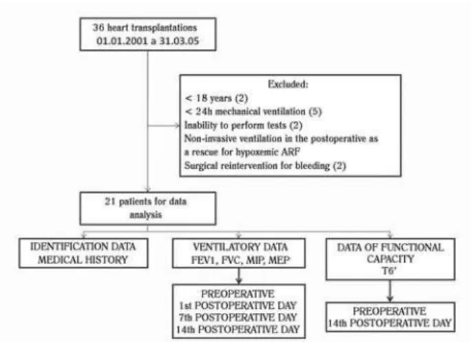
In the period aforementioned a total of 36 heart transplants were performed in the institution, of these, 21 subjects were included in the study, men and women of different ages and skin color, underwent heart transplantation, and remained on mechanical ventilation for a maximum of 24 hours, being weaned by conventional methods and aged above 18 years (Figure 1).
Were used as a criterion for exclusion from the study all patients who presented with neurological sequelae postoperatively (ischemic or hemorrhagic stroke) patients who did not have ability to perform spirometry, manovacuometer and six-minute test [19], those who needed noninvasive ventilation in the postoperative or those who were reintubated until the fourteenth postoperative day and patients who underwent surgical reintervention for bleeding problems postoperatively.
transplantation at the Institute of Cardiology - University Cardiology Foundation, from 1/1/2001 to 3/31/2005. Each patient was assessed during his visit at the outpatient heart transplant clinic (and quarterly) or during the hospital stay,
on the 1st postoperative day (until 24 hours after extubation),
7th and 14th postoperative day, as the evaluation form used by Physiotherapy Service of IC-FUC. These assessments, outpatient and inpatient, on the physical and lung capacity of these patients are routinely performed by the department of physiotherapy. All patients in this study underwent conventional techniques of chest physiotherapy (slow and abrupt manual chest compression, vibration, ventilatory patterns, active exercises of upper and lower limbs and deambulation) on the day of admission until discharge from hospital, with three to four sessions daily in the postoperative unit and two to three sessions per day in the inpatient unit.
Statistical analysis
Continuous variables were described by mean and standard deviation. Categorical variables were described using frequency tables with proportions.
It was also used analysis of variance for repeated measures in order to compare the changes in pulmonary
function test between pre-, 1st, 7th and 14th postoperative days.
The Tukey-Kramer test was used for multiple comparisons. In all comparisons it was considered an alpha level of 0.05.
RESULTS
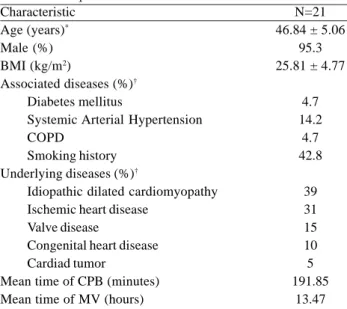
The sample consisted of 21 patients. Of these, 20 (95.3%) were men. The average age was 46.84 ± 15.06 years,
mean BMI was 25.81 ± 4.77 kg/m2. The characteristics of
the sample can be seen in Table 1. Of the 21 patients, one (4.7%) had diagnosis of diabetes mellitus, three (14.2%), arterial hypertension, one (4.7%), chronic pulmonary disease and nine (42.8%) had a history of smoking. No patients were current smokers (<60 days) in the sample. The mean CPB time was 191.85 minutes and mechanical ventilation, 13.47 hours. As for the underlying disease, eight (39%) patients had idiopathic dilated cardiomyopathy, seven (31%), ischemic heart disease, three (15%), valvular disease, two (10%), congenital heart disease and one (5%) cardiac tumor.
Changes in forced expiratory volume in one second
(FEV1) on pre-, 1st, 7th and 14th postoperative days are shown
in Figure 2. The preoperative FEV1 was 2.39 ± 0.84 l/sec, in
the 1st postoperative day was 1.32 ± 0.42 l/sec (decrease of
44.8% compared to preoperative period). The values on
the 7th postoperative day were 2.03 ± 0.77 l/sec (increase of
53.8% over the 1st postoperative day). On the 14th day after
surgery, was of 2.35 ± 0.8 l/sec (increase of 78% over the 1st
postoperative day).
Fig 1 - Flowchart of inclusion and evaluation of the sample’s patients NIV - Non-invasive ventilation; ARI - acute respiratory failure; FEV1 - forced expiratory volume in 1 second, FVC - Forced vital capacity, MIP - maximum inspiratory pressure, MEP - maximum expiratory pressure; T6’ - six-minute walk test
Data Collection
Changes in forced vital capacity (FVC) on pre-, 1st, 7th
and 14th postoperative days are shown in Figure 3. The
mean preoperative FVC was 2.79 ± 0.83 l/min. On the 1st
postoperative day, one can observe that the FVC in the group was 1.64 ± 0.51 l/min (decrease of 41.2% compared to
preoperatively). On the 7th postoperative day, the FVC was
2.43 ± 0.79 l/min (increase of 48.1% compared to the 1st
postoperative day). On the 14th postoperative day, the
average FVC was 2.79 ± 0.8 l/min (increase of 70.1%
compared to the 1st postoperative day).
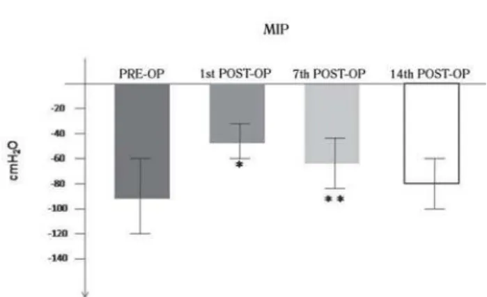
The mean value for maximum inspiratory pressure (MIP)
preoperatively was -88.85 ± 29.28 cmH2O, whereas on the
1st postoperative day the mean value of MIP was -45.95 ±
15.38 cmH2O (decrease of 48.3% compared to
preoperatively). The mean value of MIP on the 7th
postoperative day was -66.15 ± 30.79 cmH2O (increase of
43.9% compared to the 1st postoperative day). The average
value of MIP on the 14th postoperative day was -80.5 ±
26.42 cmH2O (increase of 75.1% compared to the 1st
postoperative day) - Figure 4.
The mean maximum expiratory pressure (MEP)
preoperatively was 122.7 ± 42.02 cmH2O. The mean value of
MEP in the first postoperative day was 65.95 ± 22.95 cmH2O
(decrease of 46.3% compared to preoperative period). The
mean MEP on the 7th postoperative day was 89.85 ± 28.12
cmH2O (increase of 36.2% on the 1st postoperative day).
The mean MEP on the 14th postoperative day was 107.4 ±
31.03 cmH2O, (increase of 62.8% on the 1st postoperative
day) - Figure 5.
A useful functional capacity measured using the 6-minute walk test (T6’) can be found in Figure 6. The mean distance covered on the T6’ on the preoperative period of
the group was 341.72 ± 53.99 m, and on the 14th
post-operative period was 380.15 ± 45.35 meters (increase of 11.2% compared to the preoperative period).
Table 1. Sample characteristics Characteristic
Age (years)* Male (%) BMI (kg/m2)
Associated diseases (%)†
Diabetes mellitus
Systemic Arterial Hypertension COPD
Smoking history
Underlying diseases (%)†
Idiopathic dilated cardiomyopathy Ischemic heart disease
Valve disease
Congenital heart disease Cardiad tumor
Mean time of CPB (minutes) Mean time of MV (hours)
N=21 46.84 ± 5.06
95.3 25.81 ± 4.77
4.7 14.2
4.7 42.8
39 31 15 10 5 191.85
13.47
Values described in * mean ± standard deviation and proportions †. BMI - body mass index, COPD - chronic obstructive pulmonary disease, CPB - cardiopulmonary bypass; MV - mechanical ventilation
Fig. 2 - Comparison of forced expiratory volume in 1 second (FEV1) over 14 postoperative days.
Data are expressed as mean and standard deviation. * P ≤ 0.001 when compared to other situations.
l/sec - liters per second; PRE-OP - preoperative; 1st POSTOP
-first day postoperative; 7th POST-OP - seventh postoperative day
– 14th POST-OP - fourteenth postoperative day
Fig. 3 - Comparison of Forced Vital Capacity (FVC) over 14 postoperative days
* P ≤ 0.001 when compared to other situations, ** P ≤ 0.001 when compared to preoperative and 14th postoperative day. Data are
expressed as mean and standard deviation.
l/min - liters per minute; PRE-OP - preoperative; 1st POSTOP
-first postoperative day; 7th POST-OP - seventh postoperative day,
DISCUSSION
Changes in pulmonary function occur in all patients after hours of surgical procedure [20,21]. Decreases of 44.8% in FEV1 and 41.2% in FVC were observed from the
preoperative period to the 1st postoperative day. According
Meyers et al. [20], pulmonary volumes (FEV1, FVC)
Fig. 4 - Comparison of Maximum Inspiratory Pressure (MIP) over 14 postoperative days
* P < 0.001 when compared to other situations, ** P < 0.001 when compared to preoperative and 14th postoperative day. Data
are expressed as mean and standard deviation.
cmH2O - cm of water; PRE-OP - preoperative; 1st POST-OP - first
postoperative day; 7th POST-OP - seventh postoperative day, 14th
POST-OP - fourteenth postoperative day
Fig. 5 - Comparison of maximal expiratory pressure (MEP) over 14 postoperative days
* P ≤ 0.001 when compared to other situations, ** P ≤ 0.001 when compared to preoperative and 14th postoperative day. Data
are expressed as mean and standard deviation.
cmH2O - cm of water; PRE-OP - preoperative; 1st POST-OP - first
postoperative day; 7th POST-OP - seventh postoperative day, 14th
POST-OP - fourteenth postoperative day
Fig. 6 - Comparison Test Walk 6 minutes (T6’) in the pre- and 14th
postoperative day
* P ≤ 0.001 when compared to preoperative.
PRE-OP - preoperative; 14th POST-OP - fourteenth postoperative
day
decreased postoperatively [22], with the maximum decrease
in the 1st postoperative day, returning close to preoperative
levels at 5 days postoperatively [20]. Morsch et al. [21] demonstrated in patients undergoing coronary artery bypass surgery, a statistically significant reduction in FEV1 and FVC when comparing the periods of pre- and sixth postoperative day, occurring the same with values of ventilatory muscle strength (MIP and MEP).
We had similar results when comparing these variables
in the preoperative period with the 1st postoperative day,
with decrease of 48.3% of MIP and 43.6% of MEP. Unlike the latter author, we observed an increase in pulmonary
volumes in FEV1 (53.8% on 7th and 78% at 14th postoperative
days), FVC (48.1% on 7th day and 70.1% on 14th postoperative
day), MIP (43.9% on 7th and 75.1% on 14th postoperative
days) and MEP (36.2% on 7th and 62.8% on 14th
postoperative days) compared to 1st postoperative day.
These increases in the values probably occur because of perceptual improvement in dyspnea due to improvement of cardiac pump function. Another factor of great importance is that these individuals were monitored routinely and the tests also are influenced by specific maneuvers to be performed and the will of the patient to collaborate in performing the movements and efforts really with the maximum [23].
Another important factor to be emphasized is that patients with heart failure have limited physical activity due to fatigue and dyspnoea [24] and that this is due to deconditioning of respiratory muscles and increased ventilatory work during hyperpneia. In this group it was observed that, after replacing the failing heart, the individuals recovered values of inspiratory muscle strength and increased in 11.2% the useful functional capacity on
14th postoperative day, viewed through the 6MWT. This is
probably due to the better cardiovascular function and better blood supply to skeletal muscles and better cardiac dynamics [25].
Postoperative patients of cardiac transplantation have improved quality of life. However, often present physical deconditioning, atrophy and muscle weakness and reduced aerobic capacity, resulting in part from inactivity and preoperative factors such as differences in body surface donor/recipient and denervation of the heart [26]. Regular physical activity has an important role post-transplant and should be started early for the restoration of physical capacity, enabling to patients to back to perform most of their daily activities and also recreation.
Possible mechanisms for this improvement are increased peripheral metabolism, primarily due to better extraction of oxygen and hemodynamic changes, including increased heart rate, cardiac output, endothelial function and reduction of neurohormonal activity. Moreover, the breathing efficiency is also improved during exercise [26]. These individuals showed that when implementing a basic program of mobilization and respiratory physiotherapy, they were able to recover the values of lung volumes and capacities and to improve useful functional capacity.
The improvement in physical function observed in patients undergoing cardiac transplantation is also attributed to the psychological status [27,28], because as a function of reduced functional capacity in general and “fear of dyspnea” these individuals enter into a cyclic process of inactivity and reclusion. Although in our study we have not performed any form of assessment of this psychological status, we observed significant improvement in ventilatory muscle function and functional capacity by means of the spirometric, manovacuometer measurements and walk test, which can also be attributed, besides the physical work performed, by improvement of motivation of these patients. Another factor of great importance is that the six-minute walk test, when performed in patients with preoperative heart transplant, has proven to be a safe method and its performance may be associated with mortality in these patients evaluated [29]. In performing the walking test preoperatively we did not quantify normality in relation to age, gender and body mass index of the patient, but we observed significant increase in distance traveled in the postoperative of 11.2% compared to preoperative, which
REFERENCES
1. Areosa CM, Almeida DR, Carvalho AC, Paola AA. Avaliação de fatores prognósticos da insuficiência cardíaca em pacientes encaminhados para avaliação de transplante cardíaco. Arq Bras Cardiol. 2007;88(6):667-73.
2. Dinkhuysen JJ. Transplante cardíaco ortotópico bicaval/ bipulmonar. . Rev Bras Cir Cardiovasc. 2003;18(3):268-72.
3. Haykowsky M, Eves N, Figgures L, McLean A, Koller M, Taylor D, et al. Effect of exercise training on VO2 peak and left ventricular systolic function in recent cardiac transplant recipients. Am J Cardiol. 2005;95(8):1002-4.
4. Atasever A, Bacakoglu F, Uysal FE, Nalbantgil S, Karyagdi T, Guzelant A, et al. Pulmonary complications in heart transplant recipients. Transplant Proc. 2006;38(5):1530-4.
5. Sociedade Brasileira de Cardiologia. I Diretrizes da Sociedade Brasileira de Cardiologia para Transplante Cardíaco. Arq Bras Cardiol. 1999 Sep;73(Suppl. 5):5-57.
6. Freitas HF, Nastari L, Mansur AJ, Bocchi EA, Moreira LF, Bacal F, et al. Dinâmica da avaliação de pacientes para transplante cardíaco ou cardiomioplastia. Arq Bras Cardiol. 1994;62(4):233-7.
predicts the functional improvement that can be attributed to improvement in cardiac pump.
CONCLUSION
7. Arcêncio L, Souza MD, Bortolin BS, Fernandes ACM, Rodrigues AJ, Évora PRB. Cuidados pré e pós-operatórios em cirurgia cardiotorácica: uma abordagem fisioterapêutica. Rev Bras Cir Cardiovasc. 2008;23(3):400-10.
8. Renault JA, Costa-val R, Rosseti MB. Fisioterapia respiratória na disfunção pulmonar pós-cirurgia cardíaca. Rev Bras Cir Cardiovasc. 2008;23(4):562-9.
9. Renault JA, Costa-val R, Rosseti MB, Houri Neto M. Comparação entre exercícios de respiração profunda e espirometria de incentivo no pós-operatório de cirurgia de revascularização do miocárdio. Rev Bras Cir Cardiovasc. 2009;24(2):165-72.
10. Braunwald E. Biomarkers in heart failure. N Engl J Med. 2008;358(20):2148-59.
11. Leguisamo CP, Kalil RAK, Furlani AP. A efetividade de uma proposta fisioterapêutica pré-operatória para cirurgia de revascularização do miocárdio. Rev Bras Cir Cardiovasc. 2005;20(2):134-41.
12. Khan MA, Hussain SF. Pre-operative pulmonary evaluation. J Ayub Med Coll Abbottabad. 2005;17(4):82-6.
13. Filsoufi F, Rahmanian PB, Castillo JG, Chikwe J, Adams DH. Predictors and early and late outcomes of respiratory failure in contemporany cardiac surgery. Chest. 2008;133(3):713-21.
14. Groeneveld AB, Jansen EK, Verheij J. Mechanisms of pulmonary dysfunction after on-pump and off-pump cardiac surgery: a prospective cohort study. J Cardiothorac Surg. 2007;2(11):1-7.
15. Mattner F, Fischer S, Weissbrodt H, Chaberny IF, Sohr D, Gottlieb J, et al. Post-operative nosocomial infections after lung and heart transplantation. J Heart Lung Transplant. 2007;26(3):241-9.
16. Charlson ME, Isom OW. Care after coronary-artery bypass surgery. N Engl J Med. 2003;348(15):1456-63.
17. Kavanagh T, Mertens DJ, Shephard RJ, Beyene J, Kennedy J, Campbell R, et al. Long-term cardiorespiratory results of exercise training following cardiac transplantation. Am J Cardiol. 2003;91(2):190-4.
18. Grady KL, Naftel DC, Young JB, Pelegrin D, Czerr J, Higgins R, et al. Patterns and predictors of physical functional
disability at 5 to 10 years after heart trransplantation. J Heart Lung Transplant. 2007;26(11):1182-91.
19. Sociedade Brasileira de Pneumologia e Tisiologia. Diretrizes para testes de função pulmonar. J Pneumol. 2002;28(supl 3):S1-S82.
20. Meyers JR, Lembeck L, O’Kane H, Baue AE. Changes in functional residual capacity of the lung after operation. Arch Surg. 1975;110(5):576-83.
21. Morsch KT, Leguisamo CP, Camargo MD, Coronel CC, Mattos W, Ortiz LDN, et al. Perfil ventilatório dos pacientes submetidos à cirurgia de revascularização do miocárdio. Rev Bras Cir Cardiovasc. 2009;24(2):180-7.
22. Berrizbeitia LD, Tessler S, Jacobowitz IJ, Kaplan P, Budzilowicz L, Cunningham JN. Effect of sternotomy and coronary bypass surgery on postoperative pulmonary mechanics: comparison of internal mammary and saphenous vein bypass grafts. Chest. 1989;96(4):873-6.
23. Syabbalo N. Assessment of respiratory muscle function and strength. Postgrad Med. J. 1998;74(870):208-15.
24. Dall’Ago P, Chiappa GR, Guths H, Stein R, Ribeiro JP. Inspiratory muscle training in patients with heart failure and inspiratory muscle weakness: a randomized trial. J Am Coll Cardiol. 2006;47(4):757-63.
25. Salles AF, Oliveira Fº JA. Adaptações do exercício pós-transplante cardíaco. Arq Bras Cardiol. 2000;75(1):79-84.
26. Guimarães GV, D’Ávila VM, Chizzola PR, Bacal F, Stolf N, Bocchi EA. Reabilitação física no transplante de coração. Rev Bras Med Esporte. 2004;10(5):408-11.
27. Saeed I, Rogers C, Murday A, Steering Group of the UK Cardiothoracic Transplant Audit. Health-related quality of life after cardiac transplantation: results of a UK National Survey with Norm-based Comparisons. J Heart Lung Transplant. 2008;27(6):675-81.
28. Helito RAB, Branco JNR, D’Innocenzo M, Machado RC, Buffolo E. Qualidade de vida nos candidatos a transplante do coração. Rev Bras Cir Cardiovasc. 2009;24(1):50-7.