CLINICAL DISCRIMINATORS BETWEEN
ACUTE BRAIN HEMORRHAGE AND INFARCTION
A practical score for early patient identification
Ayrton R. Massaro
1, Ralph L. Sacco
2, Milberto Scaff
1, J.P. Mohr
3ABSTRACT - New treatments for acute stroke require a rapid triage system, which minimizes treatment delays and maximizes selection of eligible patients. Our aim was to create a score for assessing the probability of brain hemorrhage among patients with acute stroke based upon clinical information. Of 1805 patients in the Stroke Data Bank, 1273 had infarction (INF) and 237 had parenchymatous hemorrhage (HEM) verified by CT. INF and HEM discriminators were determined by logistic regression and used to create a score. ROC curve was used to choose the cut-point for predicting HEM (score £ 2), with sensitivity of 76% and specificity of 83%. External validation was done using the NOMASS cohort. Although the use of a practical score by emergency personnel cannot replace the gold-standard brain image differentiation of HEM from INF for thrombolytic therapy, this score can help to select patients for stroke trials and pre-hospital treatments, alert CT scan technicians, and warn stroke teams of incoming patients to reduce treatment delays.
KEY WORDS: cerebrovascular disorders, cerebral hemorrhage, cerebral ischemia, stroke, diagnosis.
Características clínicas diferenciais entre hemorragia e infarto cerebral: uma escala prática para identificação precoce do paciente
RESUMO - Novas perspectivas no tratamento do acidente vascular cerebral (AVC) requerem um método de triagem rápido para seleção dos pacientes. Nosso objetivo foi criar uma escala com informações clínicas simples para diferenciar hematoma intra-parenquimatoso (HEM) entre os pacientes com AVC. Estudamos 1.273 pacientes com AVC isquêmico (INF) e 237 com HEM do Stroke Data Bank. Variáveis independentes para o diagnóstico de INF e HEM foram determinadas pela análise de regressão logística e utilizadas para criar uma escala. Através da curva ROC foi escolhido o nível de corte para discriminar HEM (£ 2 ), com sensibilidade de 76%, especificidade de 83%. Foi realizada validação externa utilizando os pacientes do estudo NOMASS. Embora o uso de uma escala de fácil aplicação pelas equipes de emergência não possa substituir os métodos de imagem na diferenciação entre INF e HEM para a indicação de trombolítico, a escala proposta pode ser útil para selecionar pacientes para estudos clínicos e tratamento pré-hospitalar, alertar técnicos de tomografia e as equipes médicas sobre a chegada de pacientes, contribuindo para reduzir atrasos cruciais no tratamento.
PALAVRAS-CHAVE: acidente vascular cerebral, hemorragia, isquemia.
1Department of Neurology, Faculdade de Medicina da Universidade de São Paulo, São Paulo, Brazil; 2School of Public Health
(Epidemiol-ogy) and the Sergievsky Center, Columbia University, New York, USA; 3Department of Neurology, Neurological Institute of
Columbia-Presbyterian Medical Center, New York, USA. This work was partially supported by grants from the National Institute of Neurological Disorders and Stroke R01 NS 27517 and R01 NS 29993.
Received 8 August 2001, received in final form 17 October 2001. Accepted 25 October 2001.
Ayrton Roberto Massaro, MD - Department of Neurology, Federal University of São Paulo - Rua Botucatu 740 - 04023-900 São Paulo SP - Brazil - E-mail: [email protected]
New treatments for acute stroke require a rapid triage system, which minimizes treatment delays and maximizes selection of eligible patients1. Despite
emergency physicians ability to accurately identify patients with acute stroke2,3, the differentiation
be-tween acute brain hemorrhage (HEM) and infarct (INF) is more difficult and can only be confirmed by brain imaging. Time taken to obtain a CTmay delay stroke treatment4,5 ,therefore a pre-hospital
screen-ing stroke score which can help to differentiate INF and HEM patients may become a rational approach for acute stroke management.
Although there are some stroke score systems available2,6-8 ,the Guy’s Hospital Stroke (GHS) score2
and the Siriraj Hospital Stroke (SHS) score7 are the
only two that have been largely validated9-11, and
Our aim was to create a score for assessing the probability of HEM among patients with acute stroke based upon simple, clinical information available prior to hospitalization which could be used by para-medics and emergency personnel.
METHOD
The Stroke Data Bank (SDB) was a prospective obser-vational study, which collected acute care, clinical, and laboratory data on 1805 patients with stroke. The SDB entry criteria included patients with a sudden, nonconvul-sive, focal neurological deficit persisting beyond 24 hours. Half the patients were admitted within 12 hours after stroke onset. The SDB excluded those patients in whom the stroke was coexistent with other severe illness such as tumor, hematological disease or head injury.
The collaborative study involved the Biometry and Field Studies Branch of the National Institute of Neurological Disorders and Stroke as the statistical coordinating cen-ter, and four academic hospital centers: Boston Univer-sity, Boston; Michael Reese Hospital and Medical Center, Chicago; University of Maryland, Baltimore; and the Neu-rological Institute of Columbia-Presbyterian Medical Cen-ter, New York. A full description of the SDB can be found elsewhere12.
Strokes were classified by causal mechanism into the following categories: (1) brain infarction (INF) which in-cluded infarction due to large artery atherosclerosis, lacu-ne, cardioembolism, infarction with tandem arterial pa-thology and infarction of undetermined cause13; (2) par-enchymatous hemorrhage (HEM); (3) subarachnoid hem-orrhage; and (4) stroke from other causes. Each patient with acute stroke was examined by one of the SDB inves-tigators and CT scan was used to confirm the diagnosis. The time from stroke onset to first CT scan ranged from 10 to 27 hours after onset12. For this analysis, all patients enrolled in the SDB with HEM and INF were eligible, while subarachnoid hemorrhage, and those patients classified as stroke from other causes, such as inflammatory arteriopathies and sickle cells disease, were excluded. Sub-arachnoid hemorrhage has a distinct clinical presentation. Whenever a patient arrives at the emergency room with headache, vomiting, decreased consciousness and nuchal rigidity without a focal neurological deficit suggesting subarchnoid hemorrhage, CT will be necessary. Even if CT is unremarkable, further investigation is usually required to rule out this condition.
The variables that were analyzed and compared in the HEM and INF groups included: (1) demographics: age, race, gender; (2) historical stroke risk factors: medical history of hypertension defined by prior hypertension in the phy-sician notes or if the old chart revealed a systolic blood pressure ³ 160mmHg and diastolic ³ 95mmHg or if the patient was taking anti-hypertensive treatment prior to the admission; diabetes classified as treated with diet alone, oral agents, or insulin; prior symptomatic stroke or transient ischemic attack (TIA); cardiovascular disease
con-sisted of the following: myocardial infarction, valvular heart disease, atrial fibrillation, other arrhythmia, angina, con-gestive failure, and claudication; alcohol intake within 24 hours of onset; (3) clinical features at onset: neurological symptoms or signs which presented upon awakening; se-vere headache with no features suggestive of migraine; seizures; vomiting; focal neurological deficit; coma within minutes of the onset of stroke and attributed to the ef-fect of the stroke itself rather than metabolic causes; (4)
initial neurological examination: level of consciousness (alert, lethargic, stupor, coma); motor and sensory defi-cits; extraocular movements and visual field abnormali-ties; ataxia; hemineglect; aphasia and presence of cervical bruit; (5) clinical parameters: first systolic and diastolic blood pressure measurements after admission to the hos-pital and initial Glasgow Coma Scale score. CT scan was performed to confirm the clinical diagnosis in all patients, but was not analyzed in this study, since the aim was to determine clinical predictors of the diagnosis.
and compared the classification of HEM and INF to the SDB score.
This study was approved by the National Institute of Neurological Disorders and Stroke and the local ethics committee.
RESULTS
Of 1805 patients with acute stroke enrolled in the SDB, 1273 were classified as INF and 237 as HEM. We excluded 243 patients with diagnosis of sub-arachnoid hemorrhage and only 52 who were
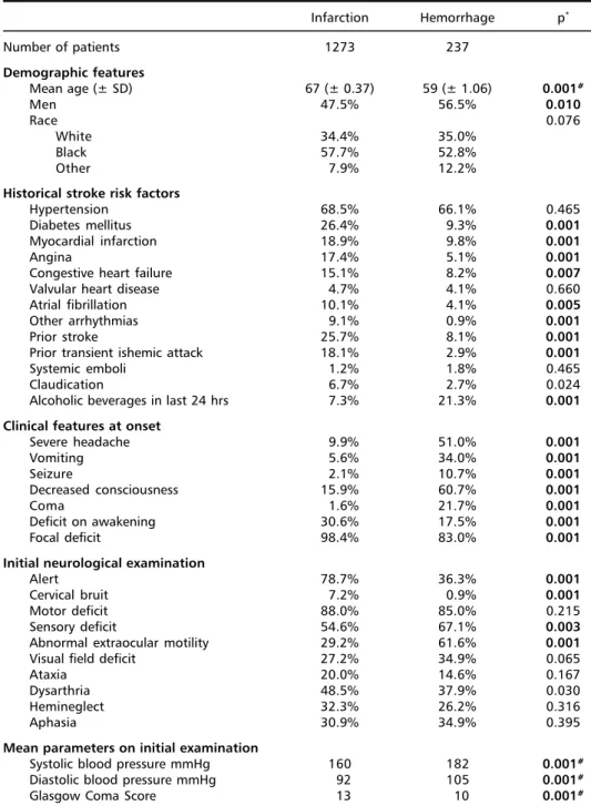
clas-sified as stroke from other causes. Patients with INF were older and less likely to be men than those with HEM (Table 1). There were no detectable ethnic dif-ferences between the 2 groups. Among stroke risk factors, hypertension was the most frequent in both groups. History of cardiovascular disease, diabetes, and TIA or stroke were more frequent among the INF group, whereas recent consumption of alcoholic beverages was more frequent in HEM patients. Among the clinical features presenting at onset, headache, vomiting, seizure, and decreased consciousness or
Table 1. Comparison of clinical features between acute brain hemorrhage and infarction in the Stroke Data Bank.
Infarction Hemorrhage p*
Number of patients 1273 237
Demographic features
Mean age (± SD) 67 (± 0.37) 59 (± 1.06) 0.001#
Men 47.5% 56.5% 0.010
Race 0.076
White 34.4% 35.0%
Black 57.7% 52.8%
Other 7.9% 12.2%
Historical stroke risk factors
Hypertension 68.5% 66.1% 0.465
Diabetes mellitus 26.4% 9.3% 0.001
Myocardial infarction 18.9% 9.8% 0.001
Angina 17.4% 5.1% 0.001
Congestive heart failure 15.1% 8.2% 0.007
Valvular heart disease 4.7% 4.1% 0.660
Atrial fibrillation 10.1% 4.1% 0.005
Other arrhythmias 9.1% 0.9% 0.001
Prior stroke 25.7% 8.1% 0.001
Prior transient ishemic attack 18.1% 2.9% 0.001
Systemic emboli 1.2% 1.8% 0.465
Claudication 6.7% 2.7% 0.024
Alcoholic beverages in last 24 hrs 7.3% 21.3% 0.001 Clinical features at onset
Severe headache 9.9% 51.0% 0.001
Vomiting 5.6% 34.0% 0.001
Seizure 2.1% 10.7% 0.001
Decreased consciousness 15.9% 60.7% 0.001
Coma 1.6% 21.7% 0.001
Deficit on awakening 30.6% 17.5% 0.001
Focal deficit 98.4% 83.0% 0.001
Initial neurological examination
Alert 78.7% 36.3% 0.001
Cervical bruit 7.2% 0.9% 0.001
Motor deficit 88.0% 85.0% 0.215
Sensory deficit 54.6% 67.1% 0.003
Abnormal extraocular motility 29.2% 61.6% 0.001
Visual field deficit 27.2% 34.9% 0.065
Ataxia 20.0% 14.6% 0.167
Dysarthria 48.5% 37.9% 0.030
Hemineglect 32.3% 26.2% 0.316
Aphasia 30.9% 34.9% 0.395
Mean parameters on initial examination
Systolic blood pressure mmHg 160 182 0.001#
Diastolic blood pressure mmHg 92 105 0.001#
Glasgow Coma Score 13 10 0.001#
coma were significantly more frequent in HEM pa-tients. For those reporting deficits upon awakening or focal deficit at onset, the subtype was predomi-nantly INF. Patients with HEM had more sensory and extraocular motility abnormalities on initial neuro-logical examination, and greater mean blood pres-sures and lower level of consciousness meapres-sures by mean Glasgow Coma Scale score on the admission examination.
Six independent clinical variables distinguished INF from HEM by logistic regression: age greater than 55 years (Odds ratio - OR =1.7; 95% Confidence In-terval-CI=1.1-2.4), history of angina (OR=3.4; CI=1.6-7.0), prior stroke or TIA (OR=3.1; CI=1.9-4.9), diabetes (OR=2.5; CI=1.4-4.3), deficit upon awakening (OR=1.8; CI=1.1-2.3), and presentation with focal deficit (OR=4.7; CI=2.4-8.9). Whereas, HEM was more likely among men (OR=1.6; CI=1.1-2.3) and those presenting with severe headache (OR=5.0; CI=3.3-7.5), vomiting (OR=3.7; CI=2.2-5.8), coma or decreased consciousness (OR=16.9; CI=8.0-37.0), or an initial blood pressure greater than 200/120mmHg (OR=3.8; CI=2.5-5.6). HEM was
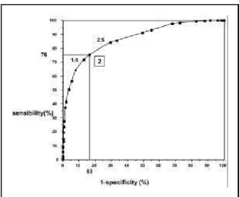
also favored if more than one of these clinical discri-minators were unmeasurable (OR=2.9; CI=1.8-4.6). The latter blood pressure category was chosen to maximize the discrimination between INF and HEM. Based on our final model, a score for the clinical diagnosis of INF or HEM was created ranging from -10 to +-10 (Fig 1). We applied our model to the SDB cohort to create a ROC curve which shows the rela-tionship between the sensitivity and 1-specificity for the diagnosis of HEM at various scores and can be used to select the best score cutpoint (Fig. 2). The more the curve is closer to the top left corner the better the sensitivity and the specificity. In the SDB cohort, the cutpoint which maximized sensitivity and specificity appeared to be 2.0. HEM was the most likely diagnosis with scores of 2.0 or lower (to -10), and INF was more likely with values of greater than 2.0 (to +10). Scores suggestive of HEM (-10 to 2.0) led to the correct classification of 76% of the HEM cases and misclassified only 17% of the infarct as HEM (sensitivity=76%; specificity=83%; positive predictive value for HEM=46%; negative predictive value for HEM=95%).
No Yes Unknown Score
Patient is more than 55 years old 0 +1.0 U
Patient is a man 0 -1.0
Has patient ever had a history of:
TIA or Stroke 0 +2.0 U
Angina 0 +2.0 U
Diabetes 0 +1.5 U
At the time of onset was there:
Focal deficit 0 +2.5 U
Deficit presented on awakening 0 +1.0 U
Patient is not alert 0 -3.0 U
Severe headache 0 -2.0 U
Vomiting 0 -1.5 U
Initial blood pressure higher than 200/120 mHg 0 -1.5
More than one of previous questions answered unknown 0 -1.0
TOTAL SCORE
rating -10 to +2
> 2 to + 10
BRAIN HEMORRHAGE BRAIN INFARCT
In the external validation of this score in the sepa-rate NOMASS cohort, the same score cutpoint of 2.0 led to the correct classification of 68% of the HEM cases and misclassified only 24% of the infarcts as HEM. Using the only other stroke score which can be used in the emergency room setting, the Siriraj Hospital Stroke score, led to a sensitivity of 24% and specificity of 97.4% for the classification of HEM cases from the NOMASS cohort. Among the 58 HEM patients in the NOMASS cohort the SHS score led to the correct diagnosis in only 14 patients.
DISCUSSION
Although the management of acute stroke re-quires institution of therapy within hours of stroke onset16,17, recent surveys have demonstrated that
there is often a delay among stroke patients in their presentation to medical personnel5,18-21. Part of the
explanation may be based upon inadequate public and physician education of the signs and symptoms of acute stroke as was apparent for myocardial infarc-tion in the past22. Treatment delays may be improved
by better recognition of stroke symptoms, rapid pa-tient transportation systems, and immediate emer-gency medical evaluation of the suspected stroke patient20,23-25. Emergency medical services (EMS)
per-sonnel are often the first medical contact for pa-tients presenting with stroke26, since most stroke
events occur at home27. They represent a crucial link
in the early triage and management of acute stroke patients. An early notification of stroke subtype by EMS to the receiving hospital may minimize the time
delay to treatment by alerting and mobilizing the appropriate stroke-treatment personnel according to the possible stroke mechanism (HEM or INF).
The SDB practical classification may be a new tool to be used in the management of acute stroke pa-tients. It was designed to use simple variables, which could be easily answered by EMS personnel. It has a reasonable sensitivity and specificity for differenti-ating HEM from INF in internal and external valida-tion cohorts and it also permits the choice of differ-ent cutpoints to increase the score accuracy. In addi-tion, clinical stroke subtype diagnosis with our score method appeared more accurate than the diagnosis made by the patient’s physician at bedside prior to revealing the results of the CT in other studies28.
Using our scoring system the clinician’s may improve their clinical skills to differentiate HEM from INF29.
It is well known that some risk factors are spe-cific to either HEM or INF30-32. We have also detected
demographic and stroke risk factor differences be-tween patients presenting with HEM or INF. Hemor-rhage has been observed in higher frequency in younger patients and in men33-35. Hypertension is a
potent risk factor for both brain infarction and hem-orrhage, however, as found by us, often does not discriminate between the two groups. In our study, as found by others33,36, the level of the admitting
blood pressure was more elevated in brain hemor-rhage patients. Stroke patients with prior hyperten-sion usually have the highest blood pressure levels compared to normotensives37. Diabetes and coronary
artery disease were strongly associated with INF and they have been established as risk factors for brain infarction in numerous studies31,32,38. History of TIA
and stroke have more often preceded a brain infarc-tion than hemorrhage and contributed to the recur-rence of infarction35,36.
Clinical features at onset may also help to discri-minate between brain infarction and hemorrhage36.
The high frequency of severe headache at onset in brain hemorrhage has also been reported by oth-ers33,35,36,39. The occurrence of headache varies by
stro-ke subtype and may be an important indicator of stroke mechanism39-41. Vomiting, decreased
con-sciousness and coma were also found to be more predictive of hemorrhage as have been observed in other clinical series33,35,36. In our study, the occurrence
of a deficit upon awakening was more predictive of INF than HEM. Previous reports have identified a cir-cadian rhythm to the onset of brain infarction42.
Other clinical scores have been developed to help discriminate brain infarction from hemorrhage. The
Kyushu University score included 16 clinical items of various different weights to arrive at the stroke sub-type6. Other than the items mentioned in this
analy-sis, this score included conjugated eye deviation, anisocoria, light reflex, corneal reflex, speech disor-der, neck stiffness, sensory deficit, and cerebrospi-nal fluid. Six factors were found to predict hemorrha-ge among a sample of cases in Rochester, NY: coma, vomiting, severe headache, marked hypertension (systolic blood pressure greater than 220 mmHg), new hyperglycemia (serum glucose level greater than 170 g/dL), and warfarin therapy43. Unfortunately, this
model included results of testing that could cause treatment delays. Besson et al.8 developed a scoring
system to identify nonhemorrhagic infarct patients based on 8 variables that included history of hyper-lipidemia, alcohol consumption, plantar response and atrial fibrillation on admission using electrocar-diogram. Although it gives a high positive predicti-ve value, this scoring system cannot be applicable as a pre-hospital screening score to all stroke pa-tients. The Guy’s Hospital Stroke score included 8 variables that were obtained not only by clinical his-tory but also using clinical examination and chest X-rays2. In addition, some variables can only be
calcu-lated 24 hours after the stroke, such as level of con-sciousness and diastolic blood pressure, so it can-not be used in acute stroke treatment trials. This score achieved a sensitivity for the diagnosis of hem-orrhage of 81% and 88% in patients from Oxford and London, respectively9.The Siriraj Hospital Stroke
score is simpler and can be calculated immediately after stroke at bedside7. This score uses five variables:
level of consciousness, vomiting, headache, diastolic blood pressure, and atheroma markers. The valida-tion study of the Siriraj Hospital Stroke score in Thai-land revealed higher sensitivities for supratentorial hemorrhage (89.3%) and infarction (93.2%)7. The
difference in prevalence of hemorrhagic stroke may explain the lower diagnostic accuracy of the Siriraj Hospital Stroke score in the NOMASS cohort. More recent evaluations of the Guy’s Hospital Stroke and Siriraj Hospital Stroke scores conclude that such scor-ing systems are not useful in routine clinical prac-tice44,45, although they may be used in large scale
epidemiological studies9,10.
The limitations of the scoring system should be understood. It is not possible to achieve total sepa-ration of all patients with HEM from those with INF using clinical features, particularly in those cases with small deep HEM that mimic the clinical features of INF. Due to this fact, some scoring systems2,7 have
created an uncertain group that will effectively in-crease the sensitivity and specificity of the final cal-culation45. The use of an uncertain group, however,
will not achieve the goal of the acute stroke score which is to classify all incoming patients with acute stroke. If a large percentage of the patients fall into the uncertain category, then the utility of the score is weakened.
The variables needed to calculate a stroke score should be available when management decisions are being made and take into account the possibility of missing information. There are diagnostic problems caused by the lack of complete information in some patients when the GHS and SHS scores were used44,45.
For this reason, we designed a model to be perfor-med by non-neurologists with minimal reliance on any laboratory testing and most of the information could be obtained by direct observation, interview-ing the patient or the family members, and mea-surement of blood pressure. Our score could be cal-culated easily and a presumptive diagnosis could be made prior to the CT scan. Most of the inaccuracies to differentiate HEM from INF arise in the diagnosis of HEM, because many of the variables used in these scoring systems (level of consciousness at onset, early headache, vomiting) discriminate in favor of hem-orrhage. If a patient or relative cannot give a clear description of the symptom at the stroke onset, the score will tend to overestimate the likelihood of INF. Taking into account all these considerations, our score has included a new variable to account for the inability to ascertain specific information (one or more missing variable).
In conclusion, the use of our stroke score system as a screening score by emergency personnel can help select patients for stroke treatments, alert CT scan technicians, and warn teams of incoming pa-tients to reduce treatment delays. New variables may be identified in further studies, which certainly will optimize our scoring system to differentiate acute brain hemorrhage from infarction. At present, we are not advocating the use of this score to replace the gold-standard brain image differentiation of HEM from INF for thrombolytic therapy. However, as we develop other safe and effective acute stroke treat-ments, such as the recently recommended use of early aspirin for suspected acute ischemic stroke when CT scan in unavailable46, the SDB score may
become a useful tool in acute stroke triage settings.
REFERENCES
1. Adams HP Jr, Brott TG, Furlan AJ, et al. Guidelines for thrombolytic therapy for acute stroke: a supplement to the guidelines for the man-agement of patients with acute ischemic stroke. A statement for healthcare professionals from a special writing group of the Stroke Council, American Heart Association. Stroke 1996;27:1711-1718. 2. Allen CMC. Clinical diagnosis of the acute stroke syndrome. Q J Med
1983;52:515-523.
3. Kothari RU, Brott T, Broderick JP, et al. Emergency physicians: accu-racy in the diagnosis of stroke. Stroke 1995;26:2238-2241.
4. Barsan WG, Brott TG, Olinger CP, et al. Identification and entry of the patient with acute cerebral infarction. Ann Emerg Med 1988;17:1192-1195.
5. Timerding BL, Barsan WG, Hedges JR, et al. Stroke patient evaluation in the emergency department before pharmacologic therapy. Am J Emerg Med 1989;7:11-15.
6. Hatano S. Variability of the diagnosis of stroke by clinical judgement and by a scoring method. Bull WHO 1976;54:533-540.
7. Poungvarin N, Viriyavejakul A, Komontri C. Siriraj stroke score and validation study to distinguish supratentorial intracerebral haemorrha-ge from infarction. BMJ 1991;302:1565-1567.
8. Besson G, Robert C, Hommel M, et al. Is it clinically possible to distin-guish nonhemorrhagic infarct from hemorrhagic stroke? Stroke 1995;26:1205-1209.
9. Sandercock PAG, Allen CMC, Corston RN, et al. Clinical diagnosis of intracranial haemorrhage using Guy’s Hospital score. BMJ 1985;291: 1675-1677.
10. Celani MG, Ceravolo MG, Duca E, et al. Was it infarction or haemor-rhage? A clinical diagnosis by means of the Allen score. J Neurol 1992; 239:411-413.
11. Celani MG, Righetti E, Migliacci R, et al. Comparability and validity of two clinical scores in the early differential diagnosis of acute stroke. BMJ 1994;308:1674-1676.
12. Foulkes MA, Wolf PA, Price TR, et al. The Stroke Data Bank: design, methods, and baseline characteristics. Stroke 1988;19:547-554. 13. Mohr JP, Barnett HFM. Classification of ischemic strokes. In Barnett
HJM, Mohr JP, Stein BM, Yatsu FM (eds.) Stroke: pathophysiology, diagnosis, and management. New York: Churchill Livingstone, 1986:281-291.
14. Gan R, Sacco RL, Gu Q, et al. Lacunes, lacunar syndromes, and the lacunar hypothesis: the Northern Manhattan experience. Neurology 1997;48:1204-1211.
15. Sacco RL, Boden-Albala B, Gan R, et al. Stroke incidence among White, Black and Hispanic residents of an urban community: the Northern Manhattan Stroke Study. Am J Epidemiol 1998;147:259-268. 16. The National Institute of Neurological Disorders and Stroke rt-PA
Stroke Study Group. Tissue plasminogen activator for acute ischemic stroke. N Engl J Med 1995; 333:1581-1587.
17. Broderick JP, Adams HP Jr, Barsan W et al. Guidelines for the manage-ment of spontaneous intracerebral hemorrhage: a statemanage-ment for healthcare professionals from a special writing group of the Stroke Council, American Heart Association. Stroke 1999;30:905-915. 18. Alberts MJ, Bertels C, Dawson DV. An analysis of time of presentation
after stroke. JAMA 1990;263:65-68.
19. Herderscheê D, Limburg M, Hijdra A, et al. Timing of hospital admis-sion in a prospective series of stroke patients. Cerebrovasc Dis 1991;1:165-167.
20. Bratina P, Greenberg L, Pasteur W, et al. Current emergency depart-ment managedepart-ment of stroke in Houston, Texas. Stroke 1995;26:409-414. 21. Fogelholm R, Murros K, Rissanen A, et al. Factors delaying hospital
admission after acute stroke. Stroke 1996;27:398-400.
22. Leitch JW, Birbara T, Freedman B, et al. Factors influencing the time from onset of chest pain to arrival at hospital. Med J Aust 1989;150:6-10. 23. Kothari R, Hall K, Brott T, et al. Early stroke recognition: developing an out-of-hospital NIH Stroke Scale. Acad Emerg Med 1997;4:986-990. 24. Dávalos A, Castillo J, Martinez-Vila E, for the Cerebrovascular Dis-eases Study Group of the Spanish Society of Neurology. Delay in neu-rological attention and stroke outcome. Stroke 1995;26:2233-2237. 25. Jørgensen HS, Nakayama H, Reith J, et al. Factors delaying hospital
admission in acute stroke: the Copenhagen Stroke Study. Neurology 1996;47:383-387.
26. Kothari R, Barsan W, Brott T, et al. Frequency and accuracy of prehospital diagnosis of acute stroke. Stroke 1995;26:937-941. 27. Kelly-Hayes M, Wolf PA, Kase CS, et al. Temporal patterns of stroke
onset: the Framingham Study. Stroke 1995;26:1343-1347.
28. von Arbin M, Britton M, de Faire U, et al. Accuracy of bedside diagno-sis in stroke. Stroke 1981;12: 288-293.
29. Chimowitz MI, Logigian EL, Caplan LR. The accuracy of bedside neu-rological diagnoses. Ann Neurol 1990;28:78-85.
30. Davis PH, Dambrosia JM, Schoenberg BS, et al. Risk factors for ischemic stroke: a prospective study in Rochester, Minnesota. Ann Neurol 1987;22:319-327.
31. Jørgensen HS, Nakayama H, Raaschou HO, et al. Intracerebral hemor-rhage versus infarction: stroke severity, risk factors, and prognosis. Ann Neurol 1995;38:45-50.
32. Sacco RL, Benjamin EJ, Broderick JP, et al. Risk factors. AHA confer-ence proceedings. Stroke 1997;28:1507-1517.
33. Aring CD, Merritt HH. Differential diagnosis between cerebral hemor-rhage and cerebral thrombosis: a clinical and pathologic study of 245 cases. Arch Intern Med 1935;56:435-456.
34. Harrison MJG. Clinical distinction of cerebral haemorrhage and cere-bral infarction. Postgrad Med J 1980;56:629-632.
35. Bogousslavsky J, van Melle G, Regli F. The Lausanne Stroke Registry: analysis of 1000 consecutive patients with first stroke. Stroke 1988; 19:1083-1092.
36. Mohr JP, Caplan LR, Melski JW, et al. The Harvard Cooperative Stroke Registry: a prospective registry. Neurology 1978;28:754-762. 37. Britton M, Carlsson A. Very high blood pressure in acute stroke. J
In-tern Med 1990; 228:611-615.
38. Mortel KF, Meyer JS, Sims PA, et al. Diabetes mellitus as a risk factor for stroke. South Med J 1990;83:904-911.
39. Melo TP, Pinto AN, Ferro JM. Headache in intracerebral hematomas. Neurology 1996;47:494-500.
40. Gorelick PB, Hier DB, Caplan LR, et al. Headache in acute cerebrovas-cular disease. Neurology 1986;36:1445-1450.
41. Jørgensen HS, Jespersen HF, Nakayama H, et al. Headache in stroke: the Conpenhagen Stroke Study. Neurology, 1994;44;1793-1797. 42. Gallerani M, Manfredini R, Ricci L, et al. Chronobiological aspects of
acute cerebrovascular disease. Acta Neurol Scand 1993;87:482-487. 43. Panzer RJ, Feibel JH, Barker WH, et al. Predicting the likelihood of
hemorrhage in patients with stroke. Arch Intern Med 1985;145:1800-1803.
44. Weir CJ, Murray GD, Adams FG, et al. Poor accuracy of stroke scoring systems for differential clinical diagnosis of intracranial haemorrhage and infarction. Lancet 1994;344:999-1002.
45. Hawkins GC, Bonita R, Broad JB, et al. Inadequacy of clinical scoring systems to differentiate stroke subtypes in population-based studies. Stroke 1995;26:1338-1342.