RESEARCH ARTICLE
Cite this:Med. Chem. Commun.,
2016,7, 345
Received 6th October 2015, Accepted 9th December 2015
DOI: 10.1039/c5md00459d
www.rsc.org/medchemcomm
Rubrolide analogues and their derived lactams as
potential anticancer agents
†‡
U. A. Pereira,
aT. A. Moreira,
aL. C. A. Barbosa,*
abC. R. A. Maltha,
aI. S. Bomfim,
cS. S. Maranhão,
cM. O. Moraes,
cC. Pessoa
cdand F. W. A. Barros-Nepomuceno
eSeven β-aryl-substituted γ-alkylidene-γ-lactone analogues of rubrolides were synthesized from mucochloric acid and converted into their correspondingγ-hydroxy-γ-lactams (76–85%) by a reaction with isobutylamine and propylamine. Further dehydration of theγ-hydroxy-γ-lactams led to the corresponding (Z)- and (E)-γ-alkylidene-γ-lactams (23–45%). All compounds were fully characterized by spectroscopic methods. These 14 compounds, together with 32 other rubrolide analogues, were assayed against four human tumor cell lines (HL-60, leukaemia; HCT-116, colon; SF-295, central nervous system; and OVCAR-8, ovarian). Of the 46 compounds assayed, 7 caused a large reduction in cell viability (% RCV>80%) in the tested cell lines and the most active compounds had halogen substituents on the aromatic ring. Com-pounds10aand14iwere the most active (RC50= 3.00 and 3.58μM, respectively) against HL-60 and were not cytotoxic to L929 normal cells at the concentrations tested (RC50>50μM). To further understand the mechanism underlying the cytotoxicity of10aand14i, studies involving DNA fragmentation, cell cycle anal-ysis, phosphatidyl serine externalization and mitochondrial depolarization were performed in the HL-60 cells, using doxorubicin as a positive control. The results indicated that the cytotoxicity of10aand14i
involved the induction of cell death by apoptosis. The cell cycle analysis showed that 14icaused the accumulation of cells in the G0/G1 phase at 2.5 and 5μM.
Introduction
Transformation of a single cell into a tumorigenic phenotype is defined as cancer, where the balance between cell prolifera-tion and cell death is disrupted. As a result of cancer, uncontrolled growth occurs, with potential cell metastasis.1
Cancer is the leading cause of death in modern society. The International Agency for Research on Cancer estimated 14.1 million new cancer cases and 8.2 million deaths from cancers occurred in 2012,2 which makes it a major life-threatening
disease. Although enormous efforts have been dedicated to the development of new drugs for cancer treatment, there is still an urgent need to find better cures for this health problem.
For the development of more potent drugs against cancer, natural products have served as important chemical proto-types for the discovery of new molecules. Since the use of plant and microbial secondary metabolites has aided in dou-bling our life span in the 20th century, these compounds continue to be the most promising source of drug leads, es-pecially in the anticancer field.3,4
According to Newman and Cragg,5 over the time frame
from around the 1950s to 2010, the utility of natural products as sources of novel structures is still alive and well. Of the 175 small molecules approved for use as anticancer agents, 48.6% are either natural products or directly derived therefrom.
Rubrolides are a class of γ-alkylidene-γ-lactones isolated from ascidian species, as illustrated by rubrolides K (1) and M (2) (Fig. 1). Several rubrolides and synthetic analogues are endowed with a large array of bioactivities, includ-ing antibiotic, anti-inflammatory, cytotoxic,6–10 inhibition of
bacterial biofilm formation,11,12 phytotoxicity and
inhibi-tion of photosynthesis.13,14 The research work by Ortega
et al.7 showed that rubrolides I–N were active against four aDepartment of Chemistry, Federal University of Viçosa, Viçosa, MG, Brazil
bDepartment of Chemistry, Universidade Federal de Minas Gerais, Av. Pres. Antônio Carlos, 6627, CEP 31270-901, Belo Horizonte, MG, Brazil. E-mail: lcab@ufmg.br; Fax: +55 31 3899 3065; Tel: +55 31 3899 3068 cCenter for Research and Drug Development, Federal University of Ceará, Rua Coronel Nunes de Melo, 1000, CEP 60430-275, Fortaleza, CE, Brazil dOswaldo Cruz Foundation, Av. Santos Dumont, 5753, Torre Saúde, Sala 1303, Papicu, Fortaleza, CE, Brazil
eInstitute of Health Sciences, University of International Integration of the Afro-Brazilian Lusophony, Rodovia CE 060, Km 51, CEP 62785-000, Acarape, CE, Brazil
†The authors declare no competing interest.
‡Electronic supplementary information (ESI) available: Additional details on the experimental part of the synthesis and biological assays. Selected spectra are in-cluded. See DOI: 10.1039/c5md00459d
Published on 11 December 2015. Downloaded by Federal University of Ceará on 22/07/2016 13:19:27.
View Article Online
tumour cell lines, with rubrolide M (2) being the most active (ED50= 1.2 μg mL−1) against all the tumour cell lines
tested.
Other classes of natural products bearing a five or six membered heterocycle, such as the γ-hydroxy-γ-lactams (3) and γ-alkylidene-γ-lactams (4) have also received attention from the chemical community due to their biological proper-ties, including anticancer activities. The myceliothermophins A (5) and F (6), isolated from the thermophilic fungus
Myceliophthora thermophila, were cytotoxic in the concentra-tion range of 0.2–1.3 μg mL−1 to various cancer cell lines.15
Besides the lactams, some pyridazin-3IJ2H)-ones have also been highlighted as promising lead structures for the devel-opment of new cancer treatment agents.16
Accordingly and in line with the continuous effort from our group in the search for bioactive molecules with antican-cer activity,17–20in the present work, a series of rubrolide
an-alogues and their corresponding lactams, totalling 14 com-pounds, were synthesized.
These new compounds together with 32 other compounds previously synthesized,11,12 including lactones as rubrolide
analogues and their derived lactams, which had not been previously analysed for their anticancer properties, were eval-uated in the present study for their cytotoxic potential against different human cancer cell lines. Also, the anti-tumor effects of the most active derivatives were assessed using the HL-60 cell line. Therefore, studies involving DNA fragmentation, cell cycle analysis, phosphatidyl serine (PS) externalization and mitochondrial depolarization were performed with the objective of understanding their mecha-nism of action.
Results and discussion
Synthesis
The lactone 3,4-dichlorofuranone (9) was prepared from commercially available mucochloric acid (8), with a yield of 88%. The subsequent step involved an aldol condensation between 9 and aromatic aldehydes in the presence of tert -butyldimethylsilyltrifluoromethanesulfonate (TBDMSOTf) and diisopropylethylamine (DIPEA), followed by treatment of the silyl ether generated in situ with DBU. Such a reaction afforded the rubrolide analogues (10a–10h), stereoselectively formed in yields ranging from 10 to 58%. All compounds were fully characterized by detailed IR, NMR, and MS analy-ses. The1H NMR spectra of all the products showed signals
related to aromatic H ranging from 6.5 to 8.0 ppm. Although some products were obtained in low yields, no effort to opti-mize the reaction conditions was made, since at this stage, we focused our attention in obtaining the final products for biological evaluation. However, from these reactions, we ob-served that lower yields were obtained with benzaldehydes substituted with electron-withdrawing groups, while those bearing electron-donating groups led to higher yields of the desired products. The low yields in such reactions were due in part to a rearrangement that occurred in the case of alde-hydes bearing electron-withdrawing groups, resulting in cyclopentenediones in large amounts.21 According to
previ-ous studies, the compounds 10a–10h have an exocyclic double bond with a Z stereochemistry. These results are due to the steric hindrance of the β substituent in the lactone ring, as observed in the synthesis of other
γ-alkylidenebutenolides.22 While this is a possible
explana-tion, other factors can also be involved since a preferential Z
stereochemistry in the products has also been reported for similar structures withoutβsubstituent.23
In a further step, lactones 10aand10bwere treated with excess amine to produce the corresponding γ-hydroxy-γ -lactams 11a and11b, in good yields (70–84%).24 An
impor-tant feature of these compounds is the presence of a hy-droxyl, which was confirmed by the absorption bands at 3306 and 3290 cm−1 for11aand11b, respectively, in the IR
spec-trum. The hydroxylactams 11 were then dehydrated with PTSA under reflux affording γ-alkylidene-γ-lactams. As re-vealed by the TLC analysis of the reaction mixture, two prod-ucts were always formed. After purification by column chro-matography, these were identified as the Z and E isomers. The stereochemistry of the exocyclic double bond was con-firmed by NOE experiments, where decoupling of H-6 caused enhancement of the H-7/H-8/H-9/H-10 absorptions, in the case of compound13a. Such results confirm that such hydro-gen atoms are in close proximity in space, as expected for the isomerE. In general, theZ-compounds were obtained in bet-ter yields (56–71%) than theE-form (27–36%).
Previous work in this area led us to prepare compounds
14–18(Fig. 2), which were shown to be able to inhibit bacte-rial biofilm formation.12,13For the present investigation, the
synthesis was repeated to produce the rubrolide analogues Fig. 1 Structures of rubrolide K (1), rubrolide M (2), γ-hydroxy-γ
-lactams (3) and γ-alkylidene-γ-lactams (4), myceliothermophins A (5) and F (6) and pyridazin-3IJ2H)-ones (7).
(14a–14d and 14f–14k) and their corresponding γ-hydroxy-γ -lactams (15a–15e) and γ-alkylidene-γ-lactams (16a–16e and
17a–17e) and pyridazin-3IJ2H)-ones (18f,18h–18m).
Biological assay
The MTT assay is widely used in cytotoxicity analysis.25–27It
is a fast, sensitive and inexpensive method, described for the first time by Mosman (1983)28and subsequently modified by
Alleyet al.(1988).29This evaluation allows one to easily
deter-mine the cytotoxicity of a particular compound, but it does not provide any insight into the mechanism of action.30
To carry out a preliminary structure–activity relationship study, the cytotoxicity of the compounds mentioned in Scheme 1 and Fig. 2 was evaluated against three human tu-mour cell lines (HCT-116, colon; SF-295, central nervous sys-tem; and OVCAR-8, ovarian) at 25μg mL−1with 72 h of
incu-bation, using the MTT assay.
The results of the antitumor activity for the 45 compounds tested are presented in Table 1 as percentage reduction in cell viability (% RCV). Compound12bwas not evaluated due to its limited amount in our laboratory. A large reduction in cell viability was achieved when the compounds had % RCV greater than 75%. Thus, for the HCT-116 cells, 27 compounds Fig. 2 Analogues of rubrolides (14a–14d and 14f–14k) and their corresponding γ-hydroxy-γ-lactams (15a–15e), (Z)-γ-alkylidene-γ-lactams (16a–16e) and (E)-γ-alkylidene-γ-lactams (17a–17e) and pyridazin-3IJ2H)-ones (18f,18h–18m).
Scheme 1 Preparation ofγ-alkylidene-γ-lactones (10a–10h) and their correspondingγ-hydroxy-γ-lactams (11a–11b) andγ-alkylidene-γ-lactams (12a–12band13a–13b).
were very active, whereas this number of compounds was less for the OVCAR-8 cells (18) and SF-295 cells (17). Accordingly, the ones with the highest % RCV against the three tested cell lines (% RCV> 80%) were selected, totalling 7 compounds,
for determination of the concentration able to reduce cell via-bility by 50% (RC50) by the MTT test. Among the 7
com-pounds, 6 of these were lactones (10a, 10d, 10g, 14h–14j), only one was a lactam (17e) and none of them had the pyridazin-3IJ2H)-one nucleus. These results showed that the
lactones had better anticancer activity compared with the lactams examined and that the pyridazinones were ineffective against the tumour cell lines used in the current study. It is also important to note that the most active compounds had halogen substituents such as F, Cl and Br on the aromatic ring, attached at carbon 3 of the lactone or lactam ring. Moreover, only one of the 7 compounds was a
γ-alkylidenelactam. Similarly, Bellina et al.31 demonstrated that some aryl-halogenated-furanones, including rubrolide M, possess anticancer activity against three tumour cell lines (NCI-H460, lung; MCF-7, breast and SF-268, CNS), in agree-ment with our results.
The RC50data (μg mL−1) for the antitumour activity of the
7 selected compounds are presented in Table 2. In this step of the study, doxorubicin was used as the positive control and the tumour cell line HL-60 (human leukaemia) was added as the tumour biological model, which is commonly used in experimental anticancer research.32–34
After 72 h of incubation, all the compounds tested showed moderate cytotoxic activity against the four tumor cell lines used. It is worth noting that lactones 10a and 14i were the most cytotoxic, with RC50 < 4 μg mL−1 against all the cell
lines used. Analyzing each cell line separately, compound14i
was the most active against the cell lines OVCAR-8, SF-295 and HCT-116, with RC50 values ranging from 1.8 to 3.2 μg
mL−1, and compound10awas the most active against HL-60
(0.6 μg mL−1). These results are in accordance with National
Cancer Institute (NCI) protocols, where compounds exhibiting IC50values<4μg mL−1are considered active.35
As a comparison of the cytotoxicity assay results of the rubrolide analogues presented in this work with the cytotox-icity of rubrolides reported in the literature, it was found that rubrolides M and K (Fig. 1) were also active against some of the tumor cell lines tested. The best results showed that rubrolides K and M were very active against HT-29 human co-lon carcinoma (ED50= 1.2μg mL−1) among the other cancer
cell lines tested.7
The cytotoxicity of10aand14iwas also evaluated against L929 normal cells (mouse fibroblasts). The results presented in Table 2 show that the cytotoxic effects of these compounds seemed to be selective for tumour cells.
Based on these results, all subsequent experiments were conducted to understand the mechanism underlying the cyto-toxicity of compounds10aand14ion the HL-60 cell line.
Initially, the morphology of untreated and treated HL-60 cells was analysed by light microscopy after 24 h of incuba-tion. The negative control cells showed a typical non-adherent morphology (Fig. 3). On the other hand, HL-60 cells treated with 10a showed intense chromatin condensation, intranuclear vacuoles, cytoplasmic budding, reduction in the cellular and nuclear volumes, and chromatolysis, which be-came more evident for incubations at higher concentrations (Fig. 3E). Additionally, an increasing number of cells with round shape were observed, suggesting a preserved mem-brane, but smaller than a viable cell and with an intense staining, different from the viable ones (Fig. 3C and D), Table 1 Percent reduction in cell viability (% RCV) caused by compounds
10a–10h,11a, 11b, 12a, 13a,13b,14a–14d, 14f–14k,15a–15e, 16a–16e,
17a–17e,18f,18h–18mat a single concentration (25μg mL−1) against
hu-man tumour cells after 72 h of incubation, using MTT assay
Compound
Cell lines–% RCVa
HCT-116 OVCAR-8 SF-295
10a 98.27 98.99 94.12
10b 97.68 98.99 68.44
10c 76.92 98.20 92.21
10d 93.05 98.20 84.12
10e 95.81 0.00 48.84
10f 62.17 99.86 86.08
10g 97.58 93.14 92.81
10h 53.64 94.90 87.84
11a 49.89 13.42 42.06
11b 25.82 9.25 10.95
12a 97.93 54.45 48.29
13a 57.48 52.94 58.59
13b 64.83 99.07 97.74
14a 84.46 98.85 71.31
14b 97.53 98.28 75.68
14c 97.09 97.92 48.09
14d 35.84 26.18 2.02
14f 55.49 32.6 84.72
14g/14g′ 92.20 53.71 82.86
14h 91.81 96.84 80.10
14i 98.22 99.50 91.06
14j 94.87 94.68 82.06
14k 74.40 57.61 49.40
15a 78.84 21.61 67.94
15b 82.39 32.10 56.18
15c 67.25 27.00 69.20
15d 78.50 44.39 63.57
15e 92.75 98.49 78.09
16a 79.09 47.91 51.01
16b 44.22 25.49 30.25
16c 38.65 42.28 25.57
16d 77.02 20.46 61.86
16e 86.98 52.15 72.36
17a 88.80 75.07 84.72
17b 97.98 90.66 53.77
17c 75.51 55.28 79.36
17d 19.23 34.80 6.91
17e 93.44 93.39 90.65
18f 14.71 42.17 18.95
18h 97.73 53.87 73.87
18i 98.03 46.18 74.92
18i 98.03 46.18 74.92
18j 59.80 29.08 48.14
8k 38.10 27.93 47.29
18l 10.78 10.69 30.95
18m 31.84 37.42 47.34
a1%<% RCV<50% = low activity; 50%<% RCV<75% = RCV
medium activity; and 75%<% RCV<100% = high activity.
which were considered apoptotic cells. Compound 14i also promoted these apoptotic features in the HL-60 cells, but with lower intensity (Fig. 3F–H) compared to 10a. Doxorubi-cin (0.5μM) induced cell shrinkage, chromatin condensation and nuclear fragmentation in the HL-60 cell line, which are features of apoptosis (Fig. 3B).
The results reported above were confirmed by fluorescence microscopy. After 24 h of incubation, compounds 10a and
14i caused a decrease in HL-60 cell viability followed by an increase in the apoptotic and necrotic cells in a concentration-dependent manner (Fig. 4). Compound10a sig-nificantly reduced the number of viable cells just at concen-trations of 5 and 10μM with values of 17.7 and 47.3%, respec-tively. Also, apoptotic cells were observed at 5 μM (24.1%) and 10 μM (46.8%) as previously described. With respect to necrosis, it was observed only at 10 μM (12.7%). Treatment with compound 14i showed a significant reduction in cell viability at all concentrations tested. At concentrations of 2.5,
5 and 10 μM, viable cells were reduced to 67.1, 38.8 and 11.8%, respectively.
Apoptosis started occurring significantly at a low concen-tration of 2.5 μM (31.3%), and it was extensive at 10 μM (71.5%). Necrotic cells occurred just at 10μM (16.7%). These data show that compounds10aand14imay have a potential lead structure to develop new drugs to overcome cancer by in-duction of apoptosis, necrosis or necroptosis.36,37On the
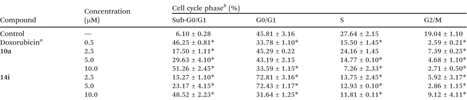
ba-sis of the morphological results suggestive of apoptoba-sis, we used flow cytometry to evaluate DNA fragmentation, cell cy-cle, PS externalization and mitochondrial depolarization in HL-60 cells incubated with compounds10aand14i. After 24 h, DNA fragmentation of the HL-60 cells was significantly in-creased by the compounds in a concentration-dependent manner (Table 3).
Doxorubicin at 0.5μM, used as a positive control, induced DNA fragmentation in 46.25% of the cells, comparable with the data from compound 10a (17.50, 29.63 and 51.26%, re-spectively) and14i (15.27, 23.17 and 48.52%, respectively) at 2.5, 5 and 10μM. Regarding cell cycle analysis (Table 3), no changes were observed after incubation with compound 10a. On the other hand, compound 14i caused accumulation of Table 2 Cytotoxic effect of the compounds synthesized on four human tumour cell lines and normal cells
Compound
RC50a(μM)
OVCAR-8 SF-295 HCT-116 HL-60 L929
10a 13.68 ± 1.68 14.68 ± 9.50 8.18 ± 5.91 3.00 ± 1.04 >50
10d 9.51 ± 1.89 29.37 ± 9.1 22.76 ± 5.63 8.98 ± 2.48 NDc
10g 11.64 ± 5 .54 32.64 ± 7.41 32.22 ± 13.95 10.01 ± 2.23 ND
14h 7.51 ± 1.41 14.12 ± 2.02 12.66 ± 1.09 5.50 ± 1.91 ND
14i 2.74 ± 1.11 4.90 ± 1.05 2.98 ± 1.84 3.58 ± 2.36 >50
14j 5.17 ± 0.22 15.44 ± 2.55 13.06 ± 0.55 9.80 ± 2.95 ND
17e 4.18 ± 0.15 8.03 ± 0.13 5.52 ± 0.66 3.46 ± 0.04 ND
Doxorubicinb 0.62 ± 0.09 0.44 ± 0.28 0.04 ± 0.03 0.02 ± 0.02 ND
aData are presented as RC
50± SEM for ovarian (OVCAR-60), central nervous system (SF-295), colon (HCT-116) and leukaemia (HL-60) tumour
cells and normal cells (L929), obtained from at least three independent experiments performed in triplicate.bDoxorubicin was used as the positive control.cNot determined.
Fig. 3 Microscopic analysis (May-Grünwald/Giemsa-stained) of the effect of10a and 14i on the HL-60 cells after 24 h of incubation. Untreated cells (A) or cells treated with10a(2.5μM, C; 5μM, D; or 10
μM, E) and14i(2.5μM, F; 5μM, G; or 10μM, H) were analysed by light microscopy (200×). Doxorubicin (0.5μM) was used as the positive con-trol (B). Black arrows show reduction in the cell volume, nuclear frag-mentation and cellular debris.
Fig. 4 Effect of10aand14ion HL-60 cell death pattern determined by acridine orange- and ethidium bromide-staining (AO/EB) after 24 h of incubation. The negative control (C) was treated with the vehicle used for diluting the test compound. Doxorubicin (0.5μM) was used as the positive control (D). The results are expressed as mean ± standard error of mean (SEM) for three independent experiments performed in triplicate (n = 2). *p < 0.05, compared to the control by ANOVA followed by Newman–Keuls test.
cells in the G0/G1 phase at 2.5 and 5μM (72.81 and 72.43%, respectively) (p< 0.05). Cell cycle arrest could have been
re-lated to an attempt by the cell to repair the DNA damage caused by14i. Since the damage appeared to be very intense, repair did not occur and apoptosis was triggered.38 The induction of apoptosis is one of the main mechanisms that inhibit cancer growth and proliferation and is used by several antitumor agents.39,40
In addition, the detection of PS externalization showed that both compounds significantly induced apoptosis (Fig. 5A) at all concentrations tested. In parallel, it was found
that10aand14iwere also able to cause mitochondrial depo-larization in a significant and concentration-dependent man-ner (Fig. 5A). After incubation with 10a, the results showed that 9.5, 19.0 and 29.0% of the cells displayed PS exter-nalization and 21.7, 37.1 and 56.2 of the cells showed mito-chondrial depolarization at 2.5, 5 and 10 μM, respectively (Fig. 5B). Compound14ipromoted PS externalization in 16.2, 33.3 and 48.1% of the cells and mitochondrial depolarization in 35.1, 47.0 and 68.0% of the cells at 2.5, 5 and 10 μM, re-spectively. All results presented herein suggested that com-pounds10aand14iinduce cell death by apoptosis in HL-60 human leukaemia cells.41
Conclusions
We synthesized new rubrolide analogues and converted them into their corresponding lactam and pyridazin-3IJ2H)-one de-rivatives. All compounds tested showed moderate cytotoxic activity against the four human cancer cell lines used. In gen-eral, the presence of Br and F substituents on the aromatic ring linked to theγposition of the lactone core increased the activity. Compared to the lactones and lactams, the pyridazin-3IJ2H)-ones did not show good activity. Moreover, compounds
10a and 14i displayed superior cytotoxicity against all cell lines tested. Interestingly, these compounds were not cyto-toxic towards normal cells at the concentrations tested. Re-garding the mechanism of action, 10a and14i induced the death of HL-60 cells by induction of apoptosis, which was demonstrated by morphological analysis (volume reduction, maintenance of cell membrane integrity and nuclear frag-mentation), DNA fragmentation and PS externalization. Additionally, compound 14i caused a large increase in the number of HL-60 cells in the G0/G1 phase. Accordingly, additional studies are needed to investigate the specific mo-lecular mechanism of cell death induction and cell cycle arrest.
Acknowledgements
We are grateful to the following Brazilian agencies: Conselho Nacional de Desenvolvimento Científico e Tecnológico (CNPq) Table 3 Effect of10aand14ion the nuclear DNA content of HL-60 cells determined by flow cytometry after 24 h of incubation
Compound
Concentration (μM)
Cell cycle phaseb(%)
Sub-G0/G1 G0/G1 S G2/M
Control — 6.10 ± 0.28 45.81 ± 3.16 27.64 ± 2.15 19.04 ± 1.10
Doxorubicina 0.5 46.25 ± 0.81
* 33.78 ± 1.10* 15.50 ± 1.45* 2.59 ± 0.21*
10a 2.5 17.50 ± 1.11* 45.29 ± 0.22 24.16 ± 1.45 7.39 ± 0.25*
5.0 29.63 ± 4.10* 43.19 ± 2.15 14.77 ± 0.10* 4.68 ± 1.10*
10.0 51.26 ± 2.45* 33.59 ± 1.15* 7.26 ± 2.33* 2.71 ± 0.50*
14i 2.5 15.27 ± 1.10* 72.81 ± 3.16* 13.75 ± 2.45* 5.92 ± 3.17*
5.0 23.17 ± 4.15* 72.43 ± 1.17* 12.93 ± 0.10* 2.86 ± 1.15*
10.0 48.52 ± 2.23* 31.64 ± 1.25* 11.81 ± 0.11* 9.12 ± 4.11*
*p<0.05, compared to the control by ANOVA followed by Dunnett's test.aDoxorubicin (0.5μM) was used as the positive control.bResults are expressed as mean ± standard error of mean (SEM) for two independent experiments performed in triplicate (n= 2).
Fig. 5 Effect of 10a and 14i on PS externalization (panel A) and mitochondrial depolarization (panel B) in the HL-60 cells by flow cy-tometry after 24 h of incubation. The negative control (C) was treated with the vehicle used for diluting the test compound. Doxorubicin (0.5
μM) was used as the positive control (D). The results are expressed as mean ± standard error of mean (SEM) for two independent experi-ments performed in triplicate (n= 2).*p<0.05, compared to the con-trol by ANOVA followed by Newman–Keuls test.
for research fellowships (CRAM, LCAB), Fundação de Amparo à Pesquisa de Minas Gerais, Coordenação de Aperfeiçoamento de Pessoal de Nível Superior (CAPES) and FINEP for financial support. Dr. A. Leyva helped with the English editing of this manuscript.
References
1 H. Seeliger, M. Guba, A. Kleespies, K.-W. Jauch and C. J. Bruns,Cancer Metastasis Rev., 2007,26, 611–621.
2 J. Ferlay, I. Soerjomataram, R. Dikshit, S. Eser, C. Mathers, M. Rebelo, D. M. Parkin, D. Forman and F. Bray, Int. J. Cancer, 2015,136, E359–386.
3 M. L. C. F. Junior, R. B. Silva, B. Mothes, A. T. Henriques and J. C. F. Moreira, Curr. Pharm. Biotechnol., 2012, 13, 235–244.
4 A. L. Demain and P. Vaishnav, Microb. Biotechnol., 2011,4, 687–699.
5 D. J. Newman and G. M. Cragg, J. Nat. Prod., 2012, 75, 311–335.
6 S. Miao and R. J. Andersen, J. Org. Chem., 1991, 56, 6275–6280.
7 M. J. Ortega, E. Zubía, J. M. Ocaña, S. Naranjo and J. Salvá,
Tetrahedron, 2000,56, 3963–3967.
8 A. N. Pearce, E. W. Chia, M. V. Berridge, E. W. Maas, M. J. Page, V. L. Webb, J. L. Harper and B. R. Copp,J. Nat. Prod., 2007,70, 111–113.
9 W. Wang, H. Kim, S.-J. Nam, B. J. Rho and H. Kang,J. Nat. Prod., 2012,75, 2049–2054.
10 J. Sikorska, S. Parker-Nance, M. T. Davies-Coleman, O. B. Vining, A. E. Sikora and K. L. McPhail, J. Nat. Prod., 2012,75, 1824–1827.
11 U. A. Pereira, L. C. A. Barbosa, C. R. A. Maltha, A. J. Demuner, M. A. Masood and A. L. Pimenta, Eur. J. Med. Chem., 2014,82, 127–138.
12 U. A. Pereira, L. C. A. Barbosa, C. R. A. Maltha, A. J. Demuner, M. A. Masood and A. L. Pimenta, Bioorg. Med. Chem. Lett., 2014,24, 1052–1056.
13 U. A. Pereira, L. C. A. Barbosa, A. J. Demuner, A. A. Silva, M. Bertazzini and G. Forlani, Chem. Biodiversity, 2015, 12, 987–1006.
14 L. C. A. Barbosa, C. R. A. Maltha, M. R. Lage, R. C. Barcelos, A. Donà, J. W. M. Carneiro, G. Forlani and J. Agric, Food Chem., 2012,60, 10555–10563.
15 Y.-L. Yang, C.-P. Lu, M.-Y. Chen, K.-Y. Chen, Y.-C. Wu and S.-H. Wu,Chem.–Eur. J., 2007,13, 6985–6991.
16 W. Malinka, A. Redzicka and O. Lozach,Farmaco, 2004,59, 457–462.
17 F. F. P. Arantes, L. C. A. Barbosa, E. S. Alvarenga, A. J. Demuner, D. P. Bezerra, J. R. O. Ferreira, L. V. Costa-Lotufo, C. Pessoa and M. O. Moraes, Eur. J. Med. Chem., 2009, 44, 3739–3745.
18 F. F. P. Arantes, L. C. A. Barbosa, C. R. A. Maltha, A. J. Demuner, P. M. Costa, J. R. O. Ferreira, L. V. Costa-Lotufo, M. O. Moraes and C. Pessoa, Eur. J. Med. Chem., 2010, 45, 6045–6051.
19 F. F. P. Arantes, L. C. A. Barbosa, C. R. A. Maltha, A. J. Demuner, P. H. Fidêncio and J. W. M. Carneiro,J. Chemom., 2011,25, 401–407.
20 Y. B. Kiran, C. D. Reddy, D. Gunasekar, C. S. Reddy, A. Leon and L. C. A. Barbosa, Eur. J. Med. Chem., 2008, 43, 885–892.
21 J. O. S. Varejão, L. C. A. Barbosa, G. A. Ramos, E. V. V. Varejão, B. King-Díaz and B. Lotina-Hennsen,J. Photochem. Photobiol., B, 2015,145, 11–18.
22 J. Boukouvalas, P. P. Beltran, N. Lachance, S. Cote, F. Maltais and M. Pouliot,Synlett, 2007, 219–222.
23 R. R. Teixeira, L. C. A. Barbosa, G. Forlani, D. Piló-Veloso and J. W. M. Carneiro, J. Agric. Food Chem., 2008, 56, 2321–2329.
24 W. K. Goh, G. Iskander, D. S. Black and N. Kumar,
Tetrahedron Lett., 2007,48, 2287–2290.
25 R. R. Teixeira, L. C. A. Barbosa, C. R. A. Maltha, M. E. Rocha, D. P. Bezerra, L. V. Costa-Lotufo, C. Pessoa and M. O. Moraes,Molecules, 2007,12, 1101–1116.
26 R. R. Reis, E. C. Azevedo, M. C. B. V. Souza, V. F. Ferreira, R. C. Montenegro, A. J. Araújo, C. Pessoa, L. V. Costa-Lotufo, M. O. Moraes, J. D. B. M. Filho, A. M. T. Souza, N. C. Carvalho, H. C. Castro, C. R. Rodrigues and T. R. A. Vasconcelos,Eur. J. Med. Chem., 2011,46, 1448–1452. 27 F. W. A. Barros, T. G. Silva, M. G. R. Pitta, D. P. Bezerra,
L. V. Costa-Lotufo, M. O. Moraes, C. Pessoa, M. A. F. B. Moura, F. C. Abreu, M. C. A. Lima, S. L. Galdino, I. R. Pitta and M. O. F. Goulart, Bioorg. Med. Chem., 2012, 20, 3533–3539.
28 T. Mosmann,J. Immunol. Methods, 1983,65, 55–63.
29 M. C. Alley, D. A. Scudiero, A. Monks, M. L. Hursey, M. J. Czerwinski, D. L. Fine, B. J. Abbott, J. G. Mayo, R. H. Shoemaker and M. R. Boyd, Cancer Res., 1988, 48, 589–601.
30 M. V. Berridge, A. S. Tan, K. D. McCoy and R. Wang,
Biochemica, 1996,4, 14–19.
31 F. Bellina, C. Anselmi and R. Rossi, Tetrahedron Lett., 2002,43, 2023–2027.
32 F. W. A. Barros, P. N. Bandeira, D. J. B. Lima, A. S. Meira, S. S. Farias, M. R. J. R. Albuquerque, H. S. Santos, T. L. G. Lemos, M. O. Morais, L. V. Costa-Lotufo and C. Pessoa,
Bioorg. Med. Chem., 2011,19, 1268–1276.
33 A. J. Araújo, A. A. de Souza, E. N. S. Júnior, J. D. B. Marinho-Filho, M. A. B. F. Moura, D. D. Rocha, M. C. Vasconcellos, C. O. Costa, C. Pessoa, M. O. Moraes, V. F. Ferreira, F. C. Abreu, A. V. Pinto, R. C. Montenegro, L. V. Costa-Lotufo and M. O. F. Goulart, Toxicol. In Vitro, 2012,26, 585–594.
34 H. I. F. Magalhães, D. V. Wilke, D. P. Bezerra, B. C. Cavalcanti, R. Rotta, D. P. Lima, A. Beatriz, M. O. Moraes, J. Diniz-Filho and C. Pessoa, Toxicol. Appl. Pharmacol., 2013,272, 117–126.
35 I. H. Hall, N. J. Peaty, J. R. Henry, J. Easmon, G. Heinisch and G. Pürstinger,Arch. Pharm., 1999,332, 115–123.
36 Y. Kaku, A. Tsuchiya, T. Kanno, T. Nakano and T. Nishizaki,
Cell. Signalling, 2015,27, 1713–1719.
37 J. Mou, A. Park, Y. Cai, J. Yuan and C. Yuan, Bioorg. Med. Chem. Lett., 2015,25, 3057–3061.
38 D. P. Maia, D. V. Wilke, J. Mafezoli, J. N. S. Júnior, M. O. Moraes, C. Pessoa and L. V. Costa-Lotufo, Chem.-Biol. Interact., 2009,180, 220–225.
39 M. Los, C. J. Burek, C. Stroh, K. Benedyk, H. Hug and A. Mackiewicz,Drug Discovery Today, 2003,8, 67–77.
40 D. R. Schultz and W. J. Harrington Jr, Semin. Arthritis Rheum., 2003,32, 345–369.
41 F. W. A. Barros, D. P. Bezerra, P. M. P. Ferreira, B. C. Cavalcanti, T. G. Silva, M. G. R. Pitta, M. C. A. Lima, S. L. Galdino, I. R. Pitta, L. V. Costa-Lotufo, M. O. Moraes, R. R. Burbano, T. N. Guecheva, J. A. P. Henriques and C. Pessoa,
Toxicol. Appl. Pharmacol., 2013,268, 37–46.