412
AEROBIC EXERCISE TRAINING INHIBITS SKELETAL
MUSCULAR APOPTOTIC SIGNALING MEDIATED
BY VEGF-VEGR2 IN SPONTANEOUSLY
HYPERTENSIVE RATS
ORIGINAL ARTICLETiago Fernandes1
Flávio de Castro Magalhães1,2
Everton Crivoi do Carmo1
Edilamar Menezes de Oliveira1
1. Laboratory of Biochemistry and Molecular Biology of the Exercise, Physical Education and Sports School, University of São Paulo-USP, São Paulo, SP, Brazil.
2. Exercise Biology Laboratory, Biological and Health Sciences School, Federal Universities of the Jequitinhonha and Mucuri Valleys, Diamantina, Minas Gerais, Brazil.
Mailing address:
Edilamar Menezes de Oliveira Escola de Educação Física e Esporte da Universidade de São Paulo Departamento de Biodinâmica do Movimento do Corpo Humano Av. Professor Mello Moraes, 65 – Butantã - 05508-900 – São Paulo, SP, Brazil
E-mail: [email protected]
EXERCISE AND SPORTS SCIENCES
ABSTRACT
Aerobic exercise training (ET) has been established as an important non-pharmacological treatment for hypertension, since it corrects the microvascular rarefaction and decreases blood pressure. Studies have shown that microvascular abnormalities are directly associated with changes in the vascular en-dothelial growth factor (VEGF) and VEGF receptor 2 (VEGFR2), as well as with imbalance of apoptotic signaling in hypertension. However, little is known about these mechanisms in hypertension. We hy-pothesized that ET restores angiogenic factors and promotes balance between anti and pro-apoptotic proteins of the Bcl-2 family, potentially contributing to revascularization and the disease regression. Twelve-week old male Spontaneously Hypertensive Rats (SHR, n=14) and Wistar Kyoto Rats (WKY, n=14) were randomly assigned into 4 groups: SHR, trained SHR (SHR-T), WKY and trained WKY (WKY-T) were studied. As expected, ten weeks of ET were effective in reducing blood pressure in SHR-T group. In addition, ET promoted resting bradycardia in trained groups (WKY-T and SHR-T), being considered an important marker of aerobic ET. ET has also corrected the capillary rarefaction in SHR-T and this response is partly due to recovery of the peripheral levels of VEGF and increase in VEGFR2 expression. Concomitantly, normalization of the apoptotic pathway was observed, with increased expression of the anti-apoptotic proteins (Bcl-2 and Bcl-x) and reduction of the pro-apoptotic protein (Bad), followed by phosphorylation of the Bad protein and decrease in the Bad/Bcl-x association. These data suggest that ET promotes peripheral revascularization in hypertension dependent on a fine balance between positive and negative regulators of angiogenesis.
Keywords: exercise training, hypertension, VEGF, apoptosis, skeletal muscle.
INTRODUCTION
Hypertension is a multifactorial syndrome characterized by high and sustained levels of blood pressure (BP) which affects approximately one billion of individuals worldwide, representing one of the most relevant risk factors in the etiology of cardiovascular diseases (CVD)1,2.
Normal cardiac output followed by increase of peripheral vascular resistance are considered the most remarkable marks of hypertension essential in adults, demonstrating that high BP is associated with damage in microcirculation3,4. Considerable data show that microvascular abnormalities such as reduction of blood flow and microvascular rarefaction are clear evidence of the dis-turb in the angiogenic process and greater destruction of vessels in hypertension3-6.
Many factors have been pointed as responsible for the endo-thelial dysfunction and by fail in the angiogenic process in hyper-tension. Among these, we can highlight the crucial role of the nitric oxide (NO)7 and the vascular endothelial growth factor (VEGF)8, as well as the balance of apoptosis stimulators and inhibitors mainly mediated by the B2 cells lymphoma family (Bcl-2)9.
VEGF is known as a key-regulating protein of the physiologi-cal angiogenesis both in the embryogenic and post-natal phases.
It also works as a powerful pro-survival factor for endothelial cells, inducing the expression of antiapoptotic proteins and inhibiting the pro-apoptotic ones8.
The Bcl-2 family has been extensively investigated for being a fa-mily of death by apoptosis inducing and repressing proteins which actively participate in the apoptosis regulation 10. The members of the Bcl-2 and Bcl-2 and Bcl-xL families inhibit apoptosis, since they prevent the release of cytochrome c and are called antiapoptotic regulators. On the other hand, Bad, Bax and Bak are pro-apoptotic proteins for inducing release of cytochrome c11. Cytochrome c li-gates to the activation fator of the pro-apoptotic 1 proteases 1 (Apaf-1) which, through ATP-dependent conformational alterations, oligomerize and makes the aptosome, a complex which will activate the pro-caspase 9. This activated pro-caspase 9 triggers the signal for activation of the caspases 3, 6 and 7 resulting in an amplified and catalytic process resulting in the apoptotic process12.
413
studies corroborated that chronic treatment with antioxidants redu-ced apoptosis of the endothelial cells in micro vessels and prevented structural rarefaction in spontaneously hypertensive rats (SHR)14.
Clinical assays and meta-analyses have shown that aerobic exer-cise training (ET) causes reduction in the BP levels in hypertensive individuals15,16. Studies with laboratory animals have also shown significant reduction in BP in SHR17-19.
Some studies point out important effects of aerobic ET on the microcirculation in SHR, such as increase in capillary density and capillary: fiber ratio in the skeletal and cardiac muscle, pro-moting complete reversion of capillary rarefaction occurred in hypertension. Moreover, aerobic exercise normalizes peripheral vascular resistance for the skeletal musculature and the arterial wall-to-lumen ratio17. Restoration of the microvascular chain may importantly contribute to the decrease in BP through reduction in peripheral vascular resistance, which has been said to be res-ponsible for hypertension in adults3,4,17-19.
Increase in blood flow to the skeletal musculature promoted by aerobic ET and consequent increase of shear stress present direct correlation with increase in release of NO and VEGF, which also contributes to flow increase, since it leads not only to endothelium--dependent vasodilatation but also to angiogenesis, being conside-red by many researchers the primary sign for vascular remodeling20. At physiological conditions, maintenance, formation and/or loss of capillaries are dependent on a thin balance between positive and negative regulators of angiogenesis9.
Mediators of cellular signaling have been identified as deter-minant in the recovery of the microvascular chain in hypertension; however, studies with non-pharmacological approach such as ET, concerning regulation of the angiogenic factors and muscular anti and pro-apoptotic proteins have not been studied. Therefore, the aim of this study is to verify: 1) the possible harm of the angiogenic factors and the imbalance of the anti and pro-apoptotic proteins associated to microvascular damage in SHR; and 2) the effect over the correction of capillary rarefaction and its cellular mediators, potentially contributing to hypertension regression.
MATERIAL AND METHODS
Experimental animals
28 SHR with 12 weeks of life, with established hypertension and 18 male Wistar Kyoto (WKY) were used as control of SHR for this study. The animals came from the Central Animal Facility of the Institute of Biomedical Sciences of the University of São Paulo (ICB-USP). The rats were weighing between 240 and 270g at the beginning of the protocol.
The animals used in this study were kept in plastic cages in groups of three or four animals per cage and separated per experi-mental group. Room temperature of the anima facility was kept between 22-24ºC, with controlled light in light/dark inverted cycle of 12 hours. Water and food were administered ad libitum.
All procedures were performed according to the Ethical Prin-ciples of Animal Experimentation by the Brazilian College of Animal Experimentation, being the research Project apoved by the Ethics Committee of the Physical Education and Sports School of the Uni-veristy of São Paulo (EEFE-USP) (# 2007/35).
Animals identification
The animals were randomly divided in four groups with seven animals per group, according to the experimental protocol: • Wistar-Kyoto rats (WKY);
• Trained Wistar-Kyoto rats (WKY-T); • Spontaneously hypertensive rats (SHR);
• Trained spontaneously hypertensive rats (SHR-T).
Aerobic physical training protocol
The swimming ET was performed according to protocol by Medeiros et al.21. The animals were trained during 10 weeks, ses-sions of 60 min, once a day, five times per week, with gradual work overload increase (weight on the tail in body weight percentage) until reaching 4% of body weight. The protocol used was charac-terized as low to moderate intensity and long duration training, being effective in promoting cardiovascular adaptations and in in-creasing muscular oxidative capacity. The rats were identified and weekly weighed for correction of training overload due to increase in body weight.
Pre ad post the ET period the animals were submitted to hemo-dynamic analyses, and subsequently they were killed by anesthesia with intraperitoneal injection of sodium pentobarbital (80mg/kg), and the necessary samples were collected and stored for histolo-gical and molecular analyses.
Evaluation of the hemodynamic responses
BP was performed pre and post ET by tail pletismography (KENT SCIENTIFIC system RTBP1001 for rats and mice, Litchfield, USA), in the four animal groups. The animals were awake, at rest and were kept under movement restriction so that the measures could be taken. In order to avoid measurement and analysis erros, the rats were submitted to a one-week period of familiarization with the technique.
The BP recording equipment consists of a rubber cuff adap-ted to the proximal region of the tail, which is connecadap-ted to the pletismograph to gradually inflate and deflate the cuff from 1 to 250/300mmHg. In a more distal region of the tail a wrist pneumatic transducer is attached for detection of the passage signals of the wrist wave of BP on the caudal artery and recorded in the signal acquisition system. This indirect BP measuring method allows quan-tify BP and heart rate (HR) throughout the protocol.
Analysis of the capillary: fiber ratio
The soleus muscle capillary:fiber ratio was assessed through the histochemical reaction for myosin ATPase in pH 10.3, as described by Sillau and Banchero22 and quantified by the analysis of 10 fields not overlapped, with amplification of 200x, randomly distributed using a morphometric computer system (Leica Quantimet 500, Cambridge, UK). In order to calculate the capillar/fiber ratio, the total number of capillaries was divided by the total number of fibers counted on the same field. Only vessels with diameter lower than 12µm were counted.
Determination of the proteins expression
414
0.3M, DTT 0.5mM, EDTA 1mM (pH 8.0), PMSF 0.3mM, NaF 10mM and cocktail of phosphatases inhibitor (1:100). The homogenate was centrifuged for 10 minutes at 4°C with 12,000rpm. The supernatant was transferred to 1.5 ml tubes and the protein concentration of the samples was analyzed in the Bradford method23. The samples were stored in freezer –80ºC until usage.
Aliquots of the homogeneized, 30μg of protein were di-luted in sample buffer (Tris-HCl pH 6.8 240mM; SDS 0.8%; β-mercaptoethanol 200mM; glycerol 40% and bromophenol blue (0.02%). Analysis of the protein levels was performed by western blotting. Thus, polyacrilamide gel electrophoresis was used (SDS-PAGE, 6-12%: depending on the protein molecular weight), which consists of the migration of loaded molecules, in a solution, derived from the application of an electrical field for minigel (Mini Pro-tean, BioRad, USA). Subsequently, the proteins were transferred to a nitrocellulose membrane (Amersham Biosciences, NJ, USA), the same way they were separated in the SDS-PAGE. The membranes were stained with Ponceau S for verification of the protein bands obtained by the electrophoresis.
In order to block unspecific ligations, the membrane was in-cubated in solution containing casein, protein which competes with ligation sites and reduces unspecific absorption of peroxi-dase conjugates.
Subsequently, the nitrocellulose membrane was incubated with the primary antibody which ligates to the protein which is tried to be detected, forming an antibody-protein complex. After having washed the membrane for removal of the non-ligated antibody, it was exposed to the secondary antibody conjugated to horseadish peroxidase (HRP), directed to specific-species portions of the pri-mary antibody.
The VEGF (Rabbit polyclonal, 1:1000, Santa Cruz, CA, USA); VEGFR1 (Rabbit polyclonal, 1:1000, Santa Cruz, CA, USA); VEGFR2 (Rabbit polyclonal, 1:1000, Santa Cruz, CA, USA); Bcl-x (Rabbit polyclonal, 1:1000, Cell Signaling Tech., MA, USA); Bcl-2 (Rabbit polyclonal, 1:1000, Cell Signaling Tech., MA, USA); Bad (Rabbit polyclonal, 1:1000, Cell Signaling Tech., MA, USA) and p-Badser112 (Rabbit polyclonal, 1:1000, Cell Signaling Tech., MA, USA) were used as primary antibodies. Subsequently, they were washed 3x10 min with TBS-T, incubated for two hours with the respective secondary antibodies (IgG anti-rabbit, anti-rabbit, anti-rabbit, anti-rabbit, anti-mouse, anti-rabbit, anti-rabbit, anti-rabbit and anti-rabbit; Amersham Biosciences, NJ, USA) conjugated to peroxidase. The complex was then detected through a chemiluminescence reaction (ECL) and the blots were visualized and quantified (number of pixels) by the Scion Image system, freely provided by the NIH (USA) via internet. The glyceraldehyde-3-phosphate dehydrogenase (GAPDH) was used as normalizer.
STATISTICAL ANALYSIS
Data were analyzed using two-way ANOVA (TF and HA as inde-pendent factors) to compare the values of the groups and Tukey test as post hoc (Statistica software, StatSoft, Inc., Tulsa, OK, USA) was also applied. A p < 0.05 of significance was adopted for all experiments. All results were presented as mean ± standard error of the mean (SEM).
RESULTS
Hemodynamic parameters: blood pressure and heart rate
The BP values expressed in mercury millimeters (mmHg) and pre and post-ET HR expressed in beats per minute (bpm) are re-presented in figure 1.
In figure 1A, ET, it can be observed that the SHR group pre-sented high levels of SBP (184 ± 3.9mmHg), DBP (150 ± 4.7mmHg) and MBP (167 ± 5.3mmHg) compared with the control groups, WKY (127 ± 3; 99 ± 2.4; 110 ± 3.2mmHg, respectively), indicating that hypertension was established. No HR alterations were found between pre-ET groups (WKY: 390 ± 12; WKY-T: 393 ± 8; SHR: 409 ± 8; SHR-T: 416 ± 6bpm).
In post-ET, represented in figure 1B, it is observed that low inten-sity and long duration swimming ET was efficient in reducing SBP, DBP and MBP of the SHR-T group (162 ± 4.4; 131 ± 2; 147 ± 7.8mmHg, respectively) compared with the SHR group (207 ± 5.5; 160 ± 2.5; 175 ± 3.6mmHg, respectively), without alteration in SBP, DBP and MBP in the control, WKY and WKY-T groups. Moreover, resting bradycardia was observed in the trained animals groups, reducing hence the HR values of these groups when compared with the groups kept sedentary at the same experimental period (post-ET- WKY: 393 ± 12; WKY-T: 322 ± 14; SHR: 407 ± 11; SHR-T: 338 ± 8bpm).
Figure 1. Hemodynamic parameters. Systolic blood pressure (SBP), diastolic blood
pressure (DBP), mean blood pressure (MBP) and resting heart rate (HR) pre ET (A) and post ET (B) of WKY and SHR rats submitted to the experimental protocol. The results were expressed as mean ± SEM. * p < 0.001 vs. WKY and WKY-T; # p < 0.01 vs. WKY and SHR; † p < 0.001 vs. SHR. ET: exercise training.
Pre ET
bpm
mmHg
A
Pós TF
bpm
mmHg
B
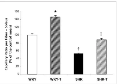
Determination of the capillary:fiber ratio in skeletal muscle
Figure 2 presents the capillar/fiber ratio through histochemical evaluation of the soleus skeletal muscle through myosin ATPase reaction. As expected, the capillary rarefaction was observed in the SHR group compared with the WKY group. On the other hand, ET
SBP DBP MBP HR
Post ET
415
was efficient in increasing 47% of the number of capillaries by analysis of the capillary/fiber ratio in the WKY-T group and corrected the capillary rarefaction in the SHR-T group when compared with the WKY group (WKY: 1.2 ± 0.06; WKY-T: 1.8 ± 0.04; SHR: 0.7 ± 0.02 and SHR-T: 1.1 ± 0.04 number of capillaries/ muscular fiber).
Figure 2. Effect of ET on the analysis of the capillary/muscular fiber ratio by the
histochemical method through myosin ATPase reaction. Data were represented as percentage of the control mean ± SEM. * p < 0.05 vs. WKY, SHR and SHR-T; † p < 0.05 vs. WKY, WKY-T and SHR-T, ‡ p < 0.05 vs. WKY-T and SHR. ET: exercise training.
Quantification of the angiogenic factors: VEGF, VEGFR1 and VEGFR2
Figure 3 illustrates the protein levels evaluated by western blot of VEGF, VEGFR1 and VEGFR2 in the soleus muscle. Compared with the sedentary WKY group, ET increased the peripheral protein levels of VEGF and VEGFR2 in group WKY-T (figures 3 – B and C, respec-tively). On the other hand, in the SHR group, the protein levels of VEGF were significantly lower compared with the protein levels of the WKY group; however, ET was able to restore the VEGF levels in the SHR-T group (figure 3B). Moreover, VEGF recovery in the SHR-T group was followed by increase in the protein levels of VEGFR2 (figure 3C). Nevertheless, no significant difference was observed in the VEGFR1 protein levels among all the studied groups (figure 3D).
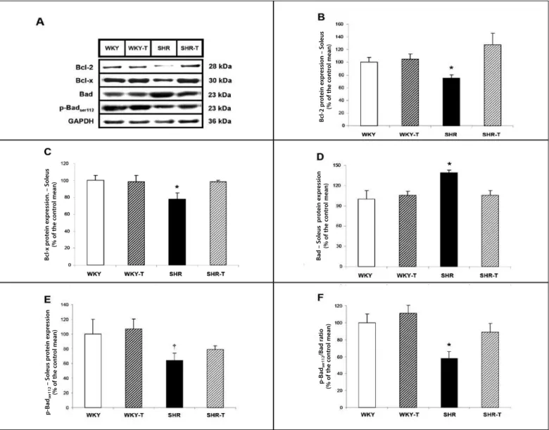
Quantification of anti and pro-apoptotic factors: family Bcl-2
Recent studies have demonstrated that the mechanisms in-volved in the inhibition of the angiogenic process are associated with increase in the expression of many genes related to apoptosis, considering significant increase in the death of endothelial cells in the skeletal muscle9.
The authors investigated key-regulatory ways which control the molecular response for apoptosis, especially the Bcl-2 family. These proteins functionally interact when acting one over the other in the control of the apoptotic process. It is mainly known that the Bcl-2 and Bcl-x proteins suppress and the Bad and p-Badser112 proteins promote apoptosis10-12.
Figure 4 presents the protein levels evaluated by western blot of Bcl-2, Bcl-x, Bad and p-Badser112 in the soleus muscle. The protein levels of the antiapoptotic proteins Bcl-2 and Bcl-x were similar between groups WKY and WKY-T (figures 4 – B and C, respectively). Moreover, no alteration in the protein levels of the pro-apopto-tic protein Bad or in its phosphorylation site in Serina 112 were
Figure 3. Effect of the aerobic ET on the skeletal muscular angiogenic factors on
hypertension. Blots representative of VEGF, VEGFR1, VEGFR2 and GAPDH of WKY, WKY-T, SHR, SHR-T (A). VEGF, VEGFR1 and VEGFR2 protein levels in the soleus muscle analyzed by western blot, respectively (B-D). The target-bands were normalized by the skeletal muscular GAPDH protein. The results were expressed as mean ± SEM. * p < 0.05 vs. WKY, SHR and SHR-T; † p < 0.05 vs. WKY, WKY-T and SHR-T; ‡ p < 0.05 vs. WKY-T and SHR; § p < 0.05 vs. WKY and SHR. ET: exercise training.
B
A
C
D
C
apillar
y R
a
tio per F
iber – S
oleus
(% of the c
on
tr
ol mean)
VEGF pr
ot
ein e
xpr
ession – S
oleus
(% of the c
on
tr
ol mean)
VEGFR1 – S
oleus
(% of the c
on
tr
ol mean)
VEGFR2 – S
oleus
(% of the c
on
tr
416
observed in group WKY-T compared with its sedentary control WKY (figures 4 – D and E, respectively). Conversely, group SHR presen-ted remarkable increase in the protein levels of the pro-apoptotic protein Bad and decrease of these levels in their phosphorylation site Serina 112, as well as of the p-Badser112/Bad ratio compared with group WKY (figures 4 – D, E and F, respectively), followed by reduction of the protein levels of the antiapoptotic proteins Bcl-2 and Bcl-x (figures 4 – B and C, respectively). Interestingly, as shown by figures 4 – A-F, ET restored the protein levels of the anti and pro-apoptotic proteins in group SHR-T, bringing them to values similar to the control group.
DISCUSSION
In the present study, the SHR model and its controls WKY were used to study the hypertension influence on the alterations in skeletal muscular microcirculation. Moreover, the effect of ET on the correction of the capillary rarefaction and the possible mechanisms involved in these alterations were evaluated, favoring hence disease regression. The main results of the study show that aerobic ET on hyper-tension: 1) reduced BP followed by resting bradycardia; 2) corrected capillary rarefaction; 3) normalized the muscular VEGF levels and
increased VEGFR2 expression; and 4) deactivated the peripheral apoptotic ways.
Imbalance in vascular tonus control observed in microcircula-tion in hypertension may be the result of excessive vasoconstric-tion causing occlusion of resistance arterioles and non-perfusion of capillaries, inducing microvascular rarefaction and decrease of conductibility in parallel to skeletal muscular microcirculation3-6. Furthermore, studies have shown that the endothelial shear stress leads to sustained release of endothelial nitric oxide synthase (eNOS) and the absence of blood flow, and of NO as well, could lead to endothelial dysfunction followed by peripheral apoptosis and later on, loss of non-perfused vessels in hypertension, promoting hence the microvascular rarefaction scenario. Thus, angiogenesis is surely an essential factor for hypertensive individuals, since it improves the blood transport to the tissues, reducing BP which is harmed by the high peripheral resistance3,4,13,19.
Considering the evidence, VEGF is considered the most im-portant regulator of angiogenesis, chemotaxis and endothelial cell survival by preventing the activation of signaling pathways of apoptosis, consequently the capillary loss8. In fact, deletion or inhibi-tion of VEGF in skeletal muscles of adult mice has shown adverse
Figure 4. Effect of the aerobic ET on the regulatory proteins of skeletal muscular apoptosis in hypertension. Blots representative of Bcl-2, Bcl-x, Bad, p-Badser112 and GAPDH of
WKY, WKY-T, SHR, SHR-T (A). Bcl-2, Bcl-x, Bad and p-Badser112 protein levels in the soleus muscle analyzed by western blot, respectively (B-F). The target-bands were normalized
by the GAPDH skeletal muscular protein. The results are expressed as mean ± SEM. * p < 0.05 vs. WKY, WKY-T and SHR-T; † p < 0.05 vs. WKY and WKY-T. ET: exercise training.
B
cl-x pr
ot
ein e
xpr
ession. – S
oleus
(% of the c
on
tr
ol mean)
B
cl-2 pr
ot
ein e
xpr
ession – S
oleus
(% of the c
on
tr
ol mean)
B
ad – S
oleus pr
ot
ein e
xpr
ession
(% of the c
on
tr
ol mean)
p
-B
ad
ser112
– S
oleus pr
ot
ein e
xpr
ession
(% of the c
on
tr
ol mean)
p
-B
ad
ser112
/B
ad r
a
tio
(% of the c
on
tr
417
effects, particularly a significant reduction in capillary density with endothelial cell apoptosis23.
It is known that the angiogenic response mediated by regular practice of aerobic physical exercise involves many mechanisms, such as increase of blood flow, triggering increase of shear stress and tension on the capillaries walls, hypoxia, activating the hypoxia-inducible factor (HIF-1α), the renin-angiotensin system, among others, which are responsible for induction of vascular growth factors, especially the VEGF24. Although the literature shows that many factors act as potential precursors of angiogenesis in the skeletal muscle, the VEGF together with the eNOS were described as the most important in the promotion of capillaries formation with aerobic ET24. Additionally, the VEGF interaction with its main angiogenic receptor, the VEGFR2, a tyrosine kinase receptor, promotes activation of a cascade of intracellular signaling mediated mainly by the PI3K/Akt way which stimulates the gene expression, such as the eNOS, committed mainly with vasodilatation, angiogenesis and survival of the endothelial cells8,24,25.
Interestingly, the VEGFR2 has shown low expression in SHR and the use of gene therapy as treatment for hypertension with use of gene transfer of VEGF was effective in improving angiogenesis harm and the endothelial proliferation in SHR26.
In fact, harm of the peripheral angiogenic factors in the SHR group observed in this investigation corroborates the data demonstrated in the literature mentioned above, since our results point to VEGF protein levels in microcirculation of SHR remarkably reduced compared with the ones from WKY, occurring in parallel to the capillary rarefaction. Thus, it seems possible that the abnormalities of the vascular structure followed by the loss of capillaries are a clear evidence of the dysfunction in the angiogenic process which makes impossible the regeneration of functional micro vessels in the skeletal muscle of hypertensive individuals.
It is known that capillary rarefaction occurs in parallel to activa-tion of the apoptotic way which negatively reflects on the angio-genesis and endothelial cellular survival in the skeletal muscle9. The Bcl-2/Bcl-x and Bad proteins are important apoptotic regulators which act inhibiting or promoting apoptosis, respectively10,27.
As mentioned, the endothelial survival signs involved by the VEGF/VEGFR2 interaction are predominantly mediated by the PI3K/AKT/eNOS way implied in the increase of antiapoptotic proteins such as Bcl-2 e Bcl-x28, inhibiting hence the activation of the caspases, consequently the apoptosis, as well as favoring angiogenesis. On the other hand, this activation cascade may inhibit the pro-apoptotic activity of Bad29. However, as we observe reduction in the VEGF levels in SHR, this control of the inducing and repressing mechanisms of apoptosis get harmed, ending in reduction in survival of the endothelial cell and promotion of microvascular rarefaction in hypertension.
Studies in the literature evidence increased apoptotic signaling of the soleus muscle in SHR compared with WKY, with increase of expression of the pro-apoptotic protein Bax, the activity of the cas-pase 3 and DNA fragmentation as well as reduction in the protein levels of Bcl-227.
In concordance with the data in the literature showing more expression of pro-apoptotic proteins and reduction of the antiapoptotic in hypertension, our results indicate that the soleus muscle of SHR
presents increase in the expression of Bad and reduction of Bcl-2 and Bcl-x. It is known that the pro-apoptotic activity of Bad is dependent of its dephosphorylation; on the other hand, inhibiting the activity of Bad represents keeping the peptide in inactive form, that is to say, phosphorylated29. Increase of the pro-apoptotic activity of Bad in SHR was confirmed, since we verified decrease of phosphorylated Bad in Ser112 and of p-Badser112/Bad ratio. Moreover, our results showed that reduction of endothelial NO synthesis followed by increase in protein expression of iNOS could contribute to moderate exchange of redox homeostasis towards a pro-oxidant status, corroborating for the activation of the microvascular pro-apoptotic signaling way since the oxidative stress mediated by hypertension harms the vasodilator response and apoptosis of the endothelial cell associated with loss of microvessels13,14,30.
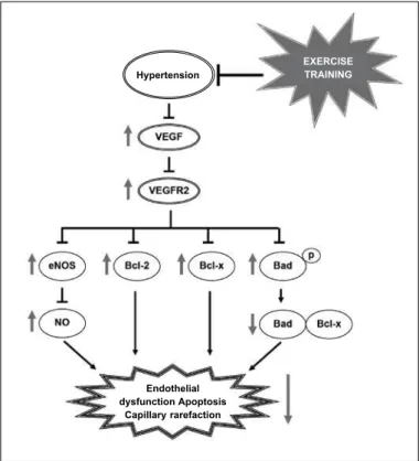
Consistent to the data in the literature, ET restored the VEGF levels followed by increase in expression of VEGFR2. Furthermore, for the first time in the literature, we showed that the levels of the antiapoptotic Bcl-2 and Bcl-x factors were normalized in hyperten-sion when induced by ET, bringing then to values similar to the ones in WKY, while the pro-apoptotic signaling was reduced with normalization of the Bad levels. It is known that the Bad transfers to the mitochondria and makes a pro-apoptotic complex with Bcl-x. The Bad heterodimerization with Bcl-x prevents the antiapoptotic effect of Bcl-x.Thus, we observed that this translocation is inhibited by the increase of the survival factors as VEGF and VEGFR2 by ET which induces Bad phosphorylation, leading to its cytosolic seques-tration, consequently avoiding the apoptotic signaling (figure 5).
Figure 5. Hypothesized signaling mechanism of cellular survival and angiogenesis
mediated by the VEGF-VEGFR2 interaction: harm to hypertension and post-ET correction. Hypertension decreased the levels of VEGF, compromising hence the signaling mediated by its interaction with VEGFR2, leading to reduction of eNOS and bioavailability of NO, as well as alteration of the survival signaling with reduction of the antiapoptotic proteins Bcl-2 and Bcl-x and increasing the pro-apoptotic activity by dephosphorylation of Bad and greater Bad-Bcl-x interaction. Interestingly, signaled by the red arrows, ET corrected these parameters mediated by hypertension, regressing the disease. ET: exercise training, p: phosphorylation.
ExERCISE TRAININg
Endothelial dysfunction Apoptosis
418
Thus, the results obtained suggest that the remaining and formation of new capillaries may be dependent on the balance between the angiogenic and apoptotic factors, since they synergetically interact to prevent microvascular abnormalities and to keep vascular homeostasis. In addition to that, increase o the protein levels of VEGF and VEGFR2 without significant alterations in the microvascular apoptotic signaling in the WKY-T group compared with the control WKY may represent a potential angiogenesis signaling way induced by aerobic ET.
ACKNOWLEDGEMENTS
To FAPESP (# 2007/56771-4 and # 2010/50048-1) and to MCT/ CNPq 14/2009 (# 480391/2009-2) and CNPq (# 307591/2009-3) for the financial support and research support.
All authors have declared there is not any potential conflict of interests concerning this article.
REFERENCES
1. Chobanian AV, Bakris GL, Black HR, Cushman WC, Green LA, Izzo JL Jr, et al. Seventh report of the Joint National Committee on Prevention, Detection, Evaluation, and Treatment of High Blood Pres-sure. Hypertension 2003;42:1206-52.
2. Pereira M, Lunet N, Azevedo A, Barros H. Differences in prevalence, awareness, treatment and control of hypertension between developing and developed countries. J Hypertens 2009;27:963-75.
3. Lévy BI, Ambrosio G, Pries AR, Struijker-Boudier HAJ. Microcirculation in hypertension: a new target for treatment? Circulation 2001;104:735-40.
4. Feihl F, Liaudet L, Waeber B, Lévy BI. Hypertension: A disease of the microcirculation?. Hypertension 2006;48:1012-7.
5. Greene AS, Tonellato PJ, Lui J, Lombard JH, Cowley AW Jr. Microvascular rarefaction and tissue vascular resistance in hypertension. Am J Physiol 1989;256:126-31.
6. Antonios TF, Singer DR, Markandu ND, Mortimer PS, MacGregor GA. Rarefaction of skin capillaries in borderline essential hypertension suggests an early structural abnormality. Hypertension 1999;34:655-8.
7. Dulak J, Józkowicz A, Dembinska-Kiec A, Guevara I, Zdzienicka A, Zmudzinska-Grochot D, et al. Nitric oxide induces the synthesis of vascular endothelial growth factor by rat vascular smooth muscle cells. Arterioscler Thromb Vasc Biol 2000;20:659-66.
8. Byrne AM, Bouchier-Hayes DJ, Harmey JH. Angiogenic and cell survival functions of vascular endo-thelial growth factor (VEGF). J Cell Mol Med 2005;9:777-94.
9. de Resende MM, Amaral SL, Munzenmaier DH, Greene AS. Role of endothelial cell apoptosis in regula-tion of skeletal muscle angiogenesis during high and low salt intake. Physiol Genomics 2006;25:325-35.
10. Borner C. The Bcl-2 protein family: sensors and checkpoints for life-or-death decisions. Mol Immunol 2003;39:615-47.
11. Haunstetter A, Izumo S. Apoptosis: basic mechanisms and implications for cardiovascular disease. Circ Res 1998;82:1111-29.
12. Slee EA. Ordering the cytochrome c-initiated caspase cascade: hierarchical activation of caspases-2, -3, -6, -7, -8, and -10 in a caspase -9-dependent manner. J Cell Biol 1999;144:281-92.
13. Gobé G, Browning J, Howard T, Hogg N, Winterford C, Cross R. Apoptosis occurs in endothelial cells during hypertension-induced microvascular rarefaction. J Struct Biol 1997;118:63-72.
14. Kobayashi N, DeLano FA, Schmid-Schönbein GW. Oxidative stress promotes endothelial cell apoptosis and loss of microvessels in the spontaneously hypertensive rats. Arterioscler Thromb Vasc Biol 2005;25:2114-21.
15. Hagberg JM, Park JJ, Brown MD. The role of exercise training in the treatment of hypertension: an update. Sports Med 2000;30:193-206.
16. Whelton SP, Chin A, Xin X, He J. Effect of aerobic exercise on blood pressure: a metaanalysis of randomized, controlled trials. Ann Intern Med 2002;136:493-503.
17. Amaral SL, Zorn TM, Michelini LC. Exercise training normalizes wall-to-lumen ratio of the gracilis muscle arterioles and reduces pressure in spontaneously hypertensive rats. J Hypertens 2000;18:1563-72.
18. Amaral SL, Silveira NP, Zorn TM, Michelini LC. Exercise training causes skeletal muscle venular growth and alters hemodynamic responses in spontaneously hypertensive rats. J Hypertens 2001;19:931-40.
19. Melo RP, Jr Martinho E, Michelini LC. Training-induced, pressure-lowering effect in SHR wide effects on circulatory profile of exercised and nonexercised muscles. Hypertension 2003;42:851-7.
20. Moyna NM, Thompson PD. The effect of physical activity on endothelial function in man. Acta Physiol Scand 2004;180:113-23.
21. Medeiros A, Oliveira EM, Gianolla R, Casarini DE, Negrão CE, Brum PC. Swimming training increases cardiac vagal activity and induces cardiac hypertrophy in rats. Braz J Med Biol Res 2004;37:1909-17.
22. Sillau AH, Banchero N. Visualization of capillaries in skeletal muscle by the ATPase reaction. Pflugers Arch 1977;369:269-71.
23. Tang K, Breen EC, Gerber HP, Ferrara NM, Wagner PD. Capillary regression in vascular endothelial growth factor-deficient skeletal muscle. Physiol Genomics 2004;18:63-9.
24. Prior BM, Yang HT, Terjung RL. What makes vessels grow with exercise training? J Appl Physiol 2004;52:1119-28.
25. Ferrara N, Gerber HP, LeCouter J. The biology of VEGF and its receptors. Nat Med 2003;9:669-76.
26. Wang H, Olszewski B, Rosebury W, Wang D, Robertson A, Keiser JA. Impaired angiogenesis in SHR is associated with decreased KDR and MT1-MMP expression. Biochem Biophys Res Commun 2004;315:363-8.
27. Quadrilatero J, Rush JW. Increased DNA fragmentation and altered apoptotic protein levels in skeletal muscle of spontaneously hypertensive rats. J Appl Physiol 2006;101:1149-61.
28. Nör JE, Christensen J, Mooney DJ, Polverini PJ. Vascular endothelial growth factor (VEGF)-mediated angiogenesis is associated with enhanced endothelial cell surivival and induction of Bcl-2 expression. Am J Pathol 1999;154:375-84.
29. Datta SR, Dudek H, Tao X, Masters S, Fu H, Gotoh Y, Greenberg ME. Akt phosphorylation of BAD couples survival signals to the cell-intrinsic death machinery. Cell 1997;91:231-41.