145
Imagens em Hematologia Rev. bras. hematol. hemoter. 2004;26(2):74-75
IMAGENS EM HEMATOLOGIA/IMAGES IN HEMATOLOGY
Priscilla M. R. Silva1
Rosemeire A. V. Bognone1
Maristela Zocca1
Katia B. B. Pagnano1
Carmen S. P. Lima2
1
Haematology and Hemotherapy Centre
2
Department of Internal Medicine
State University of Campinas, Campinas, São Paulo, Brazil
16q22 Changes in acute myeloid leukaemia
Alterações citogenéticas 16q22 na leucemia mielóide aguda
Correspondence to: Carmen Silvia Passos Lima, MD, PhD Hemocentro – Unicamp – Cidade Universitária “Zeferino Vaz” Caixa Postal 6198, Cep: 13083-970 – Campinas, SP – Brazil
Phone: + 55 19 3788-8740 – Fax: + 55 19 3788-8600 – e-mail: [email protected]
The association between structural changes of 16q and acute myeloid leukaemia (AML) with bone marrow eosinophilia was established by Arthur &
Bloomfield in 1983.1
Whereas inv(16)(p13q22) is the most common type of chromosome 16 rearrangement (8% of all cytogenetically abnormal AML ca-ses), and indeed the most common re-arrangement of any type of associated with M4 and eosinophilia, variant abnormalities – including del(16)(q22), t(16;16)(p13;q22), and translocations between 16q22 and chromosomes other than 16 – also exist.2,3
The high overall specificity of the haematologic-cytogenetic association has led the FAB Cooperative Study Group to single out M4 with abnormal eosinophilia as the separate diagnostic subgroup named M4Eo. The distinguishing morphologic features in bone marrow cells may therefore merit a more detailed description. Not only is there, in most cases an increased percentage of immature eosinophils in the bone marrow, but the morphology of individual cells is also abnormal.
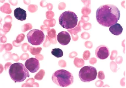
Fig. 1 – Bone marrow smear obtained from an acute myeloid leukaemia subtype M4Eo case at diagnosis showing blast cells and eosinophils with basophilic granules (Romanovsky, 1000x)
The basophilic cytoplasmatic granules are larger and more numerous than in normal immature eosinophils. In some cells, the nuclear morphology is more characteristic of the monocytic lineage, giving the impression that the cell represents a hybrid between an eosinophil and a monocyte.2,4
Changes of 16q22 region was reported in the majority of AML M4Eo subtype cases with abnormal karyotype. The few cases that were classified to other FAB subgroups also exhibited abnormal eosinophils.5 The association
between morphology and cytogenetics is so strong that one can accurately predict the result of 16q22 changes in almost every case of AML M4Eo subtype and vice versa.6
In addition, since high complete remission rate as well as its duration2 have in general been found in AML
146
Rev. bras. hematol. hemoter. 2004;26(2):74-75. Imagens em Hematologia
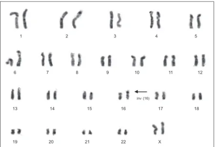
Fig. 2 – G-banded karyotype obtained from an acute myeloid leukaemia subtype M4Eo female case at diagnosis: 46,XX,inv (16)(p13;q22) The arrow shows the chromosomal abnormality
1 2 3 4 5
6 7 8 9 10 11 12
13 14 15 16 17 18
19 20 21 22 X
inv (16)
conventional chemotherapy regimens, this chromosomal abnormality has been considered as a prognostic indicator of favourable outcomes.
Herein, we present, for educational purposes, the images obtained from a bone marrow smear and karyotype (Figures 1 and 2, respectively) of a AML M4Eo subtype, a case seen at the Haematology and Haemotherapy Centre of the State University of Campinas.
References
1. Arthur DC, Bloomfield CD. Partial deletion of the long arm of chromosome 16 and bone marrow eosinophilia in acute non-lymphocytic leukemia: a new association. Blood 1983;61: 994-998.
2. Heim S, Mitelman F. Acute myeloid leukemia. In: Heim S, Mitelman F. Cancer Cytogenetics. 2nd ed. New York: Willey-Liss 1995, p.69-140.
3. Arber DA, Carter NH, Ikle D, et al. Value of combined morphologic, cytochemical, and immunophenotypic features in predicting recurrent cytogenetic abnormalities in acute myeloid leukaemia. Hum Pathol 2003;34(5):479-83.
Avaliação: Editor e dois revisores externos.
Conflito de interesse: não declarado
Recebido: 23/03/2004
Aceito após modificações: 15/05/2004
4. Sun X, Medeiros LJ, Lu D, et al. Dysplasia and high proliferation rate are common in acute myeloid leukemia with inv(16) (p13;q22). Am J Clin Pathol 2003;120(2):236-45.
5. Bernard P, Dachary D, Reiffers J, et al. Acute nonlymphocytic leukemia with marrow eosinophilia and chromosome 16 abnormalities: a report of 18 cases. Leukemia 1989;3:740-745.