147
Imagens em Hematologia Rev. bras. hematol. hemoter. 2004;26(2):147-148
IMAGENS EM HEMATOLOGIA/IMAGES IN HEMATOLOGY
Manoela M. Ortega1
Fernanda G. Pereira2
Cármino A. Souza3
Fernando F. Costa4
Carmen S. P. Lima5
Detection of 13q deletion in multiple myeloma using immunomagnetically
selected plasma cells
Detecção de deleção 13q no mieloma múltiplo utilizando plasmócitos selecionados
imunomagneticamente
Correspondence to: Carmen Silvia Passos Lima, MD, PhD
Departamento de Clínica Médica – Faculdade de Ciências Médicas – Unicamp – Cidade Universitária “Zeferino Vaz” Rua Alexandre Fleming, 181 – Distrito de Barão Geraldo
13083-970 – Campinas, SP – Brazil
Phone: + 55 19 3788-7098 – Fax: + 55 19 3289-4107 – e-mail: [email protected]
Multiple myeloma (MM) is a tumour characterised by terminally differentiated plasma cells. Since plasma cell turnover is considerably slower than that of non-malignant bone marrow cells, it is to be expected that the majority of mitotic cells in cultures from MM patients will represent normal marrow elements.1
Recent studies based on interphase fluorescence in situ hybridisation (FISH) have shown that virtually all MM patients have chromosomal abnormalities in their plasma cells.2,3 Deletions of 13q14 have been detected in 30-50%
Fig. 1 – Mature plasma cells selected from the bone marrow sample of the multiple myeloma patient using the CD138-specific antibody (Miltenyi Biotec, Germany) on a magnetic column (Miltenyi Biotec, Germany) (Romanovsky, 1200x)
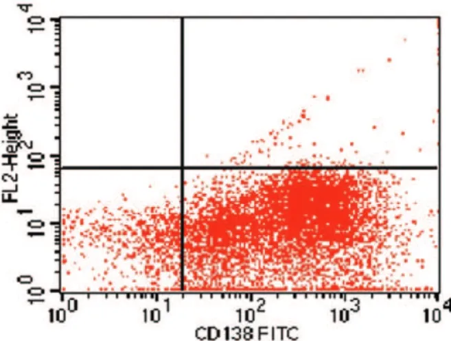
Fig. 2 – Plasma cells were selected from the bone marrow sample of the myeloma multiple patient using the CD138-specific antibody (Miltenyi Biotec, Germany) conjugated with isothiocynate fluorescein (FITC) and flow cytometry analysed (90.2% of purity)
1
Pós-graduanda (Doutoramento) do Departamento de Clínica Médica da Faculdade de Ciências Médicas da Unicamp. 2
Pós-graduanda (Mestrado) do Departamento de Clínica Médica da Faculdade de Ciências Médicas da Unicamp. 3
Professor Titular da Disciplina de Hematologia e Hemoterapia, do Departamento de Clínica Médica da Faculdade de Ciências Médicas da Unicamp. 4
Professor Titular da Disciplina de Hematologia e Hemoterapia, do Departamento de Clínica Médica da Faculdade de Ciências Médicas da Unicamp. 5
148
Rev. bras. hematol. hemoter. 2004;26(2):147-148 Imagens em Hematologia
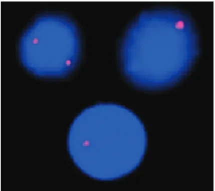
Fig. 3 – Interphase fluorescence in situ hybridisation analysis on the multiple myeloma patient’s plasma cells, using specific probe for D13S319 locus of the 13q14 region (Vysis, Downers Grove, IL, USA) (•). Normal interphase nucleus exhibiting two red hybridisation signals (right) and abnormal nuclei exhibiting heterozygous deletion of the D13S319 locus (left and middle)
of MM patients’ samples using FISH analysis,4,5 which
have been seen as a prognostic indicator of poor outcomes,5,6 as well as increased serum levels of
C-reactive protein, lactate dehydrogenase and Durie & Salmon’ stage III.7
Herein, we present, for educational purposes, the description and the images obtained from a bone marrow sample analysed by FISH of a MM case with 13q14 deletion seen at the Haematology and Haemotherapy Centre of the State University of Campinas. A 40-year-old man was referred to our service due to severe bone pain. The laboratory evaluation showed haemoglobin: 8.3g/dL, serum calcium 10.4 mg/dL (normal range: 8.6-10.0), creatinine: 2.61 mg/dL (normal range: 0.6-1.4), C-reactive protein: 17.2 mg/dL (normal range: < 0.3), lactate de-hydrogenase: 489.0 U/L (normal range: 240.0-479.0), and serum albumin: 3.4 g/dL (normal range: 3.5-5.0).
Bone marrow cytologic analysis showed 35.5% of plasma cells. X-ray revealed collapse of L4 vertebral body. The diagnosis of non-secreting MM stage IIIb was made.
FISH analysis of the 13q14 region was performed on immuno-magnetically selected plasma cells (Figures 1 and 2) according to the manufacturer ’s protocols. Heterozygous deletion of 13q14 region was detected in 30.0% of the interphase nuclei (Figure 3).
The patient died one week later due to sepsis and his very short survival gives support to the postulate that 13q14 deletion increased serum levels of C-reactive protein and lactate dehydrogenase, and stage IIIb act as predictors of unfavourable outcomes in MM.
References
1. Greipp PR, Katzmann JA, O’Fallon WM, et al. Value of beta 2-microglobulin level and plasma cell labelling as prognostic factors in patients with newly diagnosed myeloma. Blood 1988; 72:219-23.
2. Drach J, Schuster J, Nowotny H, et al. Multiple myeloma: High incidence of chromosomal aneuploidy as detected by interphase fluorescence in situ hybridization. Cancer Res 1995;55:3.854-59.
3. Perez-Simon JA, Garcia-Sanz R, Tabernero MD, et al. Prognostic value of numerical chromosome aberrations in multiple myeloma: A FISH analysis of 15 different chromosomes. Blood 1998;91(9):3.366-71.
4. Könisberg R, Zojer N, Ackermann J, et al. Predictive role of interphase cytogenetics for survival of patients with multiple myeloma. J Clin Oncol 2000;18:804-12.
5. Fonseca R, Oken M, Harrington D, et al. Deletions of chromosome 13 in multiple myeloma identified by interphase FISH usually denote large deletions of the q-arm or monosomy. Leukemia 2001;15:981-6.
6. Zojer N, Konigsberg R, Ackermann J, et al. Deletion of 13q14 remains an independent adverse prognostic variable in multiple myeloma despite its frequent detection by interphase fluorescence. Blood 2000;95(6):1.926-30.
7. Barlogie B, Shaughnessy J, Munshi N, et al. Hematology. 6th ed. New York, McGraw-Hill, 2001. p.1.279-304.
Avaliação: Editor Associado e dois revisores externos. Conflito de interesse: não declarado