119
Chest radiographs: doses and imaging quality
Radiol Bras 2007;40(2):119–122
Original Article
PATIENTS EXPOSURE AND IMAGING QUALITY IN CHEST
RADIOGRAPHS: A CRITICAL EVALUATION*
Adelaja Otolorin Osibote1
, Ana Cecília Pedrosa de Azevedo2
, Antonio Carlos Pires Carvalho3 , Helen Jamil Khoury4
, Sergio Ricardo de Oliveira5
, Marcos Otaviano da Silva6
, Carla Marchon7
OBJECTIVE: Entrance skin dose, effective dose, and imaging quality in chest radiographs of adult patients have been evaluated. MATERIALS AND METHODS: The study has been developed in eight institutions — seven public hospitals (two of them philanthropic institutions) and one private — in the cities of Agra dos Reis, Cabo Frio, Campos dos Goytacazes, Itaperuna, Niterói, Recife and Rio de Janeiro. Entrance skin dose and effective dose have been evaluated in 735 chest radiographs obtained in posteroanterior/anteroposte-rior and lateral projections. As regards imaging criteria, 44 radiographs have been evaluated. RESULTS: Variations of up to nine times in entrance skin dose, and six times in effective dose have been detected for a same type of projection. Also, significant discrepancies have been found in values resulting from radio-graphic techniques employed. Besides, imaging quality has not been good since the rate of compliance with imaging criteria was only 55%. CONCLUSION: There is a pressing need for improvement/standardization of procedures in conventional radiology; this can be achieved by implementing a quality control and assurance program in the department of radiology, including training of technicians, x-ray equipment calibration, and sensitometric control of films processors.
Keywords: Chest x-ray; Quality control; Dosimetry.
Exposição de pacientes e qualidade da imagem em radiografias de tórax: uma avaliação crítica.
OBJETIVO: Foi realizada avaliação da dose de entrada na pele, da dose efetiva e da qualidade da imagem em radiografias de tórax de pacientes adultos. MATERIAIS E MÉTODOS: O estudo realizou-se em oito hospitais, sendo sete públicos (dois filantrópicos) e um particular nos municípios de Angra dos Reis, Cabo Frio, Cam-pos dos Goytacazes, Itaperuna, Niterói, Recife e Rio de Janeiro. Foram avaliadas a dose de entrada na pele e a dose efetiva de 735 radiografias de tórax nas incidências póstero-anterior/ântero-posterior e perfil. No que se refere aos critérios de imagem, foram avaliadas 44 radiografias. RESULTADOS: Constatou-se varia-ção de até nove vezes nos valores da dose de entrada na pele e de até seis vezes na dose efetiva para um mesmo tipo de projeção. Os valores das técnicas radiográficas também apresentaram grandes discrepân-cias. A qualidade das imagens também não é boa, pois foi obtido valor médio de presença dos critérios de apenas 55%. CONCLUSÃO: Há necessidade de melhoria/padronização de procedimentos em radiologia convencional, o que pode ser atingido se for implantado um programa de controle e garantia de qualidade no setor de radiologia, incluindo o treinamento dos técnicos, a aferição do desempenho dos equipamentos emissores de radiação e o controle sensitométrico do sistema de processamento radiográfico.
Unitermos:Radiografia torácica; Controle de qualidade; Dosimetria. Abstract
Resumo
* Study developed at Fundação Oswaldo Cruz (Fiocruz), Rio de Janeiro, RJ, Brazil.
1. Physicist, Doctorship Student, Fundação Oswaldo Cruz (Fiocruz), Escola Nacional de Saúde Pública Sergio Arouca-CESTEH, Rio de Janeiro, RJ, Brazil.
2. PhD in Physics, Fundação Oswaldo Cruz (Fiocruz), Escola Nacional de Saúde Pública Sergio Arouca-CESTEH, Centro de Vigilância Sanitária da Secretaria de Estado de Saúde do Rio de Janeiro, Rio de Janeiro, RJ, Brazil.
3. MD, PhD in Medicine, Department of Radiology at Facul-dade de Medicina da UniversiFacul-dade Federal do Rio de Janeiro (UFRJ), Rio de Janeiro, RJ, Brazil.
4. PhD in Physics, Department of Nuclear Energy at Universi-dade Federal de Pernambuco (UFPE), Recife, PE, Brazil.
5. Physicist, Doctorship Student, Fundação Oswaldo Cruz (Fiocruz), Instituto Oswaldo Cruz, Rio de Janeiro, RJ, Brazil.
6. Physicist, Doctorship Student, Fundação Oswaldo Cruz (Fiocruz), Escola Nacional de Saúde Pública Sergio Arouca-CESTEH, Centro de Vigilância Sanitária da Secretaria de Estado de Saúde do Rio de Janeiro, Rio de Janeiro, RJ, Brazil.
7. MD, Service of Radiology at Hospital da Força Aérea do Galeão, Rio de Janeiro, RJ, Brazil.
Mailing address: Dra. Ana Cecília Pedrosa de Azevedo. Fun-dação Oswaldo Cruz (Fiocruz), Escola Nacional de Saúde Públi-ca Sergio ArouPúbli-ca-CESTEH. Rua Leopoldo Bulhões, 1480,
INTRODUCTION
Medical clinics and hospitals which uti-lize ionizing radiation have been searching to be in compliance with the radiological protection and quality control requirements of the Order (Portaria) No. 453/98 “Diretri-zes de proteção radiológica em radiodiag-nóstico médico e odontológico” (“Radio-logical protection guidelines in medical and odontological radiodiagnosis”), pub-lished in 1998 by the Ministry of the Health – National Sanitary Vigilance Agency(1).
In the states of Rio de Janeiro and Pernambuco, several hospitals and clinics have been the target of academic researches in the field of radiological protection and quality control in diagnostic radiology. These studies are coordinated by the Group of Radiological Protection and Quality Control of Fundação Oswaldo Cruz – Es-cola Nacional de Saúde Pública Sergio Arouca – CESTEH, and Group of Dosim-etry and Instrumentation of Universidade Federal de Pernambuco Department of Nuclear Energy.
This study presents the partial results of the interaction between these two institu-tions, with a evaluation of doses and im-ages quality in chest x-rays performed in
Manguinhos. Rio de Janeiro, RJ, Brazil, 21041-210. E-mail: [email protected]
120
Osibote AO et al.
Radiol Bras 2007;40(2):119–122 eight hospitals in seven cities in the states
of Rio de Janeiro and Pernambuco.
MATERIAL AND METHODS
This study was developed in eight hos-pitals in the following cities: Angra dos Reis, Cabo Frio, Campos dos Goytacazes, Itaperuna, Niterói, Recife and Rio de Ja-neiro. These hospitals are randomly de-nominated A, B, C, D, E, F, G e H.
Entrance skin dose (ESD) and effective dose (ED) were evaluated in 520 poster-oanterior/anteroposterior (PA/AP) and 215 lateral chest x-rays of adult patients. A par-allel critical analysis of images quality was made in 44 x-ray films, adopting the Euro-pean guidelines on quality criteria for di-agnostic radiographic images of the Com-mission of European Communities(2).
1. Doses calculation
Aiming at speeding up the process of patient dose measurement a software called DoseCal, running on Windows platform was employed(3). The DoseCal software(4) calculates the ESD, the body organ dose (BOD), and the ED, based on values of the radiographic technique employed, the x-ray tube output, and the patients’ anthropomet-ric data. The DoseCal software has been developed in the Radiological Protection Center at Hospital Saint Georges (London) and plays an essential role in the evaluation of radiation doses for a great number of patients. This software has been kindly pro-vided for the present project in Brazil.
For a correct operation of the software, it is necessary to enter the x-ray tube out-put in mGy/mAs; this data may be easily obtained with a calibrated ionization cham-ber. In the present study, we have utilized a Nero 8000-Inovision and a Radcheck Plus 06-526. Once output values, current, kilovoltage, exposure time and focus-skin distance (FSD) are known, the following equation (1) will demonstrate the ESD.
where: Output is the x-ray tube perfor-mance expressed in mGy/mAs, at 80 kV, and at a distance of 1 m normalized for 10
mAs; kV is the potential applied to the tubr (in kilovolts); mAs is the product from current × exposure time; FSD is expressed in centimeters (cm); and BSF is the back-scatter factor. The DoseCal software uti-lizes the conversion factors included in tables NRPB-SR262(5) applied for ESD, BOD and ED calculations.
2. Images criteria
Based on the premise that “the best image will provide a better diagnosis” the European Union has formed a commission to develop quality criteria for diagnostic radiographic images. Other criteria such as general principles associated with good imaging performance and guidelines on radiation dose to the patient have been in-cluded. The most recent version of this document(2) was published in 1996 (EUR 16260 EN-European Guidelines on Qual-ity Criteria for Diagnostic Radiographic Images), including criteria for imaging the chest, skull, lumbar spine, pelvis, urinary tract, and breast. These criteria were basi-cally defined considering or not the pres-ence of anatomical structures of the fo-cused region, as well as their visualization degree. The criteria are the following: vi-sualization — anatomical characteristics are detected but are not totally reproduced;
reproduction — anatomical detials are identified, but are not clearly defined; vi-sually sharp reproduction — anatomical details are clearly defined.
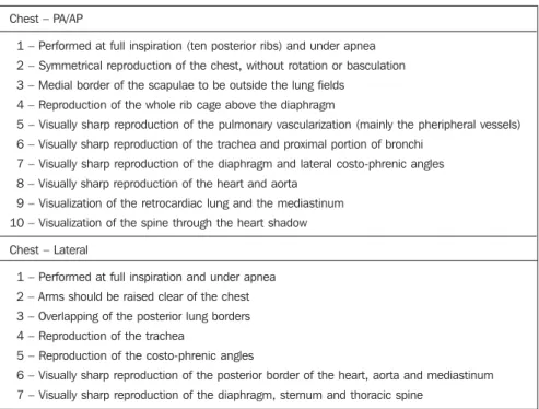
The images criteria for chest in PA/AP and lateral projections, according to the Eu-ropean Communities, are shown on Table1.
RESULTS
The Table 2 includes the statistics (mean, first and second quartiles) of ESD (in mGy) in PA/AP and lateral chest x-rays. It may be oserved that, on PA/AP projections, mean values range between 0.07 mGy (hospitals E and H) and 0.64 mGy (hospital C) (mean value, 0.24 mGy). Values on lateral projec-tion ranged between 0.14 mGy and 1.02 mGy (mean value, 0.47 mGy). As regards ED, values ranged between 0.01 mSv pitals B, D, E and H) and 0.06 mSv (hos-pital C) (mean value, 0.03 mSv) for PA/AP projections, and between 0.01 mSv and 0.7 mSv (mean value, 0.2 mSv) on lateral pro-jections.
Table 3 and Figure 1 show values of ra-diographic techniques employed and pa-tients’ anthropometric data. Mean kilovol-tage values ranged between 70 kV (hospi-tals B and C) and 93 kV (hospital E) (mean value, 78 kV) on PA/AP projections. On
ESD = Output × ( )² × ( )² ×
× mAs × BSF kV
80
100
FSD
(1)
Table 1 European Commission images criteria for diagnostic radiographic chest images in PA/AP and lateral projections.
Chest – PA/AP
1 – Performed at full inspiration (ten posterior ribs) and under apnea
2 – Symmetrical reproduction of the chest, without rotation or basculation
3 – Medial border of the scapulae to be outside the lung fields
4 – Reproduction of the whole rib cage above the diaphragm
5 – Visually sharp reproduction of the pulmonary vascularization (mainly the pheripheral vessels)
6 – Visually sharp reproduction of the trachea and proximal portion of bronchi
7 – Visually sharp reproduction of the diaphragm and lateral costo-phrenic angles
8 – Visually sharp reproduction of the heart and aorta
9 – Visualization of the retrocardiac lung and the mediastinum
10 – Visualization of the spine through the heart shadow
Chest – Lateral
1 – Performed at full inspiration and under apnea
2 – Arms should be raised clear of the chest
3 – Overlapping of the posterior lung borders
4 – Reproduction of the trachea
5 – Reproduction of the costo-phrenic angles
6 – Visually sharp reproduction of the posterior border of the heart, aorta and mediastinum
121
Chest radiographs: doses and imaging quality
Radiol Bras 2007;40(2):119–122
lateral projections, values ranged between 85 kV and 95 kV (mean value, 90 kV). As regards milliamperage, values ranged be-tween 3 mAs (hospital E) and 36 mAs (hos-pital C) (mean value, 12 mAs) on PA/AP projections. On lateral projections, the val-ues ranged between 7 mAs and 24 mAs (mean value, 15 mAs). The FSD ranged between 120 cm (hospital C) and 162 cm (hospital G) (mean value, 139 cm) on PA/ AP projections. On the lateral projections, values ranged between 109 cm and 157 cm. Patients’ mean age was 48 years for PA/AP projections, and 49 years for lateral projec-tions. Mean weight was 67 kf for PA/AP and lateral projections.
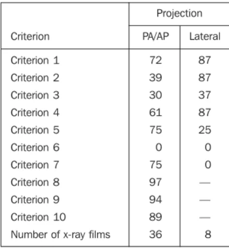
As regards image criteria, the results are shown on the Table 4 and Figura 2. The criteria with highest compliance rates were criterion 8 present in 97% of x-ray studies, and criterion 9, in 94%, both for PA/AP projections. On the other hand, criterion 6 (for both projections), and criterion 7 (for lateral projection) were absent in all the images.
DISCUSSION
ESD values demonstrate high variation among the hospitals evaluation. A differ-ence of more than nive times was found in ESD values, and more than six times in ED values. These differences reflect the dispar-ity of radiographic techniques employed in each institution. It is possible to observe that hospital C, with highest ESD value (0.64 mGy), was the one utilizing the low-est mean kilovoltage (70 kV) and highlow-est
Table 2 Statistics (mean, first and second quar-tiles) of ESD and ED on PA/AP and lateral projec-tions, for the eight hospitals.
Projection
Mean (ESD [mGy]) First quartile
Second quartile Number of x-ray films
ED (mSv)
Mean (ESD [mGy]) First quartile
Second quartile Number of x-ray films
ED (mSv)
Mean (ESD [mGy]) First quartile
Second quartile Number of x-ray films
ED (mSv)
Mean (ESD [mGy]) First quartile
Second quartile Number of x-ray films
ED (mSv)
Mean (ESD [mGy]) First quartile
Second quartile Number of x-ray films
ED (mSv)
Mean (ESD [mGy]) First quartile
Second quartile Number of x-ray films
ED (mSv)
Mean (ESD [mGy]) First quartile
Second quartile Number of x-ray films
ED (mSv)
Mean (ESD [mGy]) First quartile
Second quartile Number of x-ray films
ED (mSv) PA/AP 0.36 0.20 0.54 55 0.04 0.13 0.06 0.18 79 0.01 0.64 0.38 0.86 8 0.06 0.09 0.08 0,10 58 0.01 0.07 0.04 0.06 17 0.01 0.37 0.27 0.45 142 0.04 0.19 0.15 0.21 66 0.02 0.07 0.02 0.10 95 0.01 Lateral — — — — — — — — — — — — — — — — — — — — 0.14 0.11 0.15 13 0.01 1.02 0.77 1.20 61 0.70 0.54 0.43 0.61 61 0.05 0.18 0.08 0.20 80 0.02 Hospital A Hospital B Hospital C Hospital D Hospital E Hospital F Hospital G Hospital H
Table 3 Mean values for radiographic techniques employed in the eight hospitals, and average an-thropometric data of patients.
Projection
kV mAs
Focus-film distance (cm) Patient’s age (years)
Patient’s weight (kg)
kV mAs
Focus-film distance (cm) Patient’s age (years)
Patient’s weight (kg)
kV mAs
Focus-film distance (cm) Patient’s age (years)
Patient’s weight (kg)
kV mAs
Focus-film distance (cm) Patient’s age (years)
Patient’s weight (kg)
kV mAs
Focus-film distance (cm) Patient’s age (years)
Patient’s weight (kg)
kV mAs
Focus-film distance (cm) Patient’s age (years)
Patient’s weight (kg)
kV mAs
Focus-film distance (cm) Patient’s age (years)
Patient’s weight (kg)
kV mAs
Focus-film distance (cm) Patient’s age (years)
Patient’s weight (kg)
PA/AP 80 12 121 44 66 70 15 124 44 67 70 36 120 46 62 79 5 160 51 76 93 3 150 51 65 73 15 121 47 66 83 8 162 52 63 75 5 151 46 68 Lateral — — — — — — — — — — — — — — — — — — — — 95 7 144 50 65 85 24 109 47 66 95 16 157 53 63 85 11 153 46 68 Hospital A Hospital B Hospital C Hospital D Hospital E Hospital F Hospital G Hospital H
Table 4 Rate of presence of imaging criteria in PA/ AP and lateral projections.
Criterion Criterion 1 Criterion 2 Criterion 3 Criterion 4 Criterion 5 Criterion 6 Criterion 7 Criterion 8 Criterion 9 Criterion 10
Number of x-ray films
122
Osibote AO et al.
Radiol Bras 2007;40(2):119–122 milliamperage (36 mAs). Additionally,
hospital C employs an extremely low fo-cus-film distance (120 cm). These factors contribute for an increase in the radiation dose to patient.
Several factors also contribute for the variation of doses; the most significant are: technicians training, system of radio-graphic films processing, the luminance of the negatoscope utilized for images evalu-ation and x-ray beam filtrevalu-ation.
As regards images quality criteria, the criterion 6 in PA/AP projections (“Visually sharp reproduction of trachea and proximal portion of bronchi”) was absent in all of the images, indicating the impossibility of a visally sharp reproduction of this region in these projections. Also, criterion 6 in lat-eral projection (“Visually sharp reproduc-tion of the llposteiror border of the heart, aorta and the mediastinum”) could not be detected in any of the images. The criterion 7 (“Visually sharp reproduction of the dia-phragm, sternum and thoracic spine”), for lateral projection, also was not present in any of the images analyzed. The mean rate ofpresence of criteria was 55% (63% for PA/AP projections, and 46% for lateral projection).
Also, based on data, it could be ob-served that on PA/AP chest x-rays, the cri-teria 2 and 3 presented the lowest rate of presence. These criteria refer to the
sym-metrical reproduction of the chest and me-dial border of the scapulae to be outside of the lung fields, and therefore, to the patient positioning. This reuslt demonstrate the relevance of the necessity of technicians qualifying.
CONCLUSION
The doses standardization/reduction may be achieved by means of easy-to-implement (almost always very simple) measures. These measures should be in-cluded in a program of quality control and assurance to be implemente in every ser-vice of diagnostic radiology. The appropri-ate training of technicians, the x-ray equip-ment and automatic films processor perfor-mance, as well as the employment of high kilovoltage techniques may be extremely useful for reduction of radiation dose to patients and obtention of high quality im-ages.
As regards the quality criteria estab-lished by the European Communities, cer-tainly a x-ray in compliance with all the criteria will result in a better diagnosis. Ba-sically, a good x-ray film depends on an adequate training of the technician, who, in the absence of a radiologist, must be able to decide whether the image is adequate or not. This will be easier if the image crite-ria are known. The mean rate of presence
of the European criteria on the images in the present study (55%) demonstrates that maybe such images have not the quality re-quired for a more reliable and adequate diagnosis.
Acknowledgements
We would like to thank Centro de Vigilância Sanitária-SES/RJ, Fundação Oswaldo Cruz (Fiocruz), Universidade Federal de Pernambuco, Third World Or-ganization for Women in Science, and In-ternational Centre for Theoretical Physics-ICTP de Trieste, Italy.
REFERENCES
1. Brasil, Ministério da Saúde, Secretaria de Vigi-lância Sanitária. Diretrizes de proteção radioló-gica em radiodiagnóstico médico e odontológico. Portaria 453/98, de 1/6/1998. Brasília: Diário Ofi-cial da União 103, 2/6/1998.
2. Commission of European Communities. Euro-pean guidelines on quality criteria for diagnostic radiographic images. Report EUR 16260EN. Bruxelas: European Communities/Union, 1996. 3. Kyriou JC, Newey V, Fitzgerald MC. Patient doses in diagnostic radiology at the touch of a button. London, UK: The Radiological Protection Center, St. George’s Hospital, 2000.
4. Azevedo ACP, Mohamadain KEM, Osibote AO, Cunha ALL, Pires Filho A. Estudo comparativo das técnicas radiográficas e doses entre o Brasil e a Austrália. Radiol Bras 2005;38:343–346. 5. Hart D, Jones DG, Wall BF. Normalized organ
doses for medical x-ray examinations calculated using Monte Carlo techniques. NRPB-SR262. Chilton: NRPB, 1994.
Figure 2. Rate of presence of image quality criteria on the evaluated x-ray films.
Figure1. Mean kilovoltage employed in the studied hospitals for PA/AP and lateral chest x-rays.
K
il
o
vo
lt
a
g
e
Criterion
R
a
te
o
f
p
re
s
e
n
c
e
(%)