Guidelines on the diagnosis and management of multiple myeloma treatment:
Associação Brasileira de Hematologia e Hemoterapia e Terapia Celular
Project guidelines: Associação Médica Brasileira - 2012
Vânia Tietsche de Moraes Hungria1
Edvan de Queiroz Crusoe1
Adriana Alvarez Quero1
Manuella Sampaio1
Angelo Maiolino2
Wanderley Marques Bernardo3,4
1Faculdade de Ciências Médicas da Santa
Casa de São Paulo - FCMSCSP, São Paulo, SP, Brazil
2Universidade Federal do Rio de Janeiro –
UFRJ, Rio de Janeiro, RJ, Brazil
3Faculdade de Medicina da Universidade de
São Paulo - USP, São Paulo, SP, Brazil
4Associação Médica Brasileira - AMB, São
Paulo, SP, Brazil
Conlict-of-interest disclosure:
The authors declare no competing inancial interest
Submitted: 5/1/2013 Accepted: 6/9/2013
Corresponding author:
Vânia Tietsche de Moraes Hungria
Faculdade de Ciências Médicas da Santa Casa de São Paulo,
Departamento de Clínica Médica - FCMSCSP Rua Dr. Cesário Motta Jr. nº 112 Santa Cecília 01221-020 São Paulo, SP, Brazil
Phone: 55 11 21767293 [email protected]
www.rbhh.org or www.scielo.br/rbhh
DOI: 10.5581/1516-8484.20130050
Introduction
The guidelines project is a joint initiative of the Associação Médica Brasileira and the Conselho Federal de Medicina. It aims to bring together information in medicine to standardize conduct in order to help decision-making during treatment. The data contained in this manuscript were prepared by and are recommended by the Associação Brasileira de Hematologia, Hemoterapia e Terapia Celular. Even so, all possible conducts should be evaluated by the physician responsible for treatment depending on the patient’s setting and clinical status.
Aims
To set parameters for clinical diagnosis, evaluate severity and standardize treatment, maintenance and monitoring options for patients with multiple myeloma.
Description of the method used to gather evidence
The target audience of these guidelines is the hematologist and the objective is to contribute to decision making in the diagnosis and management of multiple myeloma.
These Guidelines were drafted by elaborating 15 clinically relevant questions related to the diagnosis and management of multiple myeloma (MM). The questions were structured using the Patient/Problem, Intervention, Compared and Outcome (PICO) system, allowing the generation of evidence search strategies in the key scientiic databases (MEDLINE/ PubMed, Lilacs, SciELO, Embase, Cochrane Library, Premedline via OVID). Moreover a manual search for evidence in dissertations (Biblioteca Digital de Teses e Dissertações do Instituto Brasileiro de Informação em Ciência e Tecnologia – BDTD/IBICT) was carried out. The evidence recovered was critically assessed using discriminatory instruments (scores) according to the category of the question: diagnostic (Quality in Diagnostic and Screening tests - QADAS) or therapeutic (JADAD for randomized clinical trials and the Newcastle Ottawa scale for non-randomized studies). After identifying potential studies to substantiate recommendations, the level of evidence and degree of recommendation were calculated using the Oxford Classiication(1).
Summary of the degree of recommendation and level of evidence: A: Major experimental and observational studies
B: Minor experimental and observational studies C: Case reports (non-controlled studies)
D: Opinion without critical evaluation based on consensus, physiological studies or animal models
Background
Multiple myeloma (MM) is a disorder characterized by abnormal clonal proliferation of plasmocytes in the bone marrow resulting in the production of monoclonal immunoglobulins associated with organic disorders(2) (D). MM accounts for 1% of all neoplastic diseases and 13% of hematologic neoplasms(2) (D).
In Brazil, there is no exact knowledge on the incidence of this disease(3) (B). The median
age at diagnosis in national studies is 60.5 years with most cases being diagnosed in the advanced stage of the disease [76.5% in Durie-Salmon (DS) Stage III](3) (B).
drugs such as thalidomide, lenalidomide and bortezomib, as well as the introduction of autologous bone marrow transplantation have changed the course of the disease, increasing survival and the quality of life of patients(2) (D).
What are the methods to conirm the diagnosis of MM?
P – Patient with symptomatic MM (more than 10% of plasma cells in the bone marrow + monoclonal protein in the blood or urine + the presence of one or more of the following symptoms: anemia, lytic bone lesions, hypercalcemia, renal failure)
I – Analysis of the myelogram, serum and urinary protein electrophoresis, immunoixation of serum and urinary proteins, measurement of serum-free light chain concentration
O – Diagnosis
The diagnostic criteria for symptomatic MM, as established by the International Myeloma Working Group (IMWG) in 2010, are (all three criteria must be met with the exception indicated)(4) (D):
- More than 10% monoclonal plasma cells present in the bone marrow
- The presence of serum or urinary monoclonal protein (except in patients with non-secretory MM, who must have more than 30% of monoclonal plasma cells in the bone marrow or plasmocytoma conirmed by biopsy(2) (D) - evidence of organic damage related to MM, speciically
using the CRAB criteria:
C: hypercalcemia – serum calcium > 11.5 mg/dL or R: renal insuficiency – serum creatinine > 2 mg/dL or creatinine clearance estimated at 40 mL/min or
A: anemia – normocytic, normochromic with hemoglobin 2 g/dL below the normal value or < 10 g/dL or
B: bone lesions: lytic lesions or severe osteopenia attributed to a proliferative disorder of plasma cells or pathological fractures.
The following should be performed to conirm these criteria in a patient with suspected MM(5) (D):
1. Anamnesis, family history and physical examination; 2. Complete blood count and morphological evaluation
using a peripheral blood smear; 3. Laboratory tests;
4. Electrophoresis and immunoixation of serum protein; 5. Quantiication by nephelometry of serum
immunoglobulin;
6. Electrophoresis and immunoixation of urinary protein; 7. Bone marrow aspiration or biopsy;
8. Cytogenetics [karyotyping in metaphase and luorescent in situ hybridization (FISH)];
9. Evaluation of x-rays of the skeleton, including the spine, pelvis, skull, femur and humerus, with magnetic resonance imaging in speciic situations;
10. Measurement of serum-free light chains (Kappa and Lambda).
The clinical history should mainly identify the symptoms related to bone pain (present in 58% of patients)(2) (D). The
family history should concentrate on close relatives diagnosed with malignant hematologic diseases, in particular lymphoma, chronic lymphocytic leukemia and plasma cell dyscrasias(5) (D).
The evaluation of the complete blood count is important because anemia is present in 73% of patients with diagnosis of MM(2) (D). As for the peripheral blood morphology, this can show speciic indings such as the presence of the rouleaux formation and circulating plasma cells(5) (D). Laboratory tests include kidney
function, liver function and the measurement of calcium levels to evaluate possible organic damage(5) (D). Changes in kidney function at the diagnosis of MM are present in 20 to 40% of cases and hypercalcemia is uncommon(2) (D).
Serum or urinary protein capillary gel or agarose gel electrophoresis is used to detect monoclonal proteins. The sensitivity of this test alone is 80.4%, with 77.9% speciicity for the diagnosis of monoclonal gammopathy(6) (B). However, quantiication of serum immunoglobulins by nephelometry, when available, should also be performed. These two tests are complementary, and evaluation by nephelometry may be particularly useful for low levels of immunoglobulins(5) (D).
Immunoixation is the gold standard method to conirm the diagnosis of MM, with the identiication of the monoclonal protein and of the light and heavy chains involved(7) (B). The sensitivity of this test alone is 41%, while its speciicity is 100%(7) (B). Serum immunoixation should also be performed
when there is hypogammaglobulinemia or when the protein electrophoresis looks normal, but there is the suspicion of MM or a related disorder(5) (D).
The dosage of immunoglobulin serum-free light chains is recommended for all patients with newly diagnosed plasma cell dyscrasia(5) (D). However, it is even more important in patients
with secretory MM, i.e. those with negative urinary and serum immunoixation and in patients that secrete small amounts of monoclonal proteins in serum or urine (oligosecretory myeloma)
(5) (D) observed in 15 to 30% of all cases of MM(8) (B).
About 6.77% of patients with B-cell malignancies (MM, light-chain amyloidosis, Waldenstrom’s macroglobulinemia and non-Hodgkin’s B-cell Lymphoma) have negative serum protein electrophoresis results, with elevated levels of serum-free light chains; 38.23% of these patients have negative serum immunoixation of clinical relevance(8) (B).
Only 80.8% of patients with positive results for urinary immunofixation, have positive serum protein electrophoresis results, 85.7% have elevated levels of serum-free light chains and 93.5% have positive results for serum immunofixation. Thus, 0.5% of patients with monoclonal gammopathy will not be diagnosed if urinary immunofixation is not performed(9) (B).
Bone marrow aspiration or biopsy conirms the diagnosis when more than 10% of clonal plasma cells are detected(5) (D).
The sensitivity of diagnosis by myelogram with morphological analysis of the cells is 79% and the speciicity is 100%(7) (B). On
using bone marrow biopsy, whenever possible, immunostaining for the CD138 antigen should be used to accurately determine the percentage of plasma cells(5) (D).
Flow cytometry is the only technique available to quantify tumor plasma cells(7) (B). CD45- cells are observed in 70.1% of symptomatic MM patients and CD19- cells in 100%. The CD56 expression in plasma cells is observed in 69% of cases, while the CD117 expression is observed in 50.6% of symptomatic MM patients(7) (B). The sensitivity of this
diagnostic test for MM in identifying abnormal plasma cells is 74%, with a sensitivity of 85%(7) (B).
Cytogenetics should be included in the initial assessment of patients with suspected MM. Despite the poor performance of this method (≤ 20%) in elucidating the diagnosis, it can provide information on prognosis. Moreover, luorescent in situ hybridization (FISH) should be carried out, preferably after screening for plasma cells, with probes that include the 17p13, t(4,14), and t(14,16) chromosome abnormalities(5) (D).
The examination of the skeleton remains the standard imaging method for diagnostic screening. This test, which is readily available at a reasonable cost, can detect the risk of long bone fractures due to lesions. The simple radiograph must include the thorax, the cervical, lumbar and thoracic regions of the spine, the upper arms, femur, skull and pelvis(2) (D). Bone lesions are present in 80% of patients at diagnosis(2) (D).
Magnetic resonance imaging (MRI) is a non-invasive technique that provides detailed information on patients with suspected spinal compression, evaluates collapsed and fractured vertebra, size of tumor mass and how it might affect the epidural space. An MRI of the spine and the pelvis is mandatory in all patients with presumed diagnosis of solitary plasmacytoma. Nuclear magnetic resonance (NMR) should also be considered in patients with asymptomatic myeloma because it may detect lesions thus predicting progression to symptomatic MM(5) (D).
The role of positron emission tomography-computed tomography PET-CT has not yet been clearly deined in MM(5) (D).
Recommendations: When MM is suspected, the following tests should be performed: complete blood count with morphological assessment of the peripheral blood, calcium and creatinine levels, and serum and urinary protein electrophoresis. On positive tests, diagnostic conirmation is achieved with serum and urinary immunoixation, bone marrow aspirate or biopsy and radiography of the skeleton.
How is the prognosis of the patient determined
(exams)?
P – Patient with symptomatic MM
I – Analysis of beta 2-microglobulin, cytogenetics and molecular studies O – Prognosis
In the era of combined treatments for MM, cytogenetic abnormalities, high-risk gene expressions and high levels of lactic dehydrogenase and beta 2-microglobulin (β2-M) are considered as the main prognostic factors associated with lower overall survival (OS) and shorter event-free survival (EFS)(10) (B).
The combined measurement of β2-M and albumin, as proposed by the International Staging System (ISS), has been validated to assess prognosis(11) (B).
Of the different MM phenotypes, the immunoglobulin A (IgA) isotype together with a genetic proile of poor prognosis reduces the OS to a median of 83 months and the EFS to a median of 49 months. Immunoglobulin D (IgD) also affects the survival with OS and EFS dropping to 84 months and 41 months, respectfully. However, because of its rare occurrence (1%), IgD does not have a signiicant impact in statistical terms even though it is strongly linked to increased lactic dehydrogenase and β2-M levels, as well as to the presence of cytogenetic and molecular abnormalities(10) (B). The OS and EFS for
the other immunoglobulin isotypes are 99 months and 58 months, respectively (p-value = 0.017 and 0.014, respectively)(11) (B).
Between 20 and 50% of patients with MM have numeric or structural chromosomal changes identiied by cytogenetics (metaphases or FISH)(12) (B).
Some gene translocations involving the immunoglobulin heavy chain (IgH), such as t(4;14) and t(14;16), give unfavorable prognosis for MM patients(13) (B); this also happens when the
difference between the values of the serum-free light chains is greater than 185. The median OS for three groups of patients is 3.97, 2.19 and 1.74 years (p-value < 0.0001) with none, one or two risk factors (serum-free light chains and one of the translocations), respectively. These indings are similar when the ratio between serum-free light chains is used rather than the difference between them(13) (B).
Other common cytogenetic changes in MM are t(11;14), del(13q14) and del(17p13). The t(11;14) is usually associated with light chain MM (p-value = 0.0021). The median EFS of patients with the t(11;14) is 25.2 months compared to 25.7 months in patients without this abnormality. Moreover, the median OS for patients with and without this translocation is 37.2 and 46.3 months, respectively (p-value = 0.78). There is no association between the t(11;14) and other biological parameters such as age, gender, hemoglobin level, β2-M, C-reactive protein, calcium, creatinine and albumin levels or the percentage of plasma cells in the bone marrow(14) (B).
For the del(13q) or t(4;14) aberrations, the time of disease progression is 5.9 months [hazard ratio (HR): 1.42; p-value = 0.09] and 8 months (HR: 1.44; p-value = 0.14), respectively(15) (B).
The EFS for patients with t(4;14) is 9.9 months compared to 25.8 months in patients without this translocation (p-value = 0.0003)
In the assessment of del(17p13), there is a signiicant reduction in survival, with the mean time to disease progression and OS being 2.2 and 4.7 months, respectively (time to disease progression: HR: 2.82; p-value = 0.001; and OS: HR: 3.23; p-value = 0.001)(15) (B).
With regards to the prognostic impact of the response to induction treatment (six cycles of combined chemotherapy), patients who have complete responses (CR) as evaluated by immunophenotyping, the measurement of serum-free light chains or immunoixation, have signiicantly better outcomes compared to those who have partial responses (PR) for 3-year EFS (90%, 69%, 60% and 35%, respectively; p-value = 0.001), time to disease progression (96%, 71%, 68% and 37%, respectively; p-value = 0.001), and increased OS (94%, 94%, 93% and 70%, respectively; p-value = 0.08)(16) (B).
The EFS evaluated by measuring serum-free light chains or by immunoixation is signiicantly longer in patients with CR than in patients with PR (p-value = 0.001). However, assessment by immunophenotyping in isolation for the EFS is an independent prognostic factor [RR: 4.1; 95% conidence interval (95% CI): 1.4-12; p-value = 0.01](16) (B).
The addition of the measurement of free light chains to a pre-existent risk stratiication model for asymptomatic MM was identiied as an important determinant of disease progression to symptomatic MM. The incorporation of this variable has enabled a calculation of the 5-year chance of progression of the different clinical stages of asymptomatic MM, DS Grades I, II and III, to symptomatic MM at 25%, 51% and 76%, respectively(17) (B).
Univariate analysis of patients after autologous bone marrow transplantation shows higher rates of relapse in patients aged > 50 years (p-value = 0.002), patients with del(13q14) (p-value = 0.006) and in patients with del(17p13) (p-value = 0.003). In multivariate analysis, only the del(13q14) (HR: 2.34; p-value = 0.03) and del(17p13) (HR: 2.24; p-value = 0.04) signiicantly inluence the incidence of relapse, while for the EFS, only age (HR: 2.8; p-value = 0.01) and del(17p13) (HR: 2.05; p-value = 0.03) are negative prognostic factors(18) (B). These data show that del(17p13) is a negative prognostic factor both to attain CR and improved EFS after autologous bone marrow transplantation. Whereas patients with the t(4;14) can have a positive response to transplantation(18) (B).
Recommendations: The dosage of β2-M and albumin should be used to evaluate prognosis in MM according to the ISS. Currently, conventional and molecular cytogenetic changes are widely recognized as prognostic factors. Evaluations by serum and urinary immunoixation and the determination of serum-free light chains after chemotherapy or bone marrow transplantation are useful to assess response to treatment.
How is the prognosis of patient determined (score)?
P – Patient with symptomatic MM
I – Durie-Salmon Staging, International Staging System O - Prognosis
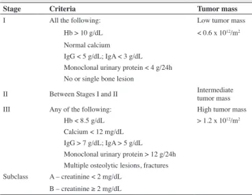
A clinical staging system called Durie-Salmon was irst
published in 1975 and is used to provide a better initial assessment and follow-up of individual patients with MM by quantifying the tumor mass using the following criteria: hemoglobin concentration, serum calcium level, levels of serum and urinary monoclonal proteins and the presence of bone lesions. Renal function is evaluated independently as it is only related to survival and not to the size of the tumor mass(19) (B). The three stages are
shown in Table 1.
Table 1 - Stages of the Durie-Salmon staging system
Stage Criteria Tumor mass
I All the following: Low tumor mass
Hb > 10 g/dL < 0.6 x 10¹²/m2 Normal calcium
IgG < 5 g/dL; IgA < 3 g/dL Monoclonal urinary protein < 4 g/24h No or single bone lesion
II Between Stages I and II Intermediate tumor mass III Any of the following: High tumor mass
Hb < 8.5 g/dL > 1.2 x 10¹²/m2 Calcium < 12 mg/dL
IgG > 7 g/dL; IgA > 5 g/dL Monoclonal urinary protein > 12 g/24h Multiple osteolytic lesions, fractures Subclass A – creatinine < 2 mg/dL
B – creatinine ≥ 2 mg/dL
On employing the DS, the median OS for patients in Stage III is 52.1 months. After 5 years, the estimate of survival in DS Stages I, II and III is 73.3%, 54.7% and 44.3%, respectively(3) (B).
More recently, under the organization of the International Myeloma Working Group, a new staging system, the ISS, based on the values of β2-M and serum albumin, has been applied to MM patients. Patients are classiied into three risk groups(11) (B):
- Stage I: β2-M < 3.5 mg/L and albumin 3.5 ≥ g/dL
- Stage II: β2-M < 3.5 mg/L and albumin < 3.5 g/dL or β2-M 3.5 to < 5.5 mg/L
- Stage III: β2-M ≥ 5.5 mg/L
With the ISS, the median OS is 62 months for Stage I, 49 to 65.5 months for Stage II and 26 to 29 months for Stage III(3,11) (B). After 5 years of follow-up, the estimated OS for Stages I, II and III is 68.2%, 52.7% and 30.4%, respectively(3) (B).
In the most advanced stage, according to the ISS, there is a higher proportion of patients with advanced age, with anemia (hemoglobin less than 10 g/dL), thrombocytopenia (platelets < 150 x 109/L), with diffuse bone marrow iniltration and poor
performance status(11) (B).
A strong correlation is not observed between cytogenetic data and the clinical stages of the ISS. The t(4; 14) occurs at a signiicantly lower incidence (p-value = 0.035) in patients in Stage I than in patients in Stage II and III (6%, 16% and 11%, respectively)(11) (B).
Considering the impact of cytogenetic abnormalities, a median OS of 42 months is observed in MM patients with any cytogenetic change, compared to 69 months for patients without cytogenetic abnormalities (p-value = 0.03)(11) (B).
Hypercalcemia, Stage III by ISS and old age are independent predictors of mortality; the variables of the DS and Stages I and II by ISS are not correlated as predictors of mortality(3) (B).
Recommendation: the two staging systems are valid in MM. However, apart from the ISS being simpler (fewer variables evaluated), it allows a better evaluation of survival for its different stages, which is not seen with the DS staging system for Stages I and II.
How to deine which patients should be treated?
P – Patient with asymptomatic MM C – with or without treatment O – OS, EFS, response rate, toxicity
The acronym ‘CRAB’ summarizes the typical clinical manifestations of MM: hypercalcemia, renal failure, anemia and bone lesions. These characteristics of MM are used to distinguish between symptomatic MM and its precursors: monoclonal gammopathy of undetermined signiicance and asymptomatic MM. This distinction is relevant not only for classiication and diagnosis, but also for therapeutic decisions; CRAB symptoms are important in the decision of when to start anticancer therapy as no improvement in survival is seen with treatment in asymptomatic MM(20) (B).
The therapy is initiated when the patient is classiied as DS Stage II with signs of progression of the disease, or as DS Stage III with any lytic lesion being seen as a symptom(21) (B). The
median time to progression from symptomatic to asymptomatic MM is one to two years(21) (B)(22) (D).
On comparing treatment with melphalan and prednisone in MM patients in two different periods, at the time of diagnosis and at the time of disease progression, there is no signiicant difference between the two groups. The estimate is 64 months for those who begin treatment immediately after diagnosis and 71 months for those who begin treatment only after disease progression [odds ratio (OR): 1.1: 95% IC: 0.57-2.42; p-value = 0.64](22) (D).
The use of bisphosphonates, while reducing the number of skeleton-related events, does not alter the progression of the disease or the OS of asymptomatic MM patients(23) (B).
Asymptomatic MM patients should be monitored every three months with complete blood count, assessment of renal function and measurement of serum calcium levels; greater attention should be given to patients with a higher risk of progression, i.e. serum monoclonal protein ≥ 3 g/dL and more than 10% of plasma cells in the bone marrow, or ≥ 95% bone marrow plasma cells with abnormal phenotypes(23) (B).
Recommendations: there is no evidence so far that asymptomatic MM patients should receive treatment at diagnosis. In these patients, the median time to progression of the disease is one to two years and treatment should only be started at the onset of symptoms (CRAB).
Is initial treatment with dexamethasone, thalidomide and cyclophosphamide superior to induction with dexamethasone and thalidomide only?
P – Patient with symptomatic MM with indication of autologous hematopoietic stem cell transplantation
I – Initial treatment
C – thalidomide + Dexamethasone (TD) / cyclophosphamide + thalidomide + dexamethasone (CTD)
O – OS, EFS, response rate, toxicity
In adult symptomatic MM patients (not previously treated, resistant or with recurrent MM) who are responsive to treatment with intravenous cyclophosphamide (500 mg/m2) or equivalent
dose (625 mg/m2) on the 1st day, oral thalidomide (100 mg) and dexamethasone (20 mg) on Days 1-4 and 8-11, the probability of survival at two years without progression of disease is 54.8%. The time to progression in patients with stable disease and those with evolution of activity is 12 months and six months, respectively. The probability of OS at two years in responsive patients is 81.7%, with no signiicant difference in relation to stable patients (67.1%). The main toxicity is neurological in 25.8% of cases; this leads to loss of adherence in 7.6% of cases(24) (B).
The 4-week treatment cycles of patients with DS Stage II/III MM who are non-responsive or relapse consists of cyclophosphamide (500 mg) on Days 1, 8 and 15, thalidomide (100 mg daily initially but increasing to 200 mg daily, if tolerated) and oral dexamethasone (40 mg) on days 1-4 and 15-18 in each cycle. With a two-year follow-up, the OS is 69.8%, with 83.8% of cases achieving PR, of which 86% are patients who had not responded to previous treatment. The main adverse effects are constipation (16%), drowsiness (6.5%), neuropathy, deep vein thrombosis (3.2%) and febrile neutropenia (4.8%)(25) (C).
MM patients who relapse or are non-responsive treated with oral cyclophosphamide (300 mg/m2 once per week) combined
with pulses of dexamethasone (40 mg/day for 4 days, once per month) and thalidomide in doses spaced out at a maximum of 300 mg/day have a mortality rate of 21% at 18 months and 78.8% CR or PR; there is no difference between refractory patients or those with recurrent MM. The OS and EFS are 73% and 34%, respectively. The main adverse events are infections, neutropenia, neuropathy, thromboembolism and constipation(26) (C).
tolerates this regimen. There may be deaths in the absence of myeloma progression (4%), sudden death (3%) due to existing heart disease, interruption of treatment due to discomfort of the patient, early death due to infection, peripheral neuropathy and drowsiness. Other possible adverse events are constipation, drowsiness, fatigue, dizziness, neutropenia, venous thrombosis and pulmonary thromboembolism. The overall response is 83% with 57% evolving with CR or PR. After six months of therapy, the overall response drops to 68% with the 2-year EFS and OS being 57% and 66%, respectively(27) (B).
The treatment of symptomatic MM patients with exclusion criteria for autologous transplantation taking cyclophosphamide (500 mg/week), thalidomide (50 mg for 4 weeks increasing by 50 mg every 4 weeks to a maximum of 200 mg/day) and dexamethasone (20 mg/day) on Days 1-4 and 15-18 of each 28-day cycle was compared to melphalan (7 mg/m2/day) and prednisolone (40 mg/day) on Days 1-4 of each 28-day cycle. There are increases in overall response by 31.2% [needed number to treat (NNT): 3] and CR by 10.7% (NNT: 9) in patients on cyclophosphamide over four years. Early mortality is statistically similar in the two forms of treatment with the most common causes of death being disease progression, infection, kidney disease and thromboembolism. The most frequent adverse effects are cytopenia, neuropathy, constipation, infection, rash and elevated levels of alkaline phosphatase. There is a higher increase in thromboembolic events in patients treated with cyclophosphamide(28) (A).
The management of symptomatic MM Patients treated with 21-day cycles of oral cyclophosphamide (500 mg/week), oral thalidomide (100 mg/day increased to 200 mg/day, if tolerated) and oral dexamethasone (40 mg/day on Days 1-4 and 12-15) was compared with oral cyclophosphamide (500 mg/week), vincristine (0.4 mg/day), intravenous doxorubicin (9 mg/m2/day) for four days and oral dexamethasone (40 mg/day on Days 1-4 and 12-15) for six cycles. There was an 11.3% increase in overall response in patients treated with thalidomide (NNT: 9) and increases in CR of 4.9% (NNT: 20) at six weeks and of 12.8% (NNT: 8) at three months. The OS and mortality were comparable in both treatments: there was a reduction in cytopenia and infections in patients treated with thalidomide, but increased constipation and drowsiness. Events of deep venous thrombosis and pulmonary thromboembolism occurred equally in both forms of treatment(29) (B).
Recommendation: For induction in the treatment of patients eligible for autologous bone marrow transplantation, there is no way to deine the effect of the addition of cyclophosphamide to the therapeutic scheme due to the absence of comparative studies of dexamethasone + thalidomide treatment
Is initial treatment with velcade, cyclophosphamide and dexamethasone superior to induction with velcade and dexamethasone?
P – Symptomatic MM patients with indication for autologous hematopoietic stem cell transplantation
I – Initial treatment
C – Velcade + dexamethasone (VD) / velcade + cyclophosphamide + dexamethasone (VCD)
O – OS, EFS, response rate, toxicity
The comparison of the treatment of MM patients on 28-day cycles of intravenous bortezomib (1.3 mg/m2 on Days 1, 4, 8, and
11), oral cyclophosphamide (300 mg/m2 on Days 1, 8, 15 and 22) and oral dexamethasone (40 mg on Days 1-4, 9-12 and 17-20) to oral lenalidomide (25 mg on Days 1-21) and dexamethasone (40 mg on Days 1-4, 9-12 and 17-20), or with lenalidomide (25 mg on Days 1-21), cyclophosphamide (300 mg/m2 on Days 1, 8 and 15) and dexamethasone (40 mg on Days 1, 8, 15 and 22) demonstrates that there is an increase in CR from 29% to 39% (NNT: 4) and PR of 30% (NNT: 4) in the patients treated with bortezomib. There is no signiicant difference in progression-free survival or OS at two years of follow-up. There is an increase in the risk of neuropathy from 38% (number needed to harm - NNH: 3) to 44% (NNH: 2) in patients treated with bortezomib(30) (B).
MM patients can be treated with eight three-week cycles of bortezomib (1.3 mg/m2 on Days 1, 4, 8 and 11), dexamethasone (40 mg on Days 1, 8 and 15) and cyclophosphamide (500 mg/m2 on Days 1,
8 and/or 15). When compared with regimens including an association of lenalinomide (15 mg on Days 1-14) or replacing cyclophosphamide by lenalinomide (25 mg), there were no signiicant differences in respect to CR, PR, progression-free survival or adverse events, whether hematological or not, during a one-year follow-up(31) (B).
In MM patients, an induction treatment of four 21-day cycles using bortezomib (1.3 mg/m2 on Days 1, 4, 8 and 11) and dexamethasone (40 mg on Days 1-4 and 8-11), when compared to a consolidation phase consisting of two cycles of dexamethasone (40 mg on Days 1-4), cyclophosphamide (700 mg/m2 on Days 1 and
2), etoposide (100 mg/m2 on Days 1 and 2) and cisplatin (25 mg/
m2 on Days 1 and 2) or compared with autologous transplantation,
presents statistically similar results regarding the CR, PR, stability and progression of the disease. The most common adverse events during induction are neuropathy, infection and constipation(32) (B).
Increases in CR by 26% (NNT: 4) and PR by 28% (NNT: 4) were observed with treatment using bortezomib was associated with both dexamethasone and cyclophosphamide compared to bortezomib and dexamethasone alone; adverse events are similar between the two forms of treatment(33) (B).
Recommendation: treatment with bortezomib, dexamethasone and cyclophosphamide has a better response compared to the use of bortezomib and dexamethasone alone with the adverse events remaining similar.
Is high-dose chemotherapy with Melphalan superior to busulfan and melphalan?
P – Symptomatic MM patients with indication for autologous hematopoietic stem cell transplantation
I – High-dose chemotherapy
In MM patients with indication for transplantation, treatment with melphalan (200 mg/m2) compared to oral busulfan (1 mg/kg
every 8 hours for 12 doses - total dose of 12 mg/kg) associated with melphalan (140 mg/m2) reduces overall response by 4% (NNH: 25), but the CR, PR, disease progression and median EFS are similar at ive years of follow up(34) (B). The time of
hospitalization is lower in patients treated with 200 mg/m2 doses however they have a 6% lower response after transplantation (NNH: 18) although the OS is similar(35) (B).
Two conditioning regimens for MM patients with indication for autologous transplantation, melphalan (200 mg/m2) or
melphalan (100 mg/m2) associated with busulfan (16 mg/kg) when compared shows a 10% increase in response (NNT: 10) with the association of drugs and greater progression-free survival at ive years, although overall mortality is similar(36) (B).
In MM patients submitted to autologous transplant previously treated with a regimen of methylprednisolone (400 mg/day), mobilization is initiated with cyclophosphamide (1500 mg/ m2), doxorubicin (90 mg/m2), vincristine (1.4 mg/m2) and oral
prednisone (80 mg/m2). After harvesting, the patients receive four
courses of vincristine (0.4 mg/day), doxorubicin (9 mg/m2/day)
and methylprednisolone (0.4 g/day). Patients who respond may be submitted to treatment with melphalan (200 mg/m2) or oral
busulfan (4 mg/kg/day) and melphalan (140 mg/m2) with the results
suggesting remission (CR and minimal residual disease)(37) (B).
Conditioning with busulfan (14 mg/kg), etoposide (60 mg/ kg) and cyclophosphamide (120 m/kg) in patients submitted to autologous transplantation for the treatment of MM when compared to conditioning using high-dose melphalan (200 mg/m2)
have different results with respect to the time of hospitalization. Treatment with high-dose melphalan reduces hospitalization by an average of four days, but there are no signiicant differences in CR, progression-free survival or OS(38) (B).
The comparison between busulfan (12 mg/kg) associated with melphalan (140 mg/m2) and melphalan alone (200 mg/m2)
as conditioning regimens for autologous transplantation in MM patients produces similar results in relation to time of hospitalization and to grafting. The high dose melphalan regimen reduces mortality by 4.9% (NNT: 20), despite of a shorter progression-free survival. In addition, the high doses need less rescue treatment after relapse or progression of the disease (NNT: 7)(39) (B).
Recommendation: there is controversy in the comparison between the results (survival and response) obtained with conditioning using busulfan associated with melphalan or high doses of melphalan; both options are employed in MM patients.
Are two successive transplants better than a single transplant?
P – Symptomatic MM patients with indication for autologous hematopoietic stem cell transplantation
I – High-dose chemotherapy
C – One or two successive transplants O – OS, EFS, response rate, toxicity
Although information aggregated from several populations and heterogeneous interventions concludes that MM patients treated with two autologous transplants do not have better OS or progression-free survival than those submitted to just one transplant, and although the response is higher, there is an increase in transplant-related mortality and consistent evidence from more homogeneous populations and appropriate interventions that suggest other conclusions(40) (B). There is evidence from
retrospective data that patients who achieve CR or PR with single or double autologous transplantations do not show signiicant differences in progression-free survival or OS(41) (B).
MM patients treated with high-dose chemotherapy do not show signiicant differences in PR or CR when single or dual autologous transplants are compared. However, the probability of progression-free survival and OS are higher in double transplant patients with increases of 10% (NNT: 10) and 21% (NNT: 5), respectively at seven years. The probability of survival is greater (32%; NNT: 3) in patients who achieve a PR after the irst transplant(42) (A).
Patients, after receiving melphalan (200 mg/m2) or melphalan
(200 mg/m2) followed by melphalan (120 mg/m2) and busulfan
(12 mg/kg) after three to six months, were compared at three years following single or double transplants. Double transplants increase the CR by 14% (NNT: 7), prolong progression-free survival at 18 months and the EFS at 12 months. Mortality is similar in the two forms of treatment(43) (A).
The comparison between double transplants (the second six months after the irst) and a single transplant in DS Stage II or III MM patients without previous treatment, demonstrates that there is a 14% increase in CR at six months (NNT: 7) with the double transplant. After three years of follow-up, the double transplant increases the overall response and progression-free survival by 20% (NNT: 5) and 28% (NNT: 4), respectively. In relation to toxicity, there is no difference between the occurrence/duration of neutropenia, thrombocytopenia or platelet transfusions comparing the two forms of treatment. In addition, 20% of the deaths that occurred in the transplants were due to toxicity and 80% due to the progression of the disease(44) (A).
The OS and progression-free survival at 10 years of follow-up of MM patients submitted to double autologous transplants are 16% (NNT: 6) and 18% (NNT: 6) higher, respectively, when compared to single transplants(45) (B).
Recommendation: Double transplants in MM patients increase survival and response when compared with a single autologous transplant particularly for patients who have achieved less than very good PR; increased toxicity, unrelated to the procedure may occur.
Is allogeneic transplant superior to autologous transplant?
P – Symptomatic MM patients with indication for hematopoietic stem cell transplantation
I – High-dose chemotherapy
On comparing autologous and allogeneic transplantations in MM patients, there is no evidence of differences in overall response or CR. In a one-year follow-up, the OS and progression-free survival were higher in patients submitted to allogeneic transplantations. However, the overall response was superior in autologous transplants (increase in 50%), as well as the progression-free survival at two years. In four years of follow-up there was a 20% increase in relapse/progression (NNH: 5) in patients submitted to autologous transplant with a 13% increase in mortality (NNH: 8). Transplant-related toxicity is higher in allogeneic transplants, with a 28% increase (NNH: 4) in a three-year follow-up. The main causes of death were hemorrhage, infection, pneumonitis, rejection and multiple organ failure(46) (B).
In MM patients followed up for ten years, allogeneic transplants have a greater progression-free survival than autologous transplants (increase of 2.1 years) and there is no signiicant difference in the OS. The toxicity at three months is higher in allogeneic transplants (15% increases; NNH: 7), but there is also a 59% increase in recurrence (NNH: 2) and a 21% increase in mortality not related to recurrence (NNH: 5)(47) (B).
After treatment comprising six cycles of chemotherapy followed by a irst autologous transplant, MM patients who fail to have CR can be submitted to a second autologous or allogeneic transplant. The CR is greater in patients submitted to allogeneic transplants (29% increase; NNT: 4), as is the toxicity (11% increase); the main causes of toxicity are infections and rejection. There is no difference in progression-free survival or in the EFS between the two forms of transplantation(48) (B).
In adult MM patients treated with allogeneic or autologous transplants, after a mean follow-up of ive years, the progression-free survival is higher (17%; NNT: 6), and there are reductions in mortality (7%; NNT: 15) and recurrence (29%; NNT: 4) in patients treated with allogeneic transplants. The CR is higher (10%; NNT: 10), mortality not related to recurrence is lower (9%; NNT: 11) and, after two years of follow up, the relapse/ progression rate is lower (29%; NNT: 4) in patients submitted to allogeneic transplants(49) (B).
Recommendation: the response and survival are higher in allogeneic compared to autologous transplants, but there is an increase in toxicity. Allogeneic transplants are not recommended outside clinical studies.
Is mobilization with cyclophosphamide and granulocyte colony stimulating factor superior to granulocyte colony stimulating factor alone?
P – Symptomatic MM patients with indication for autologous hematopoietic stem cell transplantation
I – Mobilization of hematopoietic stem cells
C – Granulocyte colony stimulating factor (Filgastrim) (10 mcg/kg/ day) cyclophosphamide + granulocyte colony stimulating factor O – Hematopoietic stem cell harvest exceeding 2.5 x 10⁶ CD34
cells/kg, toxicity
In MM DS Stage II/III patients not refractory to standard treatment, mobilization of hematopoietic stem cells in peripheral blood for autologous transplantation can be performed with the isolated use of granulocyte colony stimulating factor (G-CSF) or in combination with cyclophosphamide (4 g/m2). In relation to toxicity, 66% of patients submitted to mobilization with cyclophosphamide present with fever, bone pain and skin rash and require antibiotic therapy. In the case of G-CSF in isolation, only bone pain occurs. With respect to harvesting by apheresis, 1.47 x 10⁸/kg (range: 1.38-2.32 x 10⁸/kg) of lymphocytes and monocytes,
0.82 x 10⁴/kg (range: 0.18-13.2 x 10⁴/kg) of hematopoietic stem
cells and 1.98 x 10⁶/kg (range: 0.96-6.96 x 10⁶/kg) of CD34+
cells are obtained with the association of drugs. On the other hand, 2.44 x 10⁸/kg (range: 2.06-3.6 x 10⁸/kg) of lymphocytes
and monocytes, 0.75 x 10⁴/kg (range: 0.16-7.8 x 10⁴/kg) of
hematopoietic stem cells and 1.05 x 10⁶/kg (range: 0.32-3.4 x
106/kg) of CD34+ cells are obtained from patients with harvesting after G-CSF in isolation. The result of mobilization per patient is 7.35 x 10⁸/kg (range: 6.9-11.6 x 10⁸/kg) of lymphocytes and
monocytes, 4.1 x 10⁴/kg (range: 0.9-66 x 104/kg) hematopoietic
stem cells and 6.8 x 10⁶/kg (range: 1.8-34.8 x 10⁶/kg) of CD34+ cells with cyclophosphamide and G-CSF and 8.59 x 10⁸/kg
(range: 6.4-11.3 x 10⁸/kg) of lymphocytes and monocytes, 2.33
x 104/kg (range: 0.5-24.2 x 10⁴/kg) hematopoietic stem cells and
4.85 x 10⁶/kg (range: 2.1-10.05 x 10⁶/kg) of CD34+ cells with G-CSF alone. The times of neutrophil (> 0.5 x 10⁹/L) and platelet
(20 x 109/L) engraftment are 12 and 11 days, respectively for cyclophosphamide together with G-CSF and 11 and 13 days, respectively for G-CSF in isolation. These results support the use of the association of drugs for the cellularity of monocytes and lymphocytes and G-CSF alone for CD34+ cells; there is no signiicant difference in respect to granulocytes or neutrophil and platelet engraftment times(50) (B).
MM patients with peripheral stem cell mobilization using G-CSF at a dose of 16 µg/kg or high doses of cyclophosphamide (6 g/m2) and G-CSF (5 µg/kg) demonstrate that the isolated use of G-CSF requires three-times longer before harvesting hematopoietic stem cells, increases the frequency of hospitalization by 68% (NNH: 2) and increases transfusions of platelets by 68% (NNH: 2) and red blood cells by 31% (NNT: 3). Moreover, the incidences of pneumonia/sepsis and fever are higher, however a greater number of CD34+ cells are infused in this group of patients. The mean time to granulocyte (both > 500 x 106/L and > 2500 x 106/L) and platelet recovery (both > 50 x
109/L and > 100 x 109/L) is similar between the two forms of
mobilization, as is post-transplantation toxicity(51) (B).
In MM patients, apheresis starts, on average, 15 days after cell mobilization with the cyclophosphamide and G-CSF drug association compared to four days with G-CSF in isolation, with 80% and 94% of patients achieving 2 x 10⁶ CD34+ cells/ kg, respectively. Hematologic toxicity and febrile neutropenia are more common in patients submitted to cyclophosphamide and G-CSF mobilization. The average infusion of CD34+ is 3.8
x 106 cells/kg (range: 1.3-20.5 x 10⁶ cells/kg) and 4.0 x 10⁶ cells/
(range: 4-21 days) and 12 days (range: 10-24 days) after transplantation in patients receiving the combined regimen and G-CSF in isolation, respectively. Platelet reocvery at 20 x 109/L
is achieved on average 11 days (range: 0-27 days) and 12 days (range: 9-26 days), respectively. Reticulocyte recovery of 1% is detected, on average, 14 days (range: 10-21 days) and 14 days (range: 11-29 days), respectively. Progression of the disease is observed in fewer patients mobilized by G-CSF alone at two years of follow up, but the progression-free survival is similar between the two forms of mobilization. The main causes of death are disease progression and infection(52) (B).
Recommendation: The two methods of mobilization, cyclophosphamide plus G-CSF and G-CSF in isolation are similar with regard to eficacy and safety.
Is initial treatment with melphalan, prednisone and thalidomide superior to induction with melphalan and prednisone?
P – Symptomatic MM patients without indication for autologous hematopoietic stem cell transplantation
I – Initial treatment
C - Melphalan + prednisone (MP) / melphalan + prednisone + thalidomide (MPT)
O – OS, EFS, response rate, toxicity
The outcomes obtained in previously untreated DS Stage II or III MM patients submitted to the oral administration of melphalan (4 mg/m2) and oral prednisone (40 mg/m2) repeated
every four weeks (six cycles) in comparison to the regimen associated with thalidomide (100 mg daily for six cycles, and as maintenance therapy) demonstrate that a larger proportion of patients taking thalidomide achieved CR or PR in six months of follow-up, with a 20.7% increased response (NNT: 5). In these patients, there are also 16% reductions in progression, recurrence and death (NNT: 6); additionally there are increases in the two-year EFS of 27% (NNT: 4) and the three-year OS of 19% (NNT: 5). In patients taking thalidomide there is an increase in grade 3-4 adverse events of 23% (NNH: 5) with the most common being cardiovascular disease, hematologic disorders, thromboembolism, infections and peripheral neuropathy. The events related to death are heart failure, ventricular ibrillation, ventricular tachycardia, heart attack, pneumonia, fever of unknown origin and thromboembolism. In patients who do not use thalidomide, the death-related events are heart failure, ventricular tachycardia, infections including pneumonia and fever of unknown origin. Age over 70 years is a prognostic factor for the occurrence of grade 3-4 adverse events(53) (A).
After a three-year follow-up of patients treated with thalidomide, there is an 11.9% increase in CR (NNT: 8), 18.4% increase in very good PR (NNT: 6) and 21.3% increase the PR in general (NNT: 5). There is also, in favor of thalidomide, an increase in the time to progression of the disease, increase in progression-free survival, despite the three-year OS being similar
for the two groups. However, there still is a 33% increase in grade 3-4 adverse events (NNH: 3) in patients treated with thalidomide, without any change in the proile of these events(54) (A).
The four-year follow-up of the treatment of over 75-year-old MM patients taking melphalan (0.2 mg/kg/day) associated with prednisone (2 mg/kg/day) for 12 cycles every six weeks compared with the same regimen associated with oral thalidomide (100 mg/day) for 72 weeks demonstrates lower OS (< 15 months) and lower progression-free survival (< 6 months). However, patients treated with thalidomide have an increase in adverse events including neuropathy (NNH: 7) and neutropenia (NNH: 7)(55) (A).
Elderly MM patients (mainly over 70-year olds) can be treated with melphalan (0.25 mg/kg) and prednisone (100 mg/ day) for four days every six weeks. Additionally, the treatment can be associated to thalidomide (200 mg/day). Comparing the two forms of treatment, there is no signiicant difference in the two-year OS and progression-free survival however there is an increase in mortality with the use of thalidomide in over 75-year-old patients. Even so, there is an increase in PR and very good PR in the group treated with thalidomide (17% - NNT: 6 and 16% - NNT: 6, respectively). The quality of life improves in both forms of treatment, with no signiicant difference between them. recurrence is also similar in the two treatments, but adverse events are slightly higher in patients receiving thalidomide, with an increase in fatigue/drowsiness (4%), of granulocyte toxicity and infections (> 5%) and cardiac toxicity (> 2%); no difference is seen for thromboembolic phenomena(56) (A).
The diagnosis of MM patients who are not eligible for transplantation may lead to treatment with eight cycles of melphalan (9 mg/m2/day) and prednisone (60 mg/m2/day) of four days every
six weeks or the association of this regimen with thalidomide (100 mg/day) continuously. Analyzing the response, toxicity, disease-free survival and OS, treatment with thalidomide increases the response by 20.4% (NNT: 5) with no differences between OS or disease-free survival. There is an increase in grade 3-4 adverse events such as infections (NNH: 7) with the use of thalidomide despite 10.6% reductions in mortality (NNT: 10)(57) (A).
Recommendation: the association of thalidomide to melphalan and prednisone increases the response and survival of patients with early stage MM. However there is an increase of adverse events, and its use in over 75-year-old patients and in patients with co-morbidities should be cautious because of the impact on mortality.
Is initial treatment with melphalan, prednisone and velcade superior to induction with melphalan and prednisone?
P – Symptomatic MM patients without indication for autologous hematopoietic stem cell transplantation
I – Initial treatment
C - Melphalan + prednisone (MP) / melphalan + prednisone + velcade (MPV)
Newly diagnosed symptomatic patients, without prior treatment,
who are not candidates for transplantation receiving nine cycles of six weeks of melphalan (9 mg/m2) and prednisone (60 mg/m2) alone were compared to this regimen combined with bortezomib (1.3 mg/m2) for 54
weeks. The time to progression is greater (> 6 months) with bortezomib and the PR and the CR are also 36% (NNT: 3) and 26% higher (NNT: 4), respectively. In 16 months of follow-up, there is a 9% reduction in mortality (NNT: 12) with bortezomib, although mortality during
treatment is similar. The hematologic toxicity is similar between the two regimens, but the neuropathy, gastrointestinal symptoms, serious adverse events occur more frequently in patients treated with bortezomib(58) (A).
Of patients taking bortezomib, 47% develop peripheral neuropathy in a
mean time of 2.3 months, which is dose dependent but reaching the limit at a dose of 45 mg/m2. The neuropathy is reversible with 60% of cases resolved completely in 5.7 months(59) (B). At three years post-transplant,
there is a 35% reduction in risk of death of patients submitted to the
regimen with bortezomib. The response and adverse events are similar in both therapeutic schemes(60) (A).
Glomerular iltration, with a cut-off of 50 mL/min, is not a prognostic factor for CR. There is an increased recovery from kidney injury (50 to 60 mL/min iltration) in 10% (NNT: 10) of patients treated with bortezomib; age < 75 years and glomerular iltration rate < 30 mL/min are favorable factors for this reversal of injury. Grade 4-5 adverse events and severe events are more frequent in patients with worse renal function(61) (B).
The CR is associated to a longer time to disease progression, with the need for further treatment. The quality of the response increases during treatment with bortezomib, with the duration of CR similar during the initial (1-4) and late cycles (5-9), and among patients receiving nine versus greater than nine cycles of bortezomib(62) (B).
The prescription of bisphosphonates during treatment, progression due to worsening bone disease and the need for subsequent radiation therapy occur less with the use of bortezomib. Radiological data reveal that improvements in bone status is a prognostic factor that predicts response in patients treated with bortezomib(63) (B).
The quality of life improves with both forms of treatment (with or without bortezomib), especially for those who attain CR, with impact on health, pain and appetite depending on its duration. Lower doses of bortezomib produce better results with regards to the quality of life(64) (B).
Recommendation: the association of bortezomib to the melphalan and prednisone regimen increases the effectiveness of treatment in MM patients in addition to increasing the median OS, but also increases the adverse events.
Is post-transplant maintenance with thalidomide or with thalidomide and dexamethasone superior to no treatment?
P – Symptomatic MM patients with indication for autologous hematopoietic stem cell transplantation
I – Post-transplant maintenance
C – Thalidomide / thalidomide + dexamethasone / without maintenance
O – OS, EFS, response rate, toxicity
After treating MM patients by transplantation, maintenance treatment with oral dexamethasone in isolation (40 mg/day for four days every 28 days) or dexamethasone (at the same dose) associated with oral thalidomide (200 mg/day) for 12 months or until disease progression, demonstrates that there is no difference in the response (CR or very good PR) between the two forms of maintenance. After a follow-up of 27 months, the estimated two-year OS and progression-free survival are reduced by 15% (NNT: 7) and 34% (NNT: 3), respectively in patients treated with Dexamethasone in isolation. The comparison between responses to the two forms of treatment is not a prognostic factor for the two-year OS, but in terms of progression-free survival, patients treated with thalidomide and who obtained a response have an increased survival of 40% (NNT: 2). The association with thalidomide signiicantly increases adverse events by 25% (NNH: 4), with the most common being neuropathy (21%), which sometimes even leads to abandonment of treatment(65) (A).
Recommendation: a regimen of thalidomide and thalidomide with dexamethasone is useful in the maintenance treatment of MM patients who did not achieve a very good PR or CR after transplant
What are the best treatment options for relapsed MM patients?
P – Relapsed MM patient I – Treatment
C – Repeat the initial regimen / combinations with thalidomide / combinations with bortezomib (velcade) / combinations with lenalidomide (revlimid)
O – Response rate, duration of remission, OS
LENALIDOMIDE
by 9.6% (NNH: 11). There is a greater reduction in the dose of dexamethasone in 15.7% of patients treated with lenalinomide (NNT: 7)(66) (A).
MM patients previously treated with antimyeloma and without resistance to dexamethasone (> 200 mg) receiving lenalidomide (25 mg) and dexamethasone (40 mg) have an increase in OS of, on average, 20.6 months compared to patients taking dexamethasone alone. The most frequent adverse events are neutropenia, constipation, nausea, tremor, and dizziness. Patients treated with lenalidomide have increased rates of grade 3 neutropenia (22.7% - NNH: 5), febrile neutropenia (3.4% - NNH: 30), grade 3 or 4 thrombocytopenia (5.7% - NNH: 20), deep vein thrombosis (1.5% - NNH: 70) and pulmonary embolism (3.3% - NNH: 30)(67) (A).
Progression-free survival increases with the use of lenalinomide (6.5 months), there is an increase in the duration of the response (8.8 months), in the overall response by 38.7% (NNT: 3), in the CR by 15.3% (NNT: 7) and PR by 14.5% (NNT: 7). After 48 months of follow-up, mortality is similar between the two groups of patients but the OS is 6.4 months better with the association of lenalinomide. However, lenalinomide is associated to a 13.6% increase in grade 3 or 4 adverse events (NNH: 7), with the main ones being related to neutropenia and thrombocytopenia, thromboembolic events and peripheral neuropathy(68) (A).
In relapsed and MM patients refractory to treatment who are receiving lenalinomide (30 mg) once per day for 21 days every 28-day cycle and maintenance with dexamethasone (40 mg/day) for four days every 14 days, the overall response to lenalinomide in isolation is 25% with a duration of 23 months. The combination with dexamethasone increases the response to 29%. The OS is 28 months for patients taking just lenalinomide, with progression-free survival being 7.7 months; this increases to 8.3 months with the addition of dexamethasone. The differences with or without the addition of dexamethasone are not signiicant. The most common grade 3 and 4 adverse events are neutropenia, thrombocytopenia, anemia, fatigue, peripheral neuropathy, diarrhea and thromboembolic events; deep vein thrombosis only occurred with the association of dexamethasone(69) (A).
THALIDOMIDE
Adult MM patients, who received one to three previous treatments and who require complementary therapy due to disease progression, can be treated with dexamethasone (40 mg/day on Days 1-4, 9-12 and 17-20 in each of the irst four 28-day cycle and then on Days 1-4 for the remaining eight cycles) or with thalidomide (100 mg/day, 200 mg/day or 400 mg/day for twelve 28-day cycles). The proportion of patients treated with thalidomide (400 mg) without disease progression at one year is 18% higher when compared to patients treated with dexamethasone. There is no difference between the different doses of thalidomide in respect to overall response, and also in relation to dexamethasone at weeks 24 and 48. The duration of the response is longer in patients treated with thalidomide, independent of dose, compared to dexamethasone (> 6 months), but the progression-free survival and the OS are similar. Discontinuation for adverse events is similar among patients treated with thalidomide or dexamethasone, with the main reasons in patients treated with thalidomide being nervous system disorders,
kidney failure and psychiatric disorders; infection was the main reason in treatment with dexamethasone. The most common adverse events with thalidomide are constipation (42.1%), fatigue (23.9%), asthenia (13.9%), anemia (13.1%), dizziness (12.9%), back pain (12.1%), and nausea (11.0%), and with dexamethasone they are fatigue (22.6%), insomnia (19.4%), constipation (16.1%), edema (14.5%), diarrhea (14.5%), arthralgia (12.1%), asthenia (12.1%), anemia (11.3%), bone pain (10.5%) and bronchitis (10.5%). Grade 3 or 4 treatment-emergent adverse events were reported in 38% of patients treated with dexamethasone and 44% of those treated with thalidomide (apparently dose-related - 32% in thalidomide 100, 38% in thalidomide 200, and 60% in thalidomide 400). The incidence of grade 3 or 4 hematologic adverse events was low in all treatment groups. The most commonly reported grade 3 or 4 treatment-emergent adverse events were neutropenia (thalidomide 6% and dexamethasone 0%), anemia (thalidomide 6% and dexamethasone 4%), fatigue (thalidomide 5% and dexamethasone 2%), and pneumonia (thalidomide 4% and dexamethasone 4%). The grade 3 or 4 adverse events in thalidomide are febrile neutropenia, constipation, peripheral neuropathy, thromboembolic events and cardiac arrhythmias; there was no signiicant difference between thalidomide and dexamethasone(70) (A).
MM Patients who are not respondent or refractory to treatment with melphalan can be treated with thalidomide (50 mg once a day, increased by 50 mg every three weeks to a maximum of 200 mg per day) and dexamethasone (40 mg on Days 1-4 repeated every three weeks) or bortezomib (1.3 mg/m2 on Days 1, 4, 8 and 11 in three-week cycles) and dexamethasone (20 mg on Days 1-2, 4-5, 8-9 and 11-12). There is no difference in response between bortezomib and thalidomide despite the response time being less with bortezomib. The median progression-free survival and OS are similar between the two forms of treatment. Neurotoxicity, psychiatric reactions, infections, deep vein thrombosis, heart failure and cardiovascular events are the most common adverse events but these do not differ signiicantly between the two forms of treatment. The quality of life is also similar(71) (A).
Over 75-year-old MM patients who relapsed or failed irst-line chemotherapy can be treated with thalidomide (200 mg/day increased to a maximum dose of 800 mg/day) or the combination of thalidomide and interferon (3 MIU/m2 twice per week). The
response time is less than thalidomide alone, the duration of the response is greater and the EFS is better despite the OS being similar between the two treatments. The most frequent adverse events in the two forms of treatment are neutropenia, anemia, thrombocytopenia, constipation, drowsiness and skin rash, but grade 3 and 4 hematologic toxicity is higher with the use of thalidomide(72) (A).
BORTEZOMIB
in both forms of treatment are nausea, diarrhea, constipation, fatigue, thrombocytopenia, and neutropenia. Grade 3 or 4 adverse events are more frequent (> 16%) with the drug combination but the neuropathy, thromboembolic events, cardiovascular events and mortality due to adverse events are similar in the two forms of treatment(73) (A).
In patients previously exposed the immunomodulators, the association between doxorubicin and bortezomib increases the time to progression of the disease, but there is no difference in the OS or in the response obtained. The incidence of grade 3 or 4 adverse events such as peripheral neuropathy, neutropenia, hemorrhage, stomatitis and thromboembolic events is similar in the two forms of treatment, regardless of prior exposure to immunomodulators(74) (A).
In cases of refractoriness or recurrence after the treatment of MM patients, bortezomib (1.3 mg/m2), associated or not to
bevacizumab (15 mg/kg), can be used. There is no difference in progression-free survival, response or OS if bevacizumab is associated to treatment or not. Moreover, there is no difference between the incidence of adverse events with the most common being diarrhea, anemia, neutropenia, fatigue, respiratory infection, neuralgia, peripheral neuropathy and hypertension(75) (A).
Treatment with bortezomib (1.3 mg/m2) or dexamethasone
(40 mg) can be used in patients with progressive MM after one to three prior treatments. The result obtained is a longer time to progression with bortezomib with a 20% increase in the response rate (NNT: 5) and a 14% increase in OS (NNT: 7) in the irst year. There is no difference in bone events, infections and the common adverse events, such as peripheral neuropathy, thrombocytopenia, diarrhea, fatigue, hypercalcemia and spinal cord compression between the two forms of treatment. There is less discontinuity of treatment with the use of bortezomib (23%) although there is also an increase in grade 3 adverse events; grade 4 adverse events occur at similar frequencies in the treatment with bortezomib and dexamethasone, including medication-related death (4% and 8%, respectively) and peripheral neuropathy and cardiovascular events (13% and 5%, respectively)(76) (A).
The response to treatment with bortezomib is superior to dexamethasone in patients most at risk, with an increase of 7% (NNT: 14) in over 65-year-old patients, 6% (NNT: 16) in patients with more than one previous therapy and 4% (NNT: 25) in patients refractory to the previous therapy. The time to disease progression is higher with bortezomib treatment, as is the one-year OS. The incidence of adverse events is similar with the two forms of treatments(77) (A).
After three years of follow-up, the OS is better in patients treated with bortezomib, the one-year OS increases by 13% (NNT: 8), the response increases by 5% (NNT: 20), and the results are better than those at the start of treatment(78) (A).
Assessed by the European Organization for Research and Treatment of Cancer Quality of Life Questionnaire – Core 30 (EORTC – QLQ - C30) and the Functional Assessment of Cancer Therapy/Gynecologic Oncology Group - Neurotoxicity (NTX) of adverse events after 42 weeks of follow-up, the patients treated with bortezomib have a better overall health status, physical health, cognition, emotional function, Dyspnea and sleep symptoms than patients treated with dexamethasone(79) (A).
Recommendation: lenalinomide, thalidomide and bortezomib in different combinations are therapeutic options for refractory or recurrent MM patients
Which bisphosphonate is indicated for patients with MM and bone lesions?
P – MM patients with bone lesions I – Treatment
C – Oral clodronate / intravenous clodronate / pamidronate / zoledronic acid
O – Decrease in new skeletal events, OS
CLODRONATE
Under 75-year-old patients with more than 20% of plasma cells in the bone marrow and, if less, with evidence of spinal monoclonal plasmacytosis, detectable protein in the urine or blood, skeletal x-ray showing osteolytic lesions and no previous cytotoxic treatment except low-dose radiotherapy for pain control, on taking clodronate (1.6 g/day) for three years obtain 6.4% (NNT: 17) and 30% reductions (NNT: 3) in non-spinal and vertebral fractures, respectively(80) (A)
IBANDRONATE
Adult MM patients in DS Stage II/III, with a life expectancy of up to 12 months and at least one lytic bone lesion, can be treated with ibandronate (2 mg/month), and followed for 24 months. The time to occurrence of the irst bone event and the number of bone events and bone complications does not increase compared to patients not treated with ibandronate. Even so, no modiications of existing osteolytic lesions or in the percentage of improvement or progression of these lesions occur(81) (A)
PAMIDRONATE
In DS Stage III MM patients with at least one osteolytic lesion on a chemotherapy regime over the previous two months, with life expectancy of at least nine months, treated with pamidronate (90 mg every four weeks) in association with antimyeloma treatment, there are increases in time to the irst next bone event, to the irst pathologic fracture and to the irst need of radiation therapy. The proportion of patients with hypercalcemia is less in patients taking pamidronate, but the occurrence of spinal compression due to vertebral fracture and changes in the response of osteolytic lesions is similar to untreated patients(82) (A).