Letter to Editor
199 Rev Bras Hematol Hemoter. 2013;35(3):199-200
Does the low prevalence of bacterial contamination in random platelet concentrates
justify the use of preventive measures?
Alex Giacomini1
Elenice Stroparo2
Kárita Cláudia Freitas Lidani2
1Instituto Pasquini de Hemoterapia e
Hematologia, Curitiba, PR, Brazil
2Universidade Federal do Paraná - UFPR,
Curitiba, PR, Brazil
Conlict-of-interest disclosure:
The authors declare no competing inancial
interest
Submitted: 1/28/2013 Accepted: 3/3/2013
Corresponding author: Kárita Cláudia Freitas Lidani Hospital das Clínicas, Laboratório de Imunopatologia Molecular, Universidade Federal do Paraná - UFPR
Rua General Carneiro, 181 80060-900 Curitiba, PR, Brazil
www.rbhh.org or www.scielo.br/rbhh
DOI: 10.5581/1516-8484.20130056
Despite the growing advancement of transfusion medicine, bacterial contamination of blood components still represents a serious risk to recipients and is currently the leading cause of adverse effects(1) and the second leading cause of death related to blood transfusions(2).
It is estimated that the risk of sepsis related to platelet transfusions is 1:12.000, with a mortality rate of 26%. The rates are further increased when transfusions of platelet concentrates are obtained from multiple donors(3). Currently the Ordinance nº 1353 of June 13, 2011 is in force in Brazil; this regulation determines that 75% or more of blood products must comply with the established quality and sterility standards(4).
Given the importance of microbiological control in blood banks, the present study aimed to analyze the prevalence of positive bacterial cultures in random platelet concentrates (RPC) obtained in the period from November 2010 to April 2011 in the Instituto Pasquini de Hemoterapia e Hematologia in Curitiba, Brazil. A retrospective observational study was conducted of 409 reports of bacterial cultures on RPC, which represented a total of 3183 donations.
It is recommended that a patient requiring a platelet transfusion receives one unit for every 10 kg body weight. The Instituto Pasquini has standardized eight units of RPC for all patients except for surgical and pediatric patients. The material is prepared in a laminar low hood with prior irradiation of germicidal light for 15 minutes. After, the eight units of RPC are transferred to a 600 mL pouch, from which 10 mL are removed for blood cultures (BacT / Alert ® 3D, bioMérieux, USA) using a previously validated method(5).
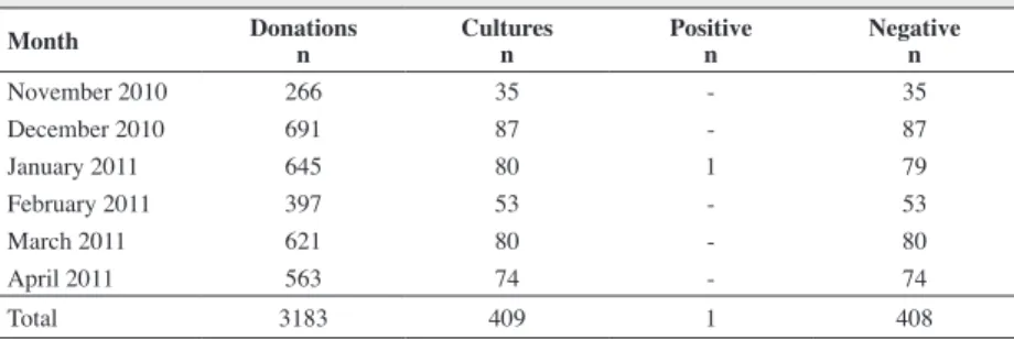
A culture in January 2011 was positive (Table 1), showing a growth of coagulase-negative Staphylococcus. This represented 1.25% of the cultures performed in January 2011 and 0.24% in the period from November 2010 to April 2011. As at least one unit of this pool was contaminated, this represents 0.15% of random platelet donors of January, and is thus well within the parameters established by Ordinance nº 1353 for the sterility of blood components(4) which determines a level of sterility higher than 75%.
Table 1 - Donors and results of blood cultures
Month Donations n
Cultures n
Positive n
Negative n
November 2010 266 35 - 35
December 2010 691 87 - 87
January 2011 645 80 1 79
February 2011 397 53 - 53
March 2011 621 80 - 80
April 2011 563 74 - 74
Total 3183 409 1 408
The rate of minimal bacterial contamination was 1:3183 donations which corresponds to 0.03%. In the United States it is estimated that bacterial contamination of random platelets is 1 in every 3000 units(6), with transfusion-related septicemia occurring in 1 of every 20,000 transfusions(7).
The most frequently agents related to platelet concentrates are gram-positive coccus,
Staphylococcus spp and Streptococcus spp(8). Ordinance nº 1353 is not speciic about the
sterility of RPC, unlike the Board Resolution (RDC) nº 153 of July 2004, repealed in December 2010, which stipulated a sterility of platelet concentrates greater than 99.5%(9).
200
Giacomini A, Stroparo E, Lidani KC
Rev Bras Hematol Hemoter. 2013;35(3):199-200 development of pathogens, especially in debilitated patients. Thus, a
strict quality control, coupled with highly sensitive techniques would allow the correct identiication of bacteria in blood products, and may contribute to reduce transfusion accidents.
References
1. Brecher ME, Hay SN. Bacterial contamination of blood components. Clin Microbiol Rev. 2005;18(1):195–204.
2. Campos TC, Pedroso CD, Dantas SC, Pinheiro FC, Costa LF, Pereira AF, et al. Monitorização da contaminação bacteriana em concentrados de plaquetas randômicas – Experiência de um banco de sangue [Abstract 696]. Rev Bras Hematol Hemoter. 2009; 31(5): 276.
3. Razouk FH, Reiche EM. Caracterização, produção e indicação clínica dos principais hemocomponentes. Rev Bras Hematol Hemoter. 2004;26(2):126-34.
4. Agência Nacional de Vigilância Sanitária. Portaria nº 1.353, de 13 de Junho de 2011. Aprova o Regulamento Técnico de Procedimentos Hemoterápicos [Internet]. Brasília: ANVISA; 2011. [cited 2012 Jun 21]. Available from: http://bvsms.saude.gov.br/bvs/saudelegis/gm/2011/ prt1353_13_06_2011.html
5. Magalhães GL, Pelisson M, Colluço AG, Koti AN, Gelinski JR, Saito M, et al. Validação do sistema BactAlert utilizado para controle microbiológico de hemocomponentes no Hemocentro Regional de Londrina [Abstract 750]. Rev Bras Hematol Hemoter. 2009;31(5):291.
6. Fiebig EW, Busch MP. Infectious disease screening. In: Roback JD. Combs MR. Grossman BJ, Hillyer CD, editors. Technical Manual. 16th ed. Bethesda: AABB; 2008. p. 241-82.
7. Mendrone AJ. Terapia transfusional no transplante de células-tronco hematopoeticas. In: Voltarelli JC, Pasquini R, Ortega ET. Transplante de células-tronco. São Paulo: Atheneu; 2009. p 653-75.
8. Rodrigues MC, Almeida DR. Avaliação de análises microbiológicas de hemocomponentes [Abstract 984]. Rev Bras Hematol Hemoter. 2011;33(2):408.
9. Brasil. Ministério da Saúde. Agência Nacional de Vigilância Sanitária. Resolução RDC n.153, de 14 de junho de 2004. Determina o Regulamento Técnico para os procedimentos hemoterápicos, incluindo a coleta, o processamento, a testagem, o armazenamento, o transporte, o controle de qualidade e o uso humano de sangue, e seus componentes, obtidos do sangue venoso, do cordão umbilical, da placenta e da medula óssea [Internet]. Brasilia, MS: 2004. [cited 2011 Sept 15]. Available from: http://portal.saude.gov.br/portal/arquivos/ pdf/resolucao_153_2004.pdf