Lab Resource: Stem Cell Line
Generation of human iPSC line from a patient with laterality defects and
associated congenital heart anomalies carrying a
DAND5
missense alteration
Fernando Cristo
a,1, José M. Inácio
a,1, Graça Rosas
a, Isabel Marques Carreira
b,c,d, Joana Barbosa Melo
b,c,d,
Luís Pereira de Almeida
e, Patrícia Mendes
f, Duarte Saraiva Martins
g, José Maio
f, Rui Anjos
g, José A. Belo
a,⁎
aStem Cells and Development Laboratory, CEDOC, NOVA Medical School, Universidade Nova de Lisboa, Lisboa, Portugal bCytogenetics and Genomics Laboratory, Faculty of Medicine, University of Coimbra, Coimbra, Portugal
cCNC.IBILI Consortium, University of Coimbra, Coimbra, Portugal
dCIMAGO - Center of Investigation on Environment Genetics and Oncobiology, Faculty of Medicine, University of Coimbra, Portugal eCNC - Center for Neurosciences & Cell Biology, University of Coimbra, Coimbra, Portugal
fDepartamento Materno-Infantil, Centro Hospital do Algarve, EPE, Faro, Portugal gHospital de Santa Cruz, Centro Hospitalar Lisboa Ocidental, Lisboa, Portugal
a b s t r a c t
a r t i c l e
i n f o
Article history:
Received 19 October 2017 Accepted 26 October 2017 Available online 31 October 2017
A human iPSC line was generated from exfoliated renal epithelial (ERE) cells of a patient affected with Congenital Heart Disease (CHD) and Laterality Defects carrying tshe variant p.R152H in theDAND5gene. The transgene-free iPSCs were generated with the human OSKM transcription factor using the Sendai-virus reprogramming system. The established iPSC line had the specific heterozygous alteration, a stable karyotype, expressed pluripotency markers and generated embryoid bodies that can differentiate towards the three germ layers in vitro. This iPSC line offers a useful resource to study the molecular mechanisms of cardiomyocyte proliferation, as well as for drug testing.
© 2017 The Authors. Published by Elsevier B.V. This is an open access article under the CC BY-NC-ND license (http://creativecommons.org/licenses/by-nc-nd/4.0/).
Resource table
Unique stem cell line identifier NMSUNLi001-A Alternative name(s) of stem cell
line
iUC-DAND5_455/10
Institution CEDOC, NOVA Medical School Contact information of distributor José A. Belo,jose.belo@nms.unl.pt Type of cell line iPSC
Origin Human
Additional origin info Age: 7 Sex: male Ethnicity: caucasian
Cell Source Exfoliated renal epithelial cells isolated from urine
Method of reprogramming Transgene free (Sendai Virus) Genetic Modification NO
Type of Modification N/A
Associated disease Heterotaxy and congenital heart disease Gene/locus rs45513495: DAND5 c.455GNA; p.R152H Method of modification No modification
Name of transgene or resistance No transgene Inducible/constitutive system N/A
Date archived/stock date January 2017 Cell line repository/bank Not applicable
Ethical approval Approved by the Ethics Committee of NOVA Medical School (Protocol N.°13/2016/CEFCM) and by the National Committee for Data Protection (CNPD, Permit N.°8694/2016).
1. Resource utility
DAND5is the human homologue of mouseCerl2/Dand5, a gene
in-volved in left-right asymmetry establishment and also in heart forma-tion. This generated iPS cell line, from a patient carrying the variant c.455GNA in theDAND5gene offers a useful resource to investigate the molecular mechanisms of cardiomyocyte proliferation, as well as for drug testing.
2. Resource details
Exfoliated renal epithelial (ERE) cells isolated from a urine sample were obtained from a 7-year old male child. The patient was clinically diagnosed with ventricular septal defect with overriding aorta, right ventricular hypertrophy and pulmonary atresia (a case of extreme
⁎ Corresponding author.
E-mail address:jose.belo@nms.unl.pt(J.A. Belo).
1 Equal authors.
(continued)
http://dx.doi.org/10.1016/j.scr.2017.10.019
1873-5061/© 2017 The Authors. Published by Elsevier B.V. This is an open access article under the CC BY-NC-ND license (http://creativecommons.org/licenses/by-nc-nd/4.0/).
Contents lists available atScienceDirect
Stem Cell Research
tetralogy of Fallot phenotype), defects that can be associated with early left-right establishment impairment. Genetically, the patient carries a heterozygous non-synonymous variant in exon 2 ofDAND5 gene (c.455GNA), causing an amino acid change of p.R152H in the functional
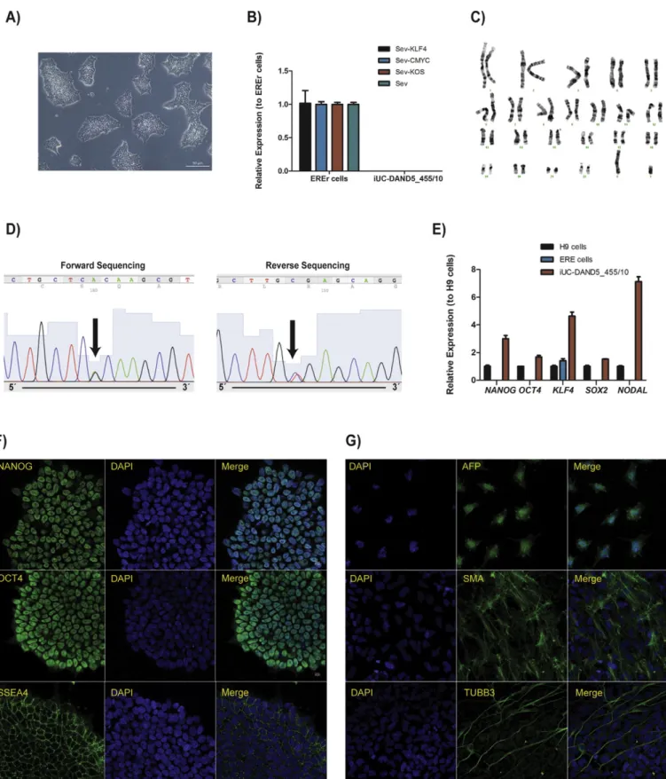
domain of the DAND5 protein.DAND5is an essential gene in the correct establishment of the laterality of visceral organs, including the heart, functioning as an inhibitor and master regular of Nodal signalling in a temporal and spatial precise way (Inacio et al., 2013; Marques et al., 2004).DAND5knockout mice display a vast array of congenital cardiac malformations associated or not with extracardiac anomalies (Marques et al., 2004). Importantly, these KO mice present thickening of the left ventricle and of the IVS due to hyperproliferation of cardiomyocytes, independent of L/R defects (Araujo et al., 2014). Our previous functional analysis of DAND5 p.R152H alteration showed a sig-nificant decrease in the function of this variant protein when compared to its wild-type counterpart. These results support a model in which the imbalance in dosage-sensitive Nodal signalling is afinal common way for laterality defects and associated CHDs and suggest a possible role of this variant in the risk of disease (Cristo et al., 2017). Moreover, it has been reported that variants in genes involved in the Nodal signalling pathway are associated with isolated cases of congenital heart defects and/or laterality defects in humans (Deng et al., 2015). In the work presented here, upon isolation of urine epithelial cells from the patient, we generated the iUC-DAND5_455/10 cell line using the CytoTune®-iPS 2.0 Reprogramming kit (Life Technologies, Invitrogen). This kit includes the reprogramming factors SOX2, OCT3/ 4, c-MYC and KLF4 and is based on a modified and non-transmissible form of Sendai virus (SeV). Seventeen days after infection, several iPSC colonies single cell-derived were picked for further expansion and char-acterization. After expansion, iUC-DAND_455/10 cell line, continued to display a typical small, round shape, and tightly packed ESC-like morphology with a high nucleus/cytoplasm ratio with prominent nucleoli (Fig. 1A). The clearance of the vectors and the exogenous reprogramming factor genes were confirmed by qPCR after
twenty-five culture passages (Fig. 1B). The clone was karyotypically normal (46, XY) after more than twenty culture passages (Fig. 1C), and DNA Sanger sequencing confirmed the presence of a c.455GNA substitution in one of the alleles of exon 2 in theDAND5gene corresponding to the R152H protein alteration (Fig. 1D). Gene expression analysis was per-formed by qPCR to confirm the expression of pluripotency markers at mRNA level, which showed that the endogenous pluripotency genes
OCT3/4,NANOG,SOX2,KLF4andNODALwere present at levels compara-ble or higher than the human embryonic stem cell line H9 (Fig. 1E). Moreover, these pluripotency genes were almost absent in ERE cells. At the protein level, immunocytochemical (ICC) analysis confirmed
the expression of self-renewal transcription factors NANOG, OCT4, and the surface marker SSEA4, (Fig. 1F), characteristic markers of pluripo-tent ES cells which illustrate the purity of the iUC-DAND_455/10 iPSC line (Table 1).
Finally, in vitro embryoid body (EB)-based differentiation followed by ICC analysis of the endodermal markerα-feto protein (AFP), the mesodermal marker smooth muscle actin (SMA) and the ectodermal marker Tubulinβ3 class III (TUBB3) confirmed the pluripotency of iPSCs and their ability to differentiate into all three germ layers (Fig. 1G).
3. Materials and methods
3.1. Ethical statement
All the experimental protocols in the present study were approved by the Ethics Committee of the NOVA Medical School (Protocol N.° 13/ 2016/CEFCM) and by the National Committee for Data Protection (CNPD, Permit N.° 8694/2016), according to European Union legislation. Written informed consent was obtained from patient guardian prior to sample collection.
3.2. Generation of iPSCs
Urine epithelial cells were collected, expanded, and reprogrammed using the 3 Sendai virus vectors included in the CytoTune-iPS 2.0 Reprogramming Kit (Life Technologies) at a 1.5 MOI (multiplicity of in-fection). After 24 h, medium was replaced with fresh RE proliferation medium and cells cultured for 7 days with medium changes every other day. On day 8, cells were passaged using TrypLE Select (Gibco, Thermo Fisher Scientific) and seeded onto a 100 mm culture dish (Corning) coated with Geltrex (Gibco, Thermo Fisher Scientific). In the next day, medium was replaced to Essential 8 (E8)flex (Gibco, Thermo Fisher Scientific) and renewed every day until hiPSC colonies appeared. 17 days after infection, individual colonies were picked and expanded, with daily renewing of the E8flex medium.
3.3. Sequencing analysis
Genomic DNA was extracted from patient hiPSCs using the Isolate Genomic DNA mini kit (BIOLINE). Subsequently, amplification by PCR of the exon 2 ofDAND5gene, containing the c.455GNA alteration,
was carried out using the primers listed inTable 2. PCR products were direct sequenced at STAB VIDA (http://www.stabvida.com/).
Table 1
Characterization and validation.
Classification Test Result Data
Morphology Photography ESC-like morphology Fig. 1, panel A
Phenotype Immunocytochemistry Staining of pluripotency markers: Oct4, Nanog, Sox2 Fig. 1, panel F qPCR Expression of pluripotency markers: NANOG, OCT3/4, SOX2, KLF4 and NODAL Fig. 1panel E Genotype Karyotype (G-banding)
and resolution
46XY, Resolution 400–500 Fig. 1, panel C
Identity Microsatellite PCR (mPCR) N/A
STR analysis 16 loci analyzed, all matching Supplementary
Fig. S1panel A Mutation analysis (IF
APPLICABLE)
Sequencing Heterozygous (GNA) Fig. 1, panel D
Southern Blot OR WGS N/A, Non-integrating reprogramming methodology
Microbiology and virology Mycoplasma Mycoplasma testing by PCR. Negative Supplementary
Fig. S1panel B Differentiation potential Embryoid body formation Proof of formation of three germ layers from Embryoid bodies:α-fetoprotein (AFP),
βIII-tubulin (TUBB3),α-smooth muscle actin (SMA).
Fig. 1, panel G
Donor screening (OPTIONAL)
HIV 1 + 2 Hepatitis B, Hepatitis C
N/A
Genotype additional info (OPTIONAL)
Blood group genotyping N/A HLA tissue typing N/A
3.4. Real-time PCR analysis
Real time PCR was carried out with Fast SYBR Green Master Mix (Ap-plied Biosystems) and the primers listed inTable 2on an Applied Biosystems® 7500 Real-Time PCR machine
3.5. Test for absence of the reprogramming Sendai vectors
ERE in reprogramming (EREr) and established hiPSC cells were test-ed for absence of the Sendai reprogramming vectors by qRT-PCR (Table 2).
3.6. Fluorescent immunocytochemistry
Undifferentiated or differentiated iUC-DAND_455/10 cells were
fixed in 4% paraformaldehyde, incubated with primary antibodies overnight at 4 °C, listed inTable 2, and then incubated with Alexa Fluor 488-conjugated secondary antibodies overnight at 4 °C. Nuclei were stained with DAPI at room temperature and cell images were ac-quired with Zeiss Axio Imager Z2 microscope (Carl Zeiss) or confocal microscopy.
3.7. In vitro differentiation potential by embryoid bodies formation assay
For the generation of embryoid bodies (EBs), iPS cells were collected and suspended in non-adherent tissue culture 100 mm dishes with E8 medium plus polyvinyl alcohol and Revitacell for 7 days. At this time, the EBs were transferred onto Geltrex-coated lummox 24-well plates (SARSTEDT) and cultured for another 14 days or longer. Then, cells werefixed with 4% formaldehyde and incubated with the indicated pri-mary antibodies specific for the three embryonic germ layers.
3.8. Karyotyping
Chromosome analysis was performed using GTG high resolution banding technique, according to standard procedures with a minimum of 10 metaphase spreads analyzed. Analysis of GTG-banded chromo-somes was performed at a resolution of 400 bands per haploid genome and karyotypes were established according to the International System for Human Cytogenetic Nomenclature (ISCN 2016).
3.9. Mycoplasma contamination detection
The absence of mycoplasma was assessed by PCR using the Primers listed inTable 2.
3.10. STR analysis
iUC-DAND_455/10 cells and the corresponding ERE cells were au-thenticated by STR analysis performed by STAB VIDA (http://www. stabvida.com/).
Supplementary data to this article can be found online athttps://doi. org/10.1016/j.scr.2017.10.019.
Author contributions
Conceived and designed the experiments: FC, JMI, JB; Diagnosis of patients: PM, JM, RA; Patient recruitment, sample collection and clinical data collection: FC, JMI, PM, JM, RA, DM; Analyzed the data: FC, JMI, GR, JB; Performed the experiments: FC, JMI, GR; Karyotype experiment and analysis: IMC, JBM, LPA; Contributed to writing the manuscript: FC, JMI and JB.
All authors read and approved thefinal manuscript.
Table 2 Reagents details.
Antibodies used for immunocytochemistry/flow-citometry
Antibody Dilution Company Cat # and RRID
Pluripotency Markers Rabbit anti-NANOG 1:200 Abcam Cat# ab21624, RRID:AB_446437 Abcam Cat# ab19857, RRID:AB_445175 Abcam Cat# ab16287, RRID:AB_778073
Rabbit anti-OCT4 1:400
Mouse anti-SSEA4 1:200
Differentiation Markers Mouse anti-Human TUBB3 1:400 Sigma-Aldrich Cat# T8660, RRID:AB_477590 Dako Cat# M0851, RRID:AB_2223500 Dako Cat# A0008,
RRID:AB_2650473 Mouse anti-Human SMA 1:600
Rabbit anti-Human AFP 1:200
Secondary antibodies Alexa Fluor 488-conjugated Donkey anti-Mouse IgG (H + L)
1:300 Jackson ImmunoResearch Labs Cat# 715–545-150, RRID:AB_2340846
Alexa Fluor 488-conjugated Donkey anti-Rabbit IgG (H + L)
1:300 Jackson ImmunoResearch Labs Cat# 711–545-152, RRID:AB_2313584
Primers
Target Forward/Reverse primer (5′-3′)
Elimination of Sendai Virus Transgenes (qPCR - TaqMan)
Sev GGATCACTAGGTGATATCGAGC/ACCAGACAAGAGTTT AAGAGATATGTATC
Sev-KLF4 TTCCTGCATGCCAGAGGAGCCC/AATGTATCGAAGGTG CTCAA
Sev-C-MYC TAACTGACTAGCAGGCTTGTCG/TCCACATACAGTCCT GGATGATGATG
Sev-KOS ATGCACCGCTACGACGTGAGCGC/ACCTTGACAATC CTGATGTGG
Pluripotency Markers (qPCR) NANOG CATGAGTGTGGATCCAGCTTG/CCTGAATAAGCAGATCCATGG
OCT3/4 GACAGGGGGAGGGGAGGAGCTAGG/CTTCCCTCCAACCAGTTGCCCCAAAC
SOX2 GGGAAATGGGAGGGGTGCAAAAGAGG/TTGCGTGAGTGTGGATGGGATTGGTG
KLF4 ACCAGGCACTACCGTAAACACA/GGTCCGACCTGGAAAATGCT
NODAL GGGCAAGAGGCACCGTCGACATCA/GGGACTCGGTGGGGCTGGTAACGTTTC
House-Keeping Genes (qPCR) GAPDH CTGGTAAAGTGGATATTGTTGCCAT/TGGAATCATATTGGAACATGTAAACC
β-actin GCAAAGACCTGTACGCCAAC/AGTACTTGCGCTCAGGAGGA
Mycoplasma detection Pair 1 CTGCAGATTGCAAAGCAAGA/CCTCCTTCTTCACCTGCTTG
Pair 2 GGCGAATGGGTGAGTAACACG/CGGATAACGCTTGCGACCTATG
Acknowledgements
We would like to thank the patient and their guardians for their gen-erous donation of the urine sample used in this study. We also would like to thank Ana Jardim for technical support in karyotype analysis. This work was supported by Fundação para a Ciência e a Tecnologia (PTDC/ BIM-MED/3363/2014). iNOVA4Health - UID/Multi/04462/2013, a pro-gramfinancially supported by Fundação para a Ciência e Tecnologia/ Ministério da Educação e Ciência, through national funds and co-funded by FEDER under the PT2020 Partnership Agreement is acknowledged.
References
Araujo, A.C., Marques, S., Belo, J.A., 2014.Targeted inactivation of Cerberus like-2 leads to left ventricular cardiac hyperplasia and systolic dysfunction in the mouse. PLoS One 9 (7), e102716.
Cristo, F., et al., 2017.Functional study of DAND5 variant in patients with congenital heart disease and laterality defects. BMC Med. Genet. 18 (1), 77.
Deng, H., Xia, H., Deng, S., 2015.Genetic basis of human left-right asymmetry disorders. Expert Rev. Mol. Med. 16, e19.
Inacio, J.M., et al., 2013.The dynamic right-to-left translocation of Cerl2 is involved in the regulation and termination of Nodal activity in the mouse node. PLoS One 8 (3), e60406.
Marques, S., et al., 2004.The activity of the Nodal antagonist Cerl-2 in the mouse node is required for correct L/R body axis. Genes Dev. 18 (19), 2342–2347.