online | memorias.ioc.fiocruz.br
Plasmodium vivax,a relatively neglected human ma-laria parasite,is a major public health challenge for Cen-tral and South America, the Middle East, CenCen-tral, South and Southeast Asia, Oceania and East Africa, where 2.85 billion people are currently at risk of infection and 70-80 million clinical cases are reported each year (Mu-eller et al. 2009, Guerra et al. 2010). The emergence of drug-resistant strains and severe (sometimes fatal) dis-ease challenges the traditional view of vivax malaria as a benign infection (Price et al. 2009). Recent epidemio-logical trends in Brazil illustrate the importance of P. vivax as a re-emerging pathogen. The annual incidence of Plasmodium falciparum (the predominant malaria parasite species between 1985-1990) decreased steadily during the 1990s, while that of P. vivax maintained an upward trend. Both P. falciparum and P. vivax are still transmitted across the Amazon Basin, with rare Plasmo-dium malariae infections. However, P. vivax now causes 85% of the 315,000 malaria cases reported in this coun-try each year (Oliveira-Ferreira et al. 2010), suggesting that this species may be less susceptible to the malaria control strategies currently used in Brazil.
Understanding the genetic structure of P. vivax is essential to accurately describing the transmission dy-namics of vivax malaria. Population genetics data are crucial, for example, to predict how fast phenotypes of interest, such as novel antigenic variants, particular relapsing patterns or drug resistance, arise and spread
in natural populations (Zilversmit & Hartl 2005). The current data on P. vivax population genetics lags behind that of P. falciparum, primarily due to the scarcity of ap-propriate genetic markers. As we cannot propagate the parasite in continuous culture in vitro (Udomsangpetch et al. 2008), the phenotypic diversity of natural human infections from P. vivax remained unexplored until the early 1990s. Advances in molecular methods, especially implementation of polymerase chain reaction (PCR)-based protocols to amplify parasite DNA, allowed for genotyping of malaria parasite samples obtained directly from patient blood, without a preceding in vitro culture step (Kimura et al. 1990). The relative paucity of genetic markers hampers further investigation into the geno-types that underlie phenogeno-types such as relapse pattern, virulence and drug resistance.
In this review, we broadly classify the available mo-lecular markers into two groups: (i) markers that are clearly under natural selection, that is, markers that map to genome regions associated with adaptive traits and (ii) markers that are neutral or nearly neutral, that is, those that are not obviously influenced by natural selection. The extensive polymorphism found in loci associated with P. vivax adaptive traits, such as those coding for surface antigens and drug-resistance (Cui et al. 2003a),
reflects the combined effects of the parasite′s population history and selective constraints imposed by the host′s
immunity and drug usage (Escalante et al. 2004). Thus, these loci do not provide much information on the P. vivax population structure. Neutral and nearly neutral markers, in contrast, may allow for unbiased estimates of genetic variation, population structure and gene flow in natural parasite populations.
When comparing different markers and parasite typ-ing systems, a clear quantitative definition of genetic diversity is required. One of the most popular methods of summarising genetic diversity levels is to report vir-Financial support: Rede Malária/PRONEX, MS/DECIT, FAPEMIG,
FAPEMAT, NIAID, NIH (R01 AI075416-04) (to MUF) + Corresponding author: [email protected] CFADB and MUF were supported by CNPq fellowships. Received 26 April 2011
Accepted 8 June 2011
Molecular markers and genetic diversity of
Plasmodium vivax
Cristiana Ferreira Alves de Brito1/+, Marcelo Urbano Ferreira2
1Laboratório de Malária, Instituto de Pesquisas René Rachou-Fiocruz, Av. Augusto de Lima 1715, 30190-002 Belo Horizonte, MG, Brasil 2Departamento de Parasitologia, Instituto de Ciências Biomédicas, Universidade de São Paulo, São Paulo, SP, Brasil
Enhanced understanding of the transmission dynamics and population genetics for Plasmodium vivax is crucial in predicting the emergence and spread of novel parasite phenotypes with major public health implications, such as new relapsing patterns, drug resistance and increased virulence. Suitable molecular markers are required for these population genetic studies. Here, we focus on two groups of molecular markers that are commonly used to analyse natural populations of P. vivax. We use markers under selective pressure, for instance, antigen-coding poly-morphic genes, and markers that are not under strong natural selection, such as most minisatellite and microsatellite loci. First, we review data obtained using genes encoding for P. vivax antigens: circumsporozoite protein, merozoite surface proteins 1 and 3α, apical membrane antigen 1 and Duffy binding antigen. We next address neutral or nearly neutral molecular markers, especially microsatellite loci, providing a complete list of markers that have already been used in P. vivax populations studies. We also analyse the microsatellite loci identified in the P. vivax genome project. Finally, we discuss some practical uses for P. vivax genotyping, for example, detecting multiple-clone infec-tions and tracking the geographic origin of isolates.
tual heterozygosity (HE) values. Virtual HE is the aver-age probability that a pair of alleles randomly obtained from the population is different. Whenever appropriate, in this review we provide HE estimates obtained using different markers for different populations.
Molecular markers under selection -Until recently, most genetic markers available for characterising natu-ral P. vivax populations were orthologues of previously identified P. falciparum antigen-coding genes. The well-characterised polymorphic regions in these genes have been extensively used to analyse genetic diversity pat-terns in P. falciparum (Roy et al. 2008). A similar ap-proach has been explored in vivax-oriented studies (Cui et al. 2003a). The following single-copy genes in P. vivax
have often been used in these studies: gam1,coding for the gametocyte antigen 1, csp,coding for the circumspo-rozoite protein (CSP), msp1 and msp3alpha,coding for the
merozoite surface proteins (MSP)-1 and 3α, respectively, ama1, coding for the apical membrane antigen 1, and dbp, coding for the Duffy binding protein (DBP) (Table I).
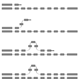
A notable feature of several P. vivax surface anti-gens (including major vaccine-candidate molecules) is tandem arrays of relatively short amino acid motifs. The CSP in P. vivax (PvCSP), an abundant antigen on the surface of sporozoites that has been extensively used as a vaccine development target, has immuno-dominant B-cell epitopes that map to central repeats between nonrepetitive sequences (Nardin & Zavala 1998). PvCSP displays two major types of nonapep-tide repeats [most commonly GDRA(D/A)GQPA and ANGA(G/D)(N/D)QPG], which define the variants known as VK210 and VK247, respectively (Arnot et al. 1985, Rosenberg et al. 1989) (Fig. 1). Both VK210 and VK247 variants are globally distributed, but geograph-ic biases have been described (Cochrane et al. 1990, Qari et al. 1993a, b). Similar repetitive sequences are found in Brazil, Southeast Asia and Papua New Guinea (Qari et al. 1991, 1992). However, markedly divergent sequence variants were found in isolates from China and North Korea (Mann et al. 1994).Although VK210 and VK247-type sequences often occur in sympatric parasite populations, there are no examples of a hybrid repeat array with both repeat types (Lim et al. 2005).
A third type of repeat unit [APGANQ(E/G)GAA], identical to that described for Plasmodium simiovale, characterises the so-called P. vivax-like parasites (Qari et al. 1993a). Although P. vivax-like CSP repeats have been found in parasite isolates across the world, its global dis-tribution remains unconfirmed (Gopinath et al. 1994). In Brazil, P. vivax-like parasites have been identified only in mixed-clone infections with VK210-type or VK247-type parasites (Machado & Póvoa 2000). More recently, com-plex mixtures of parasites harbouring several PvCSP types and co-infecting the same hosts have been demonstrated by Henry-Halldin et al. (2011) in Papua New Guinea.
The nonapeptide sequences in PvCSP are repeated ~20 times in the array. Insertions and deletions in the central repeat domain, from either sexual recombination during meiosis or intrahelical strand-slippage events during mitotic DNA replication (McConkey et al. 1990), generate novel CSP variants that may be positively
select-ed if the mutant parasites evade a host’s immunity. The central repetitive domain of P. falciparum CSP (PfCSP) contains extensive variation. This domain consists of a variable number of four-mer NANP and NVDP motif copies without breaking down the tight linkage between polymorphic sites in flanking sequences, as in interhe-lical exchanges during meiosis (Rich et al. 1997). This pattern suggests that these repeats undergo frequent in-Fig. 1: schematic representation of studied antigens: A: the circum-sporozoite protein of Plasmodium vivax (PvCSP) consists of a con-served region (light grey) flanking a repeated central region (RCR) (blank box), which contains immunodominant B-cell epitopes and is flanked by pre and post-repeat specific nonrepetitive sequences (NR) (black boxes). PvCSP displays three types of repeats [GDRA(D/A) GQPA (VK210), ANGA(G/D)(N/D)QPG (VK247)] which are the most prevalent and APGANQ[E/G]GGAA (P. vivax-like); B: the P. vivax
merozoite surface protein 1 (PvMSP-1)sequence comprises seven conserved blocks (amino acid similarity among PvMSP-1alleles, 71-85%) (light grey boxes) and six variable blocks (amino acid simi-larity, 21-34%) (black boxes), described by Putaporntip et al. (2002). For comparison, we also show the original division of PvMSP-1 into interspecies conserved blocks (ICBs) (amino acid similarity > 48% in pairwise comparisons of MSP-1 orthologues in Plasmodium falci-parum, P. vivax and Plasmodium yoelii) (light grey boxes), conserved blocks (CBs) (amino acid similarity > 50% between P. falciparum and
P. vivax, but lower in other pairwise comparisons) (dark grey boxes) and polymorphic blocks (amino acid similarity < 45%) (black boxes) (del Portillo et al. 1991). These two methods of portioning PvMSP-1 differ in that Putaporntip et al. (2002) compared intraspecific se-quences, while del Portillo et al. (1991) compared between-specific sequences. The F1, F2 and F3 were defined by Imwong et al. (2005) (horizontal bold lines); C: the P. vivax merozoite surface protein 3α
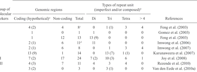
TABLE I
Genes encoding antigens used as molecular markers to study field populations of Plasmodium vivax
Antigen
Protein fragment
Population studied (n)
Totala
(n) References
PvAMA-1 Domain I BRA 105 Grynberg et al. (2008) 20 Rodrigues et al. (2005) KOR 30 Chung et al. (2003)
22 Han et al. (2002) MYA 76 Moon et al. (2010) AFR (5), CHI (5), IND (18), INDO (5),
PHI (148), PNG (23), SI (7), THAI (6)
219 Figtree et al. (2000)
Domain I, II IND 61 Thakur et al. (2008)
Whole IND 11 Rajesh et al. (2007)
SRL 23 Gunasekera et al. (2007) THAI 231 Putaporntip et al. (2009)
VEN 73 Ord et al. (2008)
PvCSP CR AFG 202 Zakeri et al. (2010)
AZE 36 Leclerc et al. (2004) BRA 48 Machado and Póvoa (2000)
45 Patil et al. (2010) 32 Santos-Ciminera et al. (2007) 155 Storti-Melo et al. (2009) Central AME, South AME, AFR, Southeast ASI, IND 126 Gopinath et al. (1994)
COL 24 Hernández-Martínez et al. (2011) GUY 61 Bonilla et al. (2006)
IND 151 Kim et al. (2006) IRA 144 Zakeri et al. (2006) KOR 632 Choi et al. (2010) MYA 116 Moon et al. (2010)
PAK 187 Zakeri et al. (2006) PAK (187), IRA (150) 337 Zakeri et al. (2010)
PNG 40 Kolakovich et al. (1996) 507 Henry-Halldin et al. (2011) THAI 90 Cui et al. (2003b)
100 Imwong et al. (2005) 171 Kain et al. (1993)
17 Rongnoparut et al. (1995) CR, NR BRA (30), PNG (14) 30 Qari et al. (1992) N, C-end PHI, CHI, SI, PNG 18 Huang et al. (1994)
Whole CHI 16 Mann et al. (1994)
PvDBP Whole COL 23 Martinez et al. (2004)
DomainII-IV South KOR 30 Kho et al. (2001)
Domain II BRA 40 Sousa et al. (2006)
122 Sousa et al. (2010) COL 20 Ampudia et al. (1996) COL (18), South KOR (14), PNG (68) 100 Cole-Tobian et al. (2002)
IRA 75 Babaeekho et al. (2009) PNG 40 Kolakovich et al. (1996) 358 Cole-Tobian et al. (2002)
Antigen
Protein fragment
Population studied (n)
Totala
(n) References
PvMSP-1 Pv200L COL 26 Valderrama-Aguirre et al. (2011) 925 pb PHI, CHI, SI, PNG 18 Cheng et al. (1993)
BL 2 BRA (28), VIET (23) 51 Hoffmann et al. (2003) ICBs 2-4 COL 20 Gutierrez et al. (2000) ICBs 4, 5 AFR (5), CHI (10), IND (21), INDO (11),
PHI (97), PNG (21), SI(6), THAI (4)
175 Figtree et al. (2000)
KOR 30 Kim et al. (2009) ICBs 5, 6
or BL 5
AZE 36 Leclerc et al. (2004) BRA 32 Santos-Ciminera et al. (2007) French GUY 120 Veron et al. (2009)
IND 25 Farooq et al. (2009) IND (9), COL (11) 20 Maestre et al. (2004)
KOR 632 Choi et al. (2010) 25 Lim et al. (2000) MYA 116 Moon et al. (2009)
PNG 40 Kolakovich et al. (1996) PAK (187), IRA (150) 337 Zakeri et al. (2010)
SRL 22 Premawansa et al. (1993) THAI 15 Putaporntip et al. (1997)
F1-F3 IND 151 Kim et al. (2006)
THAI 100 Imwong et al. (2005) F1, F3 PNG 93-108 Koepfli et al. (2009)
MSP-142 SRL 95 Dias et al. (2011)
IND 33 Thakur et al. (2008)
MSP-119 BRA 28 Soares et al. (1999)
GUY 61 Bonilla et al. (2006) C-terminal PHI (4) THAI (3) SI (8), PNG (1), South East ASI (4) 20 Pasay et al. (1995)
Whole THAI (20), BRA (8), BAN (5), South KOR (4), VAN (2), IND (1) 40 Putaporntip et al. (2002) KOR 45 Han et al. (2011) TUR 30 Zeyrek et al. (2010)
PvMSP-3α NA IND 151 Kim et al. (2006)
COL 55 Cristiano et al. (2008) French GUY 120 Veron et al. (2009)
IND 27 Prajapati et al. (2010) IRA 144 Zakeri et al. (2006) MYA 116 Moon et al. (2009)
PAK 50 Khatoon et al. (2010) PAK (187), IRA (150) 337 Zakeri et al. (2010)
PER 186 Sutton et al. (2009) PNG 28 Bruce et al. (2000) 11 Mueller et al. (2002) PNG (39), IND (4), Sudan (1), SRL (1) 45 Bruce et al. (1999)
South KOR 24 Han et al. (2004) THAI 90 Cui et al. (2003b) VEM 58 Ord et al. (2005)
Whole THAI 17 Mascorro et al. (2005)
trahelical recombination that either add or delete repeat units during mitotic DNA replication (Fig. 2), as other short repeats, such as microsatellite and minisatellite-type sequences (Levinson & Gutman 1987). Frequent mitotic recombination coupled with positive selection of new variants might accelerate sequence evolution even where meiotic recombination and outcrossing are rela-tively uncommon in malaria parasite populations (Rich et al. 2000). In length variation arrays, whether length variation affects B-cell epitope recognition is uncertain, but the conformational nature of repetitive epitopes in PfCSP (Monette et al. 2001) supports this hypothesis.
We have recently examined whether similar recom-bination mechanisms create significant variation in the PvCSP repeat arrays, which consist of longer (9-mer) repetitive motifs (Patil et al. 2010). We investigated pat-terns of sequence diversity in PvCSP encoding gene al-leles in sympatric P. vivax isolates from an area of low malaria endemicity in Brazil. In these isolates, we used single-nucleotide polymorphism typing to examine the haplotype structure of chromosome 8, where the PvC-SPencodinggene is located. We sought to determine whether the repetitive domain in the csp locus was significantly diverse under conditions of low malaria endemicity, which reduces effective meiotic recombi-nation rates in local parasites. We confirmed previous findings (Arnot et al. 1985, 1990, Qari et al. 1992, 1994, Mann et al. 1994) of substantial nucleotide sequence diversity in repeat arrays from 45 isolates. They were analysed using nine different csp alleles, each with a unique arrangement of six different nonapeptide repeat sequences, and all were of the VK210 type. Notably, different nucleotide motifs, termed repeat allotypes, coded for the same VK210-type repeat units, indicat-ing that some conservation is maintained at the amino acid level (putatively due to functional constraints), but not necessarily at the nucleotide level (Rich et al. 1997, 2000). Most repeat units in the same csp allele were ei-ther identical or nearly identical to each oei-ther, consist-ent with their recconsist-ent expansion in the repeat array. We found strong linkage disequilibrium at sites across the chromosome 8 segment flanking the csp locus, consist-ent with rare meiotic recombination in this region. This suggests that repeat array diversity may not be severely constrained by low meiotic recombination rates that are typical in areas with low malaria endemicity. New repeat variants may be readily created by nonhomolo-gous recombination events with potential implications for PvCSP-based vaccine development.
The MSP-1 and 3α are additional examples of
blood-stage vaccine-candidate antigens with repetitive arrays. The 200-kDa MSP 1 in P. vivax (PvMSP-1), originally described at the molecular level by del Portillo et al. (1991), is a major target of naturally acquired (Noguei-ra et al. 2006) and vaccine-induced immunity (Yang et al. 1999, Herrera et al. 2007). The protein contains six highly polymorphic domains (4 of them repetitive) flanked by seven fairly conserved sequences (Putaporn-tip et al. 2002) (Fig. 1). Sequence analysis has confirmed that blocks 1, 3 and 5 in PvMSP-1 are conserved at the protein level, while blocks 2, 4 and 6 are highly variable
(Kolakovich et al. 1996, Figtree et al. 2000, Hoffmann et al. 2003, Bastos et al. 2007, Farooq et al. 2009, Kim et al. 2009, Veron et al. 2009, Zakeri et al. 2010).
The extensive sequence divergence in variable do-mains of PvMSP-1 (amino acid similarity, 21-34%) has been maintained by balanced selection, most likely as a result of variant-specific immune pressure (Putaporntip et al. 2006). However, the extent to which PvMSP-1 se-quence diversity affects immune recognition for this ma-jor malaria-vaccine candidate antigen remains uncertain. To address this topic, we have recently compared natu-rally acquired antibody responses to three polymorphic domains and the highly conserved PvMSP-1 C-terminal domain in a rural, malaria-exposed Amazonian cohort. We expressed 15 recombinant proteins corresponding to PvMSP-1 variants commonly found in local parasites and showed that less than one-third had detectable IgG antibodies to at least one expressed variant of blocks 2, 6 and 10. However, 54.3% recognised the invariant C-terminal domain PvMSP-119. Although the proportion of PvMSP-1 variant responders increased substantially during subsequent acute P. vivax infections, the spe-cificity of IgG antibodies did not necessarily match the PvMSP-1 variant(s) found in infecting parasites (Bastos et al. 2007). The mechanisms underlying the limited im-mune recognition of PvMSP-1 variants that are known to circulate in a given endemic area remain unclear.
quent meiotic recombination events that shuffle differ-ent sequence types to generate new alleles are respon-sible for the mosaic structure of PvMSP-1 (Putaporntip et al. 1997, 2002). Recently, Han et al. (2011) compared PvMSP-1 alleles from re-emergent P. vivax parasites collected in the Republic of Korea over the past decade. They showed an increase in allelic diversity for variable domains of this protein, suggesting that recombination independently changed protein domains. Moreover, the greater similarity between allelic sequences within P. vivax than between P. vivax and closely related species was taken as evidence that polymorphisms in P. vivax
arose recently. This is in stark contrast to the P. falci-parum pattern, wherein different alleles are estimated to have diverged more than 25 million years ago (Polley et al. 2005). However, the cause for the large differences in this genetic history for these highly divergent parasites remains obscure. It may reflect either the significantly different sequence structure of the orthologues, differ-ent molecular functions for the gene in the two species, a different population history, or another event.
Proteolytic processing of PvMSP-1 generates a C-ter-minal fragment of 42 kDa (PvMSP-142), which is subse-quently cleaved to generate two smaller fragments with ap-parent molecular masses 33 kDa (PvMSP-133) and 19 kDa (PvMSP-119). Only the small, PvMSP-119 fragment remains at the merozoite surface during and after erythrocyte inva-sion (Holder 2009). Limited polymorphism has been ob-served in the C-terminal 19-kD PvMSP-1 fragment, with only two polymorphic sites (Soares et al. 1999, Dias et al. 2011). PvMSP-142, like PfMSP-142, exhibits extensive ge-netic polymorphism in natural infections (Escalante et al. 1998, Conway et al. 2000, Putaporntip et al. 2002, Pacheco et al. 2007, Thakur et al. 2008, Dias et al. 2011).
The P. vivaxmsp3α gene encodes a MSP with an ap-parent molecular mass ranging from 148-150 kDa. The protein contains an alanine-rich central domain that is pre-dicted to form a coiled-coil tertiary structure (Galinski et
al. 1999) (Fig. 1). The gene coding for the MSP-3α (PvMSP-3α) is highly polymorphic and its variability has been as -sessed by restriction fragment length polymorphism-PCR
analysis (Bruce et al. 1999). The suitability of PvMSP-3α
as a molecular marker has been confirmed in several geo-graphic parasite populations: Papua New Guinea (Bruce et al. 1999), Thailand (Cui et al. 2003b, Mascorro et al. 2005), Pakistan and Iran (Zakeri et al. 2006, 2010, Khatoon et al. 2010), Venezuela (Ord et al. 2005), Peru (Sutton et al. 2009), Colombia (Cristiano et al. 2008), India (Prajapati et al. 2010) and French Guiana (Veron et al. 2009). However, classifying restriction fragments according to size after electrophoresis is relatively subjective, which hampers large-scale analyses and reduces comparisons between studies. In addition, complex restriction patterns generated by mixed-clone infections may be difficult to interpret.
The P. vivax apical membrane antigen-1 (PvAMA-1) is an immunogenic, type 1 integral membrane protein, which is expressed at the apical surface of merozoites and sporozoites. It seems to play a role in erythrocyte and hepatocyte invasion (Hodder et al. 2001, Silvie et al. 2004). A number of studies have addressed polymor-phism in that PvAMA-1 encoding gene in various parts
of the world, such as Asia, Oceania and South America (Cheng & Saul 1994, Figtree et al. 2000, Han et al. 2002, Chung et al. 2003, Rodrigues et al. 2005, Gunasekera et al. 2007, Rajesh et al. 2007, Grynberg et al. 2008, Thakur et al. 2008, Moon et al. 2010). The PvAMA-1 protein includes three extracellular domains, known as domains I, II and III (Pizarro et al. 2005) (Fig. 1). Most studies show a high degree of genetic variability in do-main I (Ord et al. 2008, Thakur et al. 2008, Putaporntip et al. 2009), while the Sri Lanka domain II is the most polymorphic (Gunasekera et al. 2007). A comparison of PvAMA-1 domain I sequences from 320 worldwide iso-lates revealed a striking divergence between Old World populations and those from Brazil (Grynberg et al. 2008, Putaporntip et al. 2009). Sequence analysis of Sri Lankan isolates suggested that balancing selection maintains polymorphism at the P. vivax ama1 locus (Gunasekera et al. 2007), while no strong evidence for balancing se-lection was found in Thailand (Putaporntip et al. 2009). A number of studies have reported a substantially lower variability at this locus in P. vivax than in P. falciparum, implying that natural selection acts differently at this lo-cus in the two species (Gunasekera et al. 2007, Ord et al. 2008, Putaporntip et al. 2009).
P. vivax DBP (PvDBP) plays an important role in the formation of an irreversible junction between P. vivax
merozoites and its receptor, Duffy antigen/receptor for chemokines (DARC), at the surface of immature red blood cells (reticulocytes). This is a key step in host cell invasion (Singh et al. 2005, Galinski & Barnwell 2009). In vitro, antibodies against PvDBP inhibit DBP bind-ing to DARC and block invasion of human erythrocytes (Michon et al. 2000, Grimberg et al. 2007, Ceravolo et al. 2008). The most polymorphic segment of this protein is the erythrocyte-binding motif, a 170 amino-acid stretch located in the cysteine-rich domain II (Ranjan & Chitnis 1999, VanBuskirk et al. 2004, Hans et al. 2005) (Fig. 1 ). This segment of the protein has been used as molecular marker in genetic population studies of field isolates from Papua New Guinea (Tsuboi et al. 1994, Xainli et al. 2000), Colombia (Ampudia et al. 1996), South Korea (Kho et al. 2001), Brazil (Sousa et al. 2006, 2010) and Thailand (Gosi et al. 2008). Recently, the importance of selective diversi-fying pressure has been shown for specific regions of the protein structure (Sousa et al. 2010).
Although molecular epidemiological studies using P. vivax gam1 gene have revealed variability in P. vivax iso-lates from Sri Lanka (Snewin et al. 1995), Korea (Kho et al. 2001) and India (Prajapati et al. 2006), amplification of this gene has been associated with artefacts (Imwong et al. 2001, 2005). For this reason, P. vivax gam1 is no longerused as a reliable genetic marker.
dif-ferent endemic areas. However, little TR diversity was found within populations (Tables II, III). Rezende et al. (2009) selected the five most polymorphic loci from Leclerc’s study for their analysis of field isolates from four regions in Brazil. This set of TR markers again revealed relatively little diversity in Brazilian isolates (Table III). Most TR markers had a clearly predomi-nant allele. Moreover, these markers were inefficient in detecting multiple-clone infections and were unable to group the isolates according to their geographical ori-gin (Rezende et al. 2009). As these TR markers map to a 100-kb region in chromosome 8 with many putative protein-coding genes (including csp), natural selection is expected to affect them, even if indirectly, as a result of hitchhiking (Feng et al. 2003). Thus, these loci may not be effectively neutral given their proximity to sequences under strong diversifying or purifying selection (Leclerc et al. 2004). An alternative explanation for the low vari-ability in TR loci may include their structure, as they are relatively short arrays consisting of long (> 6 bp) repeat units. These sequences are less prone to strand slippage events (Fig. 2) than typical microsatellite-type sequenc-es consisting of long arrays of short repeat motifs.
The current markers of choice for large-scale popula-tion genetic studies in eukaryotes are highly polymorphic and short (1-6 bp-long) TRs, known as microsatellites (Schlötterer 1998). In contrast to the ~1,000 polymorphic microsatellite loci currently available for P. falciparum
typing, it was demonstrated that a few dozen microsatel-lite loci are polymorphic in P. vivax field isolates (Table II). Despite these limitations, microsatellite-based
stud-ies of the parasite have provided valuable information on
P. vivax population structure and diversity in the Ameri-cas, Asia and Africa (Ferreira et al. 2007, Karunaweera et al. 2007, 2008, Koepfli et al. 2009, Orjuela-Sánchez et al. 2009a, Gunawardena et al. 2010, Rezende et al. 2010, Van den Eede et al. 2010a, b, 2011).
The first microsatellite described for P. vivax was a dinucleotide (AT) repeat, used to identify high vari-ability in 89 isolates from Papua New Guinea (Gómez et al. 2003) (Tables II, III). Leclerc et al. (2004) reported low variability in dinucleotide microsatellite loci. In this study, only one of 13 microsatellites showed extensive variability and nine were entirely monomorphic among the eight P. vivax populations analysed (Table III). Us-ing a draft of the unpublished P. vivax genome, Imwong et al. (2006) designed primer pairs for 11 dinucleotide microsatellites and showed extensive variability in P. vivax populations from areas with intermediate to high levels of endemicity. They compared their findings with those of Leclerc et al. (2004) and explained the discrep-ancy in microsatellite diversity levels as the result of repeat array length at the loci studied. They suggested that microsatellite diversity correlates positively repeat array length, as the rate of strand-slippage events that create diversity increases exponentially with repeat ar-ray lengths. Recently, Rezende et al. (2010) corroborated this hypothesis using a set of microsatellites with di, tri and tetra nucleotide repeat units. They highlighted the importance of microsatellite loci structure in genetic diversity studies using malaria parasites. Karunaweera et al. (2007) described a new set of highly polymorphic
TABLE II
Description of molecular markers: minisatellites and microsatellites used in genetic population analysis of Plasmodium vivax field isolates
Group of molecular markers
Number of microsatellites
Genomic regions
Types of repeat unit (imperfect and/or composed)b
References Coding (hypothetical)a Non-coding Total Di Tri Tetra > 4
I 4 (2) 4 8c 0 1 (1) 3 4 Feng et al. (2003)
II 1 0 1 1 0 0 0 Gomez et al. (2003)
III 1 12 13 13 (9) 0 0 0 Feng et al. (2003) IV 2 (1) 6 11d 11 0 0 0 Imwong et al. (2006)
V 2 (1) 6 8 0 1 3 4 Imwong et al. (2007)
VI 13 (9) 1 14 0 13 (7) 1 (1) 0 Karunaweera et al. (2007) VII 7 (2) 17 24 7 (2) 10 (3) 6 1 Joy et al. (2008) VIII 4 (3) 7 11 4 3 4 0 Rezende et al. (2010) IX 3 (2) 0 3 0 3 (1) 0 0 Van den Eede et al. (2010a)
tricleotide and tetranucleotide microsatellites, which are expected to yield more accurate allele scores than dinu-cleotide markers (Anderson et al. 1999).
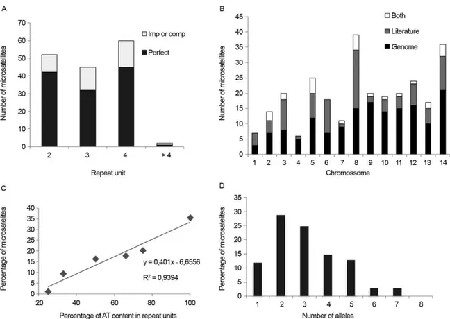
P. vivax genome sequencing allowed for further char-acterisation of a panel including ~160 microsatellites (Carlton et al. 2008). While searching for a repeat unit with 2-6 nucleotides, the authors found only one microsatel-lite locus with repeats consisting of more than four nucle-otides. Most (76%) loci had perfect repeat units (Fig. 3A), in contrast with the markers described by Karunaweera et al. (2007). All except one microsatellite were assigned to chromosomes, but their chromosome distribution was clearly heterogeneous. Chromosomes 1 and 4 had the lowest marker density, whereas chromosomes 14 and 9 had the highest density (Fig. 3B). Despite the relatively low AT content in the P. vivax genome, when compared with that of P. falciparum, P. vivax microsatellite-type repeats are particularly AT-rich (Fig. 3C). To character-ise polymorphic microsatellite markers, these loci were analysed in eight monkey-adapted P. vivax strains (Bra-zil I, Miami II, Pakchong, Panama I, Nicaragua, Thai II, Vietnam IV and Indonesia XIX). Nineteen markers were monomorphic among these samples, none were polymor-phic in all strains and the large majority of markers were polymorphic only in 2-4 strains (Fig. 3D). Further, only one-fourth of the markers shown to be polymorphic in natural P. vivax populations were also identified as poly-morphic using these eight P. vivax strains.
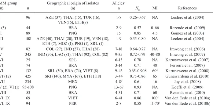
High variability in P. vivax isolates has been demon-strated using different sets of microsatellites (Table III). However, this comparison is complicated, as it is difficult to determine how much of the variation depends on the set of markers and how much is due to genetic diversity in the studied populations (Imwong et al. 2007a, Koep- Koep-fli et al. 2009). Currently, the most widely used markers are a set of 14 polymorphic microsatellites described by Karunaweera et al. (2007), which were used to analyse the population structures of P. vivax in Brazil, Vietnam, Sri Lanka, Myanmar, Ethiopia and Peru (Ferreira et al. 2007, Karunaweera et al. 2008, Gunawardena et al. 2010, Van den Eede et al. 2010a, b, 2011). Using the same set of markers, similar variability was detected in different geographical areas, with HE ranging from 0.71 in Bra-zil to 0.86 in Myanmar and Vietnam; only Peru showed a slightly lower variability (HE = 0.58). More recently, Joy et al. (2008) selected 24 microsatellites of ~160, de-scribed by Carlton et al. (2008), and used them to show local adaptation between mosquito vectors and different genetic populations of P. vivax from Mexico. There was no correlation between the number of alleles identified by Carlton et al. (2008) and the variability identified using a large number of P. vivax field isolates using the same set of markers by Joy et al. (2008). These data reinforce the uncertainty in using laboratory strains to identify poly-morphic markers (Fig. 4). Koepfli et al. (2009) showed high variability in Papua New Guinea isolates using this
TABLE III
Genetic variability analysis of Plasmodium vivax field populations using minisatellites and microsatellites as molecular markers
MM group
(n) n
Geographical origin of isolates (n)
Allelesa
n HE MI References
I 96 AZE (37), THAI (15), TUR (16), VEN(16), ETH(8)
1-8 0.26-0.67 NA Leclerc et al. (2004)
I (5) 44 BRA 2-9 0.57 0-66 Rezende et al. (2009)
II 89 PNG 15 0.85 4.5 Gomez et al. (2003)
III 108 AZE (40), THAI (20), TUR (19), VEN (18), ETH (7), MOZ (1), PNG (1), SRL (1)
1-9 0.35-0.80 NA Leclerc et al. (2004)
IV 82 COL (27), IND (27), THAI (28) 7-18 0.64-0.77 NA Imwong et al. (2006) V 345 IND (90), LAO (81), THAI (92), COL (82) 9-35 0.72-0.79 40-80 Imwong et al. (2007) VI 25 SRL 6-13 0.78 NA Karunaweera et al. (2007) VI 74 BRA 3-14 0.71 48 Ferreira et al. (2007) VI 132 SRL (50), BRA (74), VIET (8) 9-43 0.65-0.908 60b Karunaweera et al. (2008)
VI (12) 425 SRI (140), MYA (167), ETH (118) 3-44 0.75-0.86 65 Gunawardena et al. (2010) VII 234 MEX 4-8d 0.61 16 Joy et al. (2008)
V (2), VI (1) 93-108 PNG 13-67 0.93 NA Koefli et al. (2009) VIII 53 BRA 4-31 0.71 60 Rezende et al. (2010) VI, IX 69 VIET 6-41 0.86 100c Van den Eede et al. (2010a)
VI, IX 94 PER 2-8 0.58 11-70c Van den Eede et al. (2010b)
set of microsatellites and three selected from other studies (Table II). Rezende et al. (2010) described a set of 11 mi-crosatellites used to show a positive correlation between transmission levels and genetic variability in P. vivax iso-lates from geographical regions in Brazil. Taken together, these data suggest that, regardless of endemicity in the isolates’ geographical origin, most studies show that P. vivax populations are highly variable across the globe.
Practical uses for P. vivax genotyping - The vast field of infectious disease molecular epidemiology has produced a number of examples involving practical ap-plications for molecular markers in addressing issues of clear biological and public health importance. Here, we briefly review four areas of potential interest: (i) detec-tion of naturally occurring multiple-clone infecdetec-tions, (ii) after-treatment follow-up in clinical trials, (iii) between-species comparisons of genetic diversity levels and (iv) tracking of the geographic origin of infections.
Over the last two decades, molecular markers have revealed that many natural P. vivax infections comprise a complex mixture of clonal populations; that is, multiple-clone vivax infections are fairly common in human hosts (Havryliuk & Ferreira 2009). The earliest attempts to
in-vestigate the clonal diversity of P. vivax isolates in human infections focused on characterisation of phenotypes us-ing monoclonal antibodies or isoenzymes (Udagama et al. 1987, 1990, Joshi et al. 1989), an approach that required blood samples with relatively high parasitaemias. As a rule, multilocus analysis, such as microsatellite typing, detects more multiple-clone infections than single-locus analysis, and augmenting the number of markers increas-es the chance of detecting multiple clonincreas-es. For example, in 90 P. vivax isolates from Thailand, Cui et al. (2003b) found a multiple-clone infection rate of 25.6% using the
PvCSP encoding gene alone, 19.3% using PvMSP-3α
alone and 35.6% when the markers were combined. Par-ticularly surprising is the extensive clonal diversity of
P. vivax infections in areas with relatively low malaria transmission, such as Brazil, Colombia and Sri Lanka (Ferreira et al. 2007, Imwong et al. 2007b, Karunaweera et al. 2008, Orjuela-Sánchez et al. 2009a, Gunawardena et al. 2010). The evolutionary and epidemiological con-sequences of multiple-clone P. vivax infections have not been well-investigated. However, experimental rodent malaria models suggest that, for two or more genetically distinct clones in the same host, intra-host competition for limited resources may select for P. vivax traits
senting major public health challenges, such as increased virulence, transmissibility and antimalarial drug resist-ance (reviewed by Havryliuk & Ferreira 2009).
Molecular markers have also been used to type P. vivax infections after drug treatment. Reappearance of parasitaemia after drug treatment can result from either recrudescence of surviving asexual blood-stage parasites; relapse from dormant liver stages, known as hypnozoites; or new infections with unrelated parasites. Paired parasite molecular genotyping distinguishes between recrudes-cences (with the same genotype as the initial infection) and new infections (with a different genotype). Until re-cently, relapses were thought to be caused by hypnozoites that are genetically identical to the blood-stage parasites found in primary infections (Craig & Kain 1996, Kirch-gatter & del Portillo 1998). This suggested that molecular methods could easily discriminate relapses, which would have the same genotype as the primary infection, from new infections, which would have a different genotype. This view has been challenged by the recent discovery of different parasite genotypes in primary infections and relapses for 72% of P. vivax-infected patients from Thai-land, India and Myanmar (Imwong et al. 2007b). Accu-rate detection of multiple-clone P. vivax infections is even more important in light of this report. If the primary in-fection comprises multiple clones, then some of them may have been missed or partially characterised or, even worse, had their alleles combined to create artificial hap-lotypes during genotyping. Likewise, the relapsing clone could have been either missed or incorrectly typed dur-ing the primary infection, leaddur-ing to a false conclusion that different genotypes were present during the primary and relapse infections. The current consensus is that re-lapses may originate from reactivation of either the same parasite clone found in the primary bloodstream infection (homologous hypnozoites) or another, genetically different clone (heterologous hypnozoites). Our recent microsatel-lite analysis of 28 paired acute-infection and recurrence parasites from rural Amazonia revealed only two pairs of identical haplotypes (consistent with recrudescences or reactivation of homologous hypnozoites) and four pairs of related haplotypes (sharing alleles at 11-13 of 14 microsat-ellites analysed) (Orjuela-Sánchez et al. 2009a).
As the characteristics of endemic settings may in-fluence the genetic diversity of the parasite, noteworthy findings could be obtained from sympatric P. vivax and
P. falciparum isolates that are analysed using similar protocols. Two comparisons of microsatellite diversity in human malaria parasite species co-circulating in the same area in Brazil have been published. Both suggest that P. vivax infections are more diverse (that is, they comprise a higher number of alleles and virtual HE) and comprise multiple clones more often than P. falciparum
infections in rural Amazonia (Ferreira et al. 2007, Or- Or-juela-Sánchez et al. 2009b). As P. vivax currently pre-dominates in Brazil, differences in species-specific transmission levels might translate into differences in genetic diversity. However, we recently found more mi-crosatellite diversity in P. vivax compared with P. fal-ciparum populations in western Cambodia, where both species are similarly prevalent (MU Ferreira and RM Fairhurst, unpublished observations). Thus, we suggest that the higher microsatellite diversity found in P. vivax
isolates may reflect greater plasticity in microsatellite-type short repeats for this species.
Finally, standardised molecular methods applied to worldwide populations may also help characterise popu-lation or region-specific molecular barcodes. This is es-sential for tracking the geographic origin of infections. A microsatellite-based analysis of P. vivax samples from Sri Lanka, Ethiopia and Myanmar recently showed that parasites may be classified as originating from Asia or Africa with 70-80% accuracy. This suggests that micros-atellite data might be useful in predicting the origin of P. vivax parasites (Gunawardena et al.2010). Further analy-ses are required to confirm whether microsatellite-based molecular barcodes are useful for determining whether particular infections were either transmitted locally or imported into areas approaching the elimination phase.
REFERENCES
Ampudia E, Patarroyo M, Patarroyo M, Murillo L 1996. Genetic polymorphism of the Duffy receptor binding domain of Plas-modium vivax in Colombian wild isolates. Mol Biochem Para-sitol 78: 269-272.
Anderson TJC, Su XZ, Bockarie M, Lagog M, Day KP 1999. Twelve mi-crosatellite markers for characterization of Plasmodium falciparum
from finger-prick blood samples. Parasitology 119: 113-125.
Arnot DE, Barnwell JW, Tam JP, Nussenzweig V, Nussenzweig RS, Enea V 1985. Circumsporozoite protein of Plasmodium vivax: gene cloning and characterization of the immunodoninant epitope. Science 230: 815-818.
Arnot DE, Stewart MJ, Barnwell JW 1990. Antigenic diversity in Thai Plasmodium vivax circumsporozoite proteins. Mol Biochem Parasitol 43: 147-149.
Babaeekho L, Zakeri S, Djadid ND 2009. Genetic mapping of the duffy binding protein (DBP) ligand domain of Plasmodium vivax
from unstable malaria region in the Middle East. Am J Trop Med Hyg 80: 112-118.
Bastos MS, da Silva-Nunes M, Malafronte RS, Hoffmann EH, Wun-derlich G, Moraes SL, Ferreira MU 2007. Antigenic polymor-phism and naturally acquired antibodies to Plasmodium vivax
merozoite surface protein 1 in rural Amazonians. Clin Vaccine Immunol 14: 1249-1259.
Bonilla JA, Validum L, Cumming R, Palmer CJ 2006. Genetic diver-sity of Plasmodium vivax Pvcsp and Pvmsp1 in Guyana, South America. Am J Trop Med Hyg 75: 830-835.
Bruce MC, Galinski MR, Barnwell JW, Donnelly CA, Walmsley M, Alpers MP, Walliker D, Day KP 2000. Genetic diversity and dy-namics of Plasmodium falciparum and P. vivax populations in multiply infected children with asymptomatic malaria infections in Papua New Guinea. Parasitology 121: 257-272.
Bruce MC, Galinski MR, Barnwell JW, Snounou G, Day KP 1999. Poly-morphism at the merozoite surface protein-3 locus of Plasmodium vivax: global and local diversity. Am J Trop Med Hyg61: 518-525.
Carlton JM, Adams JH, Silva JC, Bidwell SL, Lorenzi H, Caler E, Crabtree J, Angiuoli SV, Merino EF, Amedeo P, Cheng Q, Coul-son RM, Crabb BS, del Portillo HA, Essien K, Feldblyum TV, Fernandez-Becerra C, Gilson PR, Gueye AH, Guo X, Kang’a S, Kooij TW, Korsinczky M, Meyer EV, Nene V, Paulsen I, White O, Ralph SA, Ren Q, Sargeant TJ, Salzberg SL, Stoeckert CJ, Sullivan SA, Yamamoto MM, Hoffman SL, Wortman JR, Gard-ner MJ, Galinski MR, Barnwell JW, Fraser-Liggett CM 2008. Comparative genomics of the neglected human malaria parasite
Plasmodium vivax. Nature 455: 757-763.
Cheng Q, Stowers A, Huang TY, Bustos D, Huang YM, Rzepczyk C, Saul A 1993. Polymorphism in Plasmodium vivax MSA1 gene the result of intragenic recombinations? Parasitology 106: 335-345.
Ceravolo I, Souza-Silva F, Fontes CJ, Braga EM, Madureira A, Kret-tli AU, Souza JM, Brito CF, Adams JH, Carvalho LH 2008. In-hibitory properties of the antibody response to Plasmodium vivax
Duffy binding protein in an area with unstable malaria transmis-sion. Scand J Immunol67: 270-278.
Cochrane AH, Nardin EH, Arruda M, Maracic M, Clavijo P, Collins WE, Nussenzweig RS 1990. Widespread reactivity of human sera with variant repeat of the circumsporozoite protein of Plasmo-dium vivax. Am J Trop Med Hyg 43: 446-451.
Cole-Tobian JL, Cortés A, Baisor M, Kastens W, Xainli J, Bockarie M, Adams JH, King CL 2002. Age-acquired immunity to a Plas-modium vivax invasion ligand, the Duffy binding protein. J Infect Dis 186: 531-539.
Conway DJ, Cavanagh DR, Tanabe K, Roper C, Mikes ZS, Sakihama N, Bojang KA, Oduola AM, Kremsner PG, Arnot DE, Green-wood BM, McBride JS 2000. A principal target of human im-munity to malaria identified by molecular population genetic and immunological analyses. Nat Med 6: 689-692.
Craig AA, Kain KC 1996. Molecular analysis of strains of Plasmo-dium vivax from paired primary and relapse infections. J Infect Dis 174: 373-379.
Cristiano FA, Pérez MA, Nicholls RS, Guerra AP 2008. Polymor-phism in the Plasmodium vivaxmsp3α gene in field samples from Tierralta, Colombia. Mem Inst Oswaldo Cruz 103: 493-496.
Cui L, Escalante AA, Imwong M, Snounou G 2003a. The genetic diversi-ty of Plasmodium vivax populations. Trends Parasitol 19: 220-226.
Cui L, Mascorro CN, Fan Q, Rzomp KA, Khuntirat B, Zhou G, Chen H, Yan G, Sattabongkot J 2003b. Genetic diversity and multiple infections of Plasmodium vivax malaria in western Thailand. Am J Trop Med Hyg 68: 613-619.
Cheng Q, Saul A 1994. Sequence analysis of the apical membrane antigen I (AMA-1) of Plasmodium vivax. Mol Biochem Parasi-tol 65: 183-187.
Choi YK, Choi KM, Park MH, Lee EG, Kim YJ, Lee BC, Cho SH, Rhie HG, Lee HS, Yu JR, Lee JS, Kim TS, Kim JY 2010. Rapid dissemination of newly introduced Plasmodium vivax genotypes in south Korea. Am J Trop Med Hyg 82: 426-432.
Chung JY, Chun EH, Chun JH, Kho WG 2003. Analysis of the Plas-modium vivax apical membrane antigen-1 gene from re-emerging Korean isolates. Parasitol Res 90: 325-329.
del Portillo HA, Longacre S, Khouri E, David PH 1991. Primary structure of the merozoite surface antigen 1 of Plasmodium vivax
reveals sequences conserved between different Plasmodium spe-cies. Proc Natl Acad Sci USA 88: 4030-4034.
Dias S, Longacre S, Escalante AA, Udagama-Randeniya PV 2011. Genetic diversity and recombination at the C-terminal fragment of the merozoite surface protein-1 of Plasmodium vivax (PvM-SP-1) in Sri Lanka. Infect Genet Evol 11: 145-156.
Escalante AA, Cornejo OE, Rojas A, Udhayakumar V, Lal AA 2004. Assessing the effect of natural selection in malaria parasites.
Trends Parasitol 20: 388-395.
Escalante AA, Lal AA, Ayala FJ 1998. Genetic polymorphism and natural selection in the malaria parasite Plasmodium falciparum.
Genetics 149: 189-202.
Farooq U, Malla N, Dubey ML 2009. Polymorphism in merozoite sur-face protein-1 gene in north and northwest Indian field isolates of
Plasmodium vivax. Indian J Med Res 130: 736-741.
Feng X, Carlton JM, Joy DA, Mu J, Furuya T, Suh BB, Wang Y, Barnwell JW, Su XZ 2003. Single-nucleotide polymorphisms and genome diversity in Plasmodium vivax. Proc Natl Acad Sci USA 100: 8502-8507.
Ferreira MU, Karunaweera ND, da Silva-Nunes M, da Silva NS, Wirth DF, Hartl DL 2007. Population structure and transmission dynamics of Plasmodium vivax in rural Amazonia. J Infect Dis
195: 1218-1226.
Figtree M, Pasay CJ, Slade R, Cheng Q, Cloonan N, Walker J, Saul A 2000. Plasmodium vivax synonymous substitution frequencies, evolution and population structure deduced from diversity in AMA 1 and MSP 1 genes. Mol Biochem Parasitol 108: 53-66.
Galinski M, Barnwell J 2009. Monkey malaria kills four humans.
Trends Parasitol 25:200-204.
Galinski MR, Corredor-Medina C, Povoa M, Crosby J, Ingravallo P, Barnwell JW 1999. Plasmodium vivax merozoite surface protein-3 contains coiled-coil motifs in an alanine-rich central domain. Mol Biochem Parasitol1011: 131-147.
Gómez JC, McNamara T, Bockarie MJ, Baird JK, Carlton JM, Zim-merman PA 2003. Identification of a polymorphic Plasmodium vivax microsatellite marker. Am J Trop Med Hyg 69: 377-389.
Gopinath R, Wongsrichanalai C, Cordón-Rosales C, Mirabelli L, Kyle D, Kain KC 1994. Failure to detect a Plasmodium vivax -like malaria parasite in globally collected blood samples. J Infect Dis 170: 1630-1633.
Gosi P, Khusmith S, Khalambaheti T, Lanar D, Schaecher K, Fukuda M, Miller S 2008. Polymorphism patterns in Duffy-binding pro-tein among Thai Plasmodium vivax isolates. Malar J 7: 112.
Grimberg BT, Udomsangpetch R, Xainli J, McHenry A, Panichakul T, Sattabongkot J, Cui L, Bockarie M, Chitnis C, Adams J, Zim-merman PA, King CL 2007. Plasmodium vivax invasion of hu-man erythrocytes inhibited by antibodies directed against the Duffy binding protein. PLoS Med 4: e337.
Grynberg P, Fontes CJF, Hughes AL, BragaEM2008. Polymorphism at the apical membrane antigen 1 locus reflects the world population history of Plasmodium vivax. BMC Evolutionary Biology 8: 123.
Gunasekera AM, Wickramarachchi T, Neafsey DE, Ganguli I, Perera L, Premaratne PH, Hartl D, Handunnetti SM, Udagama-Ran-deniya PV, Wirth DF 2007. Genetic diversity and selection at the
Plasmodium vivax apical membrane antigen-1 (PvAMA-1) locus in a Sri Lankan population. Mol Biol Evol 24: 939-947.
Gunawardena S, Karunaweera ND, Ferreira MU, Phone-Kyaw M, Pollack RJ, Alifrangis M, Rajakaruna RS, Konradsen F, Amera- singhe PH, Schousboe ML, Galappaththy GN, Abeyasinghe RR, Hartl DL, Wirth DF 2010. Geographic structure of Plasmodium vivax: microsatellite analysis of parasite populations from Sri Lanka, Myanmar, and Ethiopia. Am J Trop Med Hyg 82: 235-242.
Gutierrez A, Vicini J, Patarroyo ME, Murillo LA, Patarroyo MA 2000.
Plasmodium vivax: polymorphism in the merozoite surface protein 1 gene from wild Colombian isolates. Exp Parasitol 95: 215-219.
Han ET, Park JH, Shin EH, Choi MH, Oh MD, Chai JY 2002. Api-cal membrane antigen-1 (AMA-1) gene sequences of re-emerging
Plasmodium vivax in South Korea. Korean J Parasitol 40: 157-162.
Han ET, Song TE, Park JH, Shin EH, Guk SM, Kim TY, Chai JY 2004. Allelic dimorphism in the merozoite surface protein-3alpha in Ko-rean isolates of Plasmodium vivax. Am J Trop Med Hyg 71: 745-749.
Han ET, Wang Y, Lim CS, Cho JH, Chai JY 2011. Genetic diversity of the malaria vaccine candidate merozoite surface protein 1 gene of
Plasmodium vivax field isolates in Republic of Korea. Parasitol Res [Epub ahead of print]. Available from springerlink.com/con-tent/x085hm23431201g1/.
Hans D, Pattnaik P, Bhattacharyya A, Shakri A, Yazdani S, Sharma M, Choe H, Farzan M, Chitnis C 2005. Mapping binding residues in the Plasmodium vivax domain that binds Duffy antigen during red cell invasion. Mol Microbiol55: 1423-1434.
Havryliuk T, Ferreira MU 2009. A closer look at multiple-clone Plas-modium vivax infections: detection methods, prevalence and con-sequences. Mem Inst Oswaldo Cruz 104: 67-73.
Henry-Halldin CN, Sepe D, Susapu M, McNamara DT, Bockarie M, King CL, Zimmerman PA 2011. High-throughput molecular di-agnosis of circumsporozoite variants VK210 and VK247 detects complex Plasmodium vivax infections in malaria endemic popu-lations in Papua New Guinea. Infect Genet Evol 11: 391-398.
Hernández-Martínez MA, Escalante AA, Arévalo-Herrera M, Her-rera S 2011. Antigenic diversity of the Plasmodium vivax circum-sporozoite protein in parasite isolates of Western Colombia. Am J Trop Med Hyg 84: 51-57.
Herrera S, Corradin G, Arévalo-Herrera M 2007. An update on the search for a Plasmodium vivax vaccine. Trends Parasitol 23: 122-128.
Hodder AN, Crewther PE, Anders RF 2001. Specificity of the pro-tective antibody response to apical membrane antigen 1. Infect Immun 69: 3286-3294.
Hoffmann EHE, Ribolla PEM, Ferreira MU 2003. Genetic related-ness of Plasmodium falciparum isolates and the origin of allelic diversity at the merozoite surface protein-1 (MSP-1) locus in Bra-zil and Vietnam. Malar J2: 24.
Holder AA 2009. The carboxy-terminus of merozoite surface protein 1: structure, specific antibodies and immunity to malaria. Para-sitology 136: 1445-1456.
Huang T, Cheng Q, Allan S, Huang Y 1994. DNA sequencing of circumsporozoite protein genes of Plasmodium vivax from four different countries in west Pacific region: comparative study on the flank sequences. Zhongguo Ji Sheng Chong Xue Yu Ji Sheng Chong Bing Za Zhi 12: 85-92.
Imwong M, Nair S, Pukrittayakamee S, Sudimack D, Williams JT, Mayxay M, Newton PN, Kim JR, Nandy A, Osorio L, Carlton JM, White NJ, Day NPJ, Anderson TJ 2007a. Contrasting genetic
structure in Plasmodium vivax populations from Asia and South America. Int J Parasitol 37: 1013-1022.
Imwong M, Pukrittayakamee S, Grüner AC, Rénia L, Letourneur F, Looareesuwan S, White NJ, Snounou G 2005. Practical PCR genotyping protocols for Plasmodium vivax using Pvcs and Pvm-sp1. Malar J 4: 20.
Imwong M, Pukrittakayamee S, Looareesuwan S, Poirriez J, Pasvol G, White NJ, Snounou G 2001. Plasmodium vivax: polymerase chain reaction amplification artifacts limit the suitability of pvgam1 as a genetic marker. Exp Parasitol 99: 175-179.
Imwong M, Snounou G, Pukrittayakamee S, Tanomsing N, Kim JR, Nandy A, Guthmann JP, Nosten F, Carlton J, Looareesuwan S, Nair S, Sudimack D, Day NP, Anderson TJ, White NJ 2007b. Re-lapses of Plasmodium vivax infection usually result from activa-tion of heterologous hypnozoites. J Infect Dis 195: 927-933.
Imwong M, Sudimack D, Pukrittayakamee S, Osório L, Carlton JM, Day NPJ, White NJ, Anderson TJC 2006. Microsatellite varia-tion, repeat array length and population history of Plasmodium vivax. Mol Biol Evol 23: 1016-1018.
Joshi H, Subbarao SK, Raghavendra K, Sharma VP 1989. Plasmo-dium vivax: enzyme polymorphism in isolates of Indian origin.
Trans R Soc Trop Med Hyg 83: 179-181.
Joy DA, Gonzalez-Cerón L, Carlton JM, Gueye A, Fay M, Mc-Cutchan TF, Su XZ 2008. Local adaptation and vector-mediated population structure in Plasmodium vivax malaria. Mol Biol Evol 25: 1245-1252.
Kain KC, Brown AE, Mirabelli L, Webster HK 1993. Detection of
Plasmodium vivax by polymerase chain reaction in a field study.
J Infect Dis 168: 1323-1326.
Karunaweera ND, Ferreira MU, Hartl DL, Wirth DF 2007. Fourteen polymorphic microsatellite DNA markers for the human malaria parasite Plasmodium vivax. Mol Ecol Notes 7: 172-175.
Karunaweera ND, Ferreira MU, Munasinghe A, Barnwell JW, Col-lins WE, King CL, Kawamoto F, Hartl DL, Wirth DF 2008. Extensive microsatellite diversity in the human malaria parasite
Plasmodium vivax. Gene 410:105-112.
Khatoon L, Baliraine FN, Bonizzoni M, Malik SA, Yan GY 2010. Ge-netic structure of Plasmodium vivax and Plasmodium falciparum
in the Bannu district of Pakistan. Malar J9: 112.
Kho W, Chung J, Sim E, Kim D, Chung W 2001. Analysis of poly-morphic regions of Plasmodium vivax Duffy binding protein of Korean isolates. Korean J Parasitol 39: 143-150.
Kim JR, Imwong M, Nandy A, Chotivanich K, Nontprasert A, Tonomsing N, Maji A, Addy M, Day NP, White NJ, Pukrit-tayakamee S 2006. Genetic diversity of Plasmodium vivax in Kolkata, India. Malar J 5: 71.
Kim SH, Hwang SY, Shin JH, Moon CS, Kim DW, Kho WG 2009. Molecular genetic characterization of the merozoite surface pro-tein 1 gene of Plasmodium vivax from reemerging Korean iso-lates. Clin Vaccine Immunol 16: 733-738.
Kimura E, Mattei D, di Santi SM, Scherf A 1990. Genetic diversity in the major merozoite surface antigen of Plasmodium falciparum: high prevalence of a third polymorphic form detected in strains derived from malaria patients. Gene 91: 57-62.
Kirchgatter K, del Portillo HA 1998. Molecular analysis of Plasmodi-um vivax relapses using the MSP1 molecule as a genetic marker.
J Infect Dis 177: 511-515.
Kolakovich KA, Ssengoba A, Wojcik K, Tsuboi T, Al-Yaman F, Al- pers M, Adams JH 1996. Plasmodium vivax: favored gene frequen- cies of the merozoite surface prote1 and the multiplicity of in-fection in a malaria endemic region. Exp Parasitol 83: 11-18.
Leclerc MC, Durant P, Gauthier C, Patot S, Billote N, Menegon M, Severini C, Ayala FJ, Renaud F 2004. Meager genetic variability of the human malaria agent Plasmodium vivax. Proc Natl Acad Sci USA 101: 14455-14460.
Levinson G, Gutman GA 1987. Slipped-strand mispairing: a major mech-anisms for DNA sequence evolution. Mol Biol Evol 4: 203-221.
Lim CS, Kim SH, Kwon SI, Song JW, Song KJ, Lee KN 2000. Analysis of Plasmodium vivax merozoite surface protein-1 gene sequences from resurgent Korean isolates. Am J Trop Med Hyg 62: 261-265.
Lim CS, Tazi L, Ayala FJ 2005. Plasmodium vivax:recent world ex-pansion and genetic identity to Plasmodium simium. Proc Natl Acad Sci USA 102: 15523-15528.
Machado RLD, Póvoa MM 2000. Distribution of Plasmodium vivax
variants (VK210, VK247 and P. vivax-like) in three endemic ar-eas of the Amazon Region of Brazil and their correlation with chloroquine treatment. Trans R Soc Trop Med Hyg 94: 377-381.
Maestre A, Sunil S, Ahmad G, Mohmmed A, Echeverri M, Corredor M, Blair S, Chauhan VS, Malhotra P 2004. Inter-allelic recombi-nation in the Plasmodium vivax merozoite surface protein 1 gene among Indian and Colombian isolates. Malar J 3: 4.
Mann VH, Huang T, Cheng Q, Saul A 1994. Sequence variation in the circumsporozoite protein gene of Plasmodium vivax appears to be regionally biased. Mol Biochem Parasitol 68: 45-52.
Martinez P, Suarez CF, Cardenas PP, Patarroyo MA 2004. Plasmo-dium vivax Duffy binding protein: a modular evolutionary pro-posal. Parasitology 128: 353-366.
Mascorro CN, Zhao K, Khuntirat B, Sattabongkot J, Yan G, Escalante AA, Cui L 2005. Molecular evolution and intragenic recombina-tion of the merozoite surface protein MSP-3 alpha from the malaria parasite Plasmodium vivax in Thailand. Parasitology131: 25-35.
McConkey GA, Waters AP, McCutchan TF 1990. The generation of genetic diversity in malaria parasites. Annu Rev Microbiol 44: 479-498.
Michon P, Fraser T, Adams J 2000. Naturally acquired and vaccine-elicited antibodies block erythrocyte cytoadherence of the Plasmo-dium vivax Duffy binding protein. Infect Immun 68: 3164-3171.
Monette M, Opella SJ, Greenwood J, Willis AE, Perham RN 2001. Structure of a malaria parasite antigenic determinant displayed on filamentous bacteriophage determined by NMR spectrosco-py: implications for the structure of continuous peptide epitopes of proteins. Protein Sci 10: 1150-1159.
Moon SU, Lee HW, Kim JY, Na BK, Cho SH, Lin K, Sohn WM, Kim TS 2009. High frequency of genetic diversity of Plasmodium vivax field isolates in Myanmar. Acta Trop 109: 30-36.
Moon SU, Na BK, Kang JM, Kim JY, Cho SH, Park YK, Sohn WM, Lin K, Kim TS 2010. Genetic polymorphism and effect of natural selec-tion at domain I of apical membrane antigen-1 (AMA-1) in Plasmo-dium vivax isolates from Myanmar. Acta Tropica 114: 71-75
Mueller I, Galinski MR, Baird JK, Carlton JM, Kochar DK, Alonso PL, del Portillo HA 2009. Key gaps in the knowledge of Plasmo-dium vivax, a neglected human malaria parasite. Lancet Infect Dis 9: 555-566.
Mueller I, Kaiok J, Reeder JC, Cortés A 2002. The population struc-ture of Plasmodium falciparum and Plasmodium vivax during an epidemic of malaria in the Eastern Highlands of Papua New Guinea. Am J Trop Med Hyg 67: 459-464.
Nardin EH, Zavala F 1998. Acquired immunity to sporozoites. In IW Sherman, Malaria: parasite biology, pathogenesis, and protec-tion, ASM Press, Washington DC, p. 495-511.
Nogueira PA, Alves FP, Fernandez-Becerra C, Pein O, Santos NR, Pereira da Silva LH, Camargo EP, del Portillo HA 2006. A re-duced risk of infection with Plasmodium vivax and clinical pro-tection against malaria are associated with antibodies against the N terminus but not the C terminus of merozoite surface protein 1.
Infect Immun 74: 2726-2733.
Oliveira-Ferreira J, Lacerda MV, Brasil P, Ladislau JL, Tauil PL, Daniel-Ribeiro CT, 2010. Malaria in Brazil: an overview. Malar J 9: 115.
Ord R, Polley S, Tami A, Sutherland CJ 2005. High sequence diversi-ty and evidence of balancing selection in the PvMSP3 alpha gene of Plasmodium vivax in the Venezuelan Amazon. Mol Biochem Parasitol144: 86-93.
Ord RL, Tami A, Sutherland CJ 2008. ama1 genes of sympatric Plas-modium vivax and P. falciparum from Venezuela differ signifi-cantly in genetic diversity and recombination frequency. PLoS ONE 3: e3366.
Orjuela-Sánchez P, da Silva NS, da Silva-Nunes M, Ferreira MU 2009a. Recurrent parasitemias and population dynamics of Plas-modium vivax polymorphisms in rural Amazonia. Am J Trop Med Hyg 81: 961-968.
Orjuela-Sánchez P, da Silva-Nunes M, da Silva NS, Scopel KK, Gon-çalves RM, Malafronte RS, Ferreira MU 2009b. Population dy-namics of genetically diverse Plasmodium falciparum lineages: community-based prospective study in rural Amazonia. Parasi-tology 136: 1097-1105.
Pacheco MA, Poe AC, Collins WE, Lal AA, Tanabe K, Kariuki SK, Udhayakumar V, Escalante AA 2007.A comparative study of the genetic diversity of the 42 kDa fragment of the merozoite surface protein 1 in Plasmodium falciparum and P. vivax. Infect Genet Evol 7: 180-187.
Pasay MC, Cheng Q, Rzepczyk C, Saul A 1995. Dimorphism of the C terminus of the Plasmodium vivax merozoite surface protein 1.
Mol Biochem Parasitol 70: 217-219.
Patil A, Orjuela-Sánchez P, da Silva-Nunes M, Ferreira MU 2010. Evolutionary dynamics of the immunodominant repeats of the
Plasmodium vivax malaria-vaccine candidate circumsporozoite protein (CSP). Infect Genet Evol 10: 298-303.
Pizarro JC, Vulliez-Le Normand B, Chesne-Seck ML, Collins CR, Withers-Martinez C, Hackett F, Blackman MJ, Faber BW, Re-marque EJ, Kocken CH, Thomas AW, Bentley GA 2005. Crystal structure of the malaria vaccine candidate apical membrane anti-gen 1. Science 308: 408-411.
Polley SD, Weedall GD, Thomas AW, Golightly LM, Conway DJ 2005. Orthologous gene sequences of merozoite surface protein 1 (MSP1) from Plasmodium reichenowi and P.gallinaceum con-firm an ancient divergence of P. falciparum alleles. Mol Biochem Parasitol 142: 25-31.
Prajapati SK, Joshi H, Valecha N 2010. Plasmodium vivax merozoite surface protein-3 alpha: a high-resolution marker for genetic di-versity studies. J Vec Bor Dis472: 85-90.
Prajapati SK, Verma A, Adak T, Yadav RS, Kumar A, Eapen A, Das MK, Singh N, Sharma SK, Rizvi MA, Dash AP, Joshi H 2006. Allelic dimorphism of Plasmodium vivax gam-1 in the Indian subcontinent. Malar J 5: 90.
Premawansa S, Snewin VA, Khouri E, Mendis KN, David PH 1993.