online | memorias.ioc.fiocruz.br Geographic distribution of Tachia spp - Tachia Aubl.
(Gentianaceae) was first described by the botanist Aublet in 1775 based on plants collected in French Guyana. Be-fore 1975, only the type species Tachia guianensis Aubl. was known. Recent studies show that T. guianensis oc-curs only in French Guyana and Suriname (Peters et al. 2005). Today, it is accepted that Tachia is comprised of 13 closely related understory shrubs or small tree species (Peters et al. 2005), which occur also in the wet forests of the Amazon Basin and the lower elevations of mountain-ous areas of Peru, Ecuador, Colombia, Venezuela and Brazil (7 species) and Panama (1 species) (Peters et al. 2005). Tachia grandiflora (Maguire & Weaver 1975),
studied herein, is widespread in wet tropical forests of the central and eastern part of the Amazon Basin and northwestern South America and from French Guyana and Suriname in the north to central parts of the state of Amazonas (AM) in Brazil to the west and south [see map in Peters et al. (2005)].
Traditional use of Tachia spp as antimalarials - Eth-nobotanical and ethnopharmacological publications have described the traditional uses of Tachia spp as antima-larials and febrifuges in The Guyanas, Brazil, Colombia and Peru (Milliken 1997). However, in many Brazilian (Carvalho & Krettli 1991, Brandão et al. 1992, Milliken 1997, Mors et al. 2000, Krettli et al. 2001), Colombian (Schultes & Raffauf 1990) and Peruvian (Milliken 1997) studies the plants collected are incorrectly identified as the type species of the genus, T. guianensis Aubl.
T. grandiflora - We became interested in studying the local caferana plant, T. grandiflora, based on earlier re-ports by the Dr Antoniana Krettli group (Oswaldo Cruz Foundation, state of Minas Gerais, Brazil) in which the water extract of roots of a Tachia sp. exhibited significant in vivo activity in a mouse model of malaria. Tachia spp are rare, sparsely populated plants in the Amazon forests. We initially conducted studies on the propagation of this plant from stem cuttings (Silva et al. 2006). Pio Corrêa (1926) reported that caferana extracts were toxic. Polar
doi: 10.1590/0074-0276108042013017
Financial support: CNPq (520354/99-0 & 550260/01-3), CNPq- Brazilian Malaria Network, CNPq-Bionorte Program, Bioamazonia-BASA-FEPAD Contract
TCMS , LFRS and AMP were supported by scholarships from the PIBIC/INPA, PCI/INPA and PQ/CNPq, respectively.
ESL is a member of INCT-Redoxoma (MCT/CNPq). + Corresponding author: [email protected] Received 11 December 2012
Accepted 26 March 2013
In vitro and in vivo antimalarial activity and cytotoxicity
of extracts, fractions and a substance isolated
from the Amazonian plant
Tachia grandiflora
(Gentianaceae)
Luiz Francisco Rocha e Silva1,2,3, Emerson Silva Lima2, Marne Carvalho de Vasconcellos2,
Ellen Suzany Pereira Aranha2, David Siqueira Costa1,3, Elba Vieira Mustafa1,2,
Sabrina Kelly Reis de Morais1,2, Maria das Graças Costa Alecrim4, Sergio Massayoshi Nunomura1,
Lena Struwe5, Valter Ferreira de Andrade-Neto6, Adrian Martin Pohlit1/+
1Laboratório de Princípios Ativos da Amazônia, Coordenação de Tecnologia e Inovação, Instituto Nacional de Pesquisas da Amazônia, Manaus, AM, Brasil 2Universidade Federal do Amazonas, Manaus, AM, Brasil 3Centro Universitário do Norte, Manaus, AM, Brasil 4Fundação de Medicina Tropical Doutor Heitor Vieira Dourado, Manaus, AM, Brasil 5Rutgers, The State University of New Jersey,
New Brunswick, NJ, USA 6Universidade Federal do Rio Grande do Norte, Natal, RN, Brasil
Tachia sp. are used as antimalarials in the Amazon Region and in vivo antimalarial activity of a Tachia sp. has been previously reported. Tachia grandiflora Maguire and Weaver is an Amazonian antimalarial plant and herein its cytotoxicity and antimalarial activity were investigated. Spectral analysis of the tetraoxygenated xanthone decus-satin and the iridoid aglyone amplexine isolated, respectively, from the chloroform fractions of root methanol and leaf ethanol extracts was performed. In vitro inhibition of the growth of Plasmodium falciparum Welch was evalu-ated using optical microscopy on blood smears. Crude extracts of leaves and roots were inactive in vitro. However, chloroform fractions of the root and leaf extracts [half-maximal inhibitory concentration (IC50) = 10.5 and 35.8 µg/ mL, respectively] and amplexine (IC50 = 7.1 µg/mL) were active in vitro. Extracts and fractions were not toxic to type MRC-5 human fibroblasts (IC50 > 50 µg/mL). Water extracts of the roots of T. grandiflora administered by mouth were the most active extracts in the Peters 4-day suppression test in Plasmodium berghei-infected mice. At 500 mg/ kg/day, these extracts exhibited 45-59% inhibition five to seven days after infection. T. grandiflora infusions, frac-tions and isolated substance have potential as antimalarials.
extracts of T. grandiflora were not toxic to Artemia fran-ciscana in the brine shrimp assay (Quignard et al. 2003). In another study, extracts of T. grandiflora at 500 µg/mL exhibited moderate toxicity (7-64% lethality) to larvae of Aedes aegypti (Pohlit et al. 2004). Also, extracts of T. grandiflora were highly active inhibitors of the growth of cancer tumour cell lines (Pohlit et al. 2007).
Antimalarial plants such as T. grandiflora are po-tential sources of drug leads against Plasmodium spp (Andrade-Neto et al. 2007, Schmidt et al. 2012a, b). Re-cently, we isolated the tetra-oxygenated xanthone decus-satin (1) and a rare seco-iridoid monoterpene aglycone, djalonenol (amplexine) (2), from T. grandiflora (Pohlit et al. 2012). In the present work, the in vitro and in vivo an-tiplasmodial activity and cytoxicity of the extracts, frac-tions and chemical components of the leaves and roots of the central Amazonian caferana plant, T. grandiflora, were investigated. Spectroscopic characterisation of the isolates 1 and 2 is also presented.
MATERIALS AND METHODS
Chemicals - All solvents used for extraction, parti-tioning and chromatography were fractionally distilled prior to use. Solvents for NMR were purchased from Sigma-Aldrich (St. Louis, USA).
Instrumentation - Medium pressure liquid chroma-tography (MPLC) was performed using a Büchi System with Pump model 688, Gradient Former model 687, ultraviolet visible spectroscopy and fraction collec-visible spectroscopy and fraction collec- and fraction collec-tor model 684 and a normal phase column with 40-63 µm particle size. 1H-NMR, 13C-NMR, DEPT 135, 1H-1H
COSY and HMQC spectra were acquired on a Bruker DPX 300 (300 MHz) in CDCl3/TMS or (CD3)2CO/TMS. FT-IR spectra were acquired on a Bomem model M 102 spectrometer. Electronic ionization-gas chromatogra-gas chromatogra-phy-mass spectrometry (EI-GC-MS) was performed on a Hewlett-Packard HP 5890 series gas chromatograph coupled to mass detector HP 5971 operating at an ioni-zation energy of 70 eV.
Plant material - Plant materials were collected in September and October, 2000 in National Institute for Amazonian Research’s (INPA) Campina and Adolpho Ducke Forest Reserves which are located in greater Ma- naus, AM. Voucher specimens were deposited at the INPA Herbarium under the accessions 208104 (collector AM Pohlit) and 205948 (collector AM Pohlit). Identification of the plant samples as T. grandiflora Maguire and Weaver (Gentianaceae) was corroborated by LS (co-author of the present paper). Roots and mature leaves were separately dried in the shade and ground to fine powders.
Extraction and isolation of 1 - Dried, powdered roots were continuously extracted in a Soxhlet apparatus with methanol (3 × 6 h). The extracts were combined and evaporated under vacuum using a rotary evaporator with
heat bath (≤ 40ºC) and then freeze-dried. The resulting
dry methanol extract (5 g) was dissolved in methanol-distilled water (9:1) and partitioned with hexanes, then chloroform. After total evaporation, the chloroform fraction was analysed by thin-layer chromatography (TLC) and tested positively for the presence of
alka-loids (Dragendorf reagent) and phenolic (ferric ion test) compounds. The chloroform fraction (950 mg) was sub-mitted to normal-phase column chromatography. After combination of initial fractions based on TLC profiles, 10 fractions were obtained. Fraction 8 contained yellow-ish white crystals in the form of small plates or needles of 1 (24 mg). These procedures were partially described in Pohlit et al. (2012).
Spectral analysis and identification of 1 - 1H, 13C
NMR and IR data obtained of the crystals obtained above were consistent with a structure containing a
phenolic OH (δH 13.2; 3432 cm
-1) forming an
intramo-lecular H-bond with the C=O of a xanthone (δC 181.1;
1608 cm-1). Also, three singlets (CH
3O), a pair of
dou-blets characteristic of meta-coupled aromatic Hs (J = 2 Hz) and a pair of doublets characteristic of ortho- cou-pled aromatic Hs (J = 9 Hz) were further evidence for a 1,3,7,8-tetrasubstituted xanthone. The EI-MS spectra of 1 exhibited ions m/z 302 [M+] and 287 [(M+-15), base
peak]. Comparison of these data with 1H and 13C NMR
spectral data in the literature (Tchamo et al. 2000, Dua et al. 2004) led to the identification of this compound as 1-hydroxy-3,7,8-trimethoxyxanthone whose trivial name is decussatin (1) (Fig. 1).
Extraction and isolation of 2 - Dried, powdered leaves (211 g) of T. grandiflora were macerated in ethanol (2 × 1 wk). After total evaporation, 49 g of the dry ethanol extract were dissolved in methanol-water (9:1) and this mixture was partitioned with hexanes. H2O was added to the methanol-distilled water fraction to yield a 30% water solution, which was partitioned with chloroform. Hexanes fractions were combined, rotary evaporated and then freeze-dried to yield a dry, combined hexanes frac-tion (4.35 g, 8% w/w based on ethanol extract). A similar procedure applied to the chloroform fractions yielded a dry, combined chloroform fraction (13.62 g, 27.8%). Dry chloroform fraction (10 g) was dissolved in chloroform and chromatographed on a column of silica gel 60 (Mer-ck, 0.063-0.200 mm, h × d = 10 × 7 cm). The column was eluted first with hexanes, then with a gradient of increas-ing polarity of acetone in chloroform. After combination based on TLC, five fractions resulted. Fraction 2 (2 g) was chromatographed on a column of silica gel 60 (h × d = 30 × 4.5 cm) using a gradient of increasing polarity
of acetone in chloroform. After combining, nine frac-tions were obtained. Fraction 7 (323 mg) was separated by MPLC on a column of silica gel (h × d = 25.0 × 2.15 cm) using hexanes, then hexanes-chloroform-isopropa-nol (80:16:4) and finally chloroform-isopropahexanes-chloroform-isopropa-nol (80:20). Six fractions were obtained and fraction 5 (101 mg) was separated on a column of Sepak silica (Varian, h × d = 30 × 2 cm) eluted with hexanes, then a gradient of hexanes-diethyl ether-isopropanol and finally isopropanol. Three fractions were obtained and fraction 3 (46 mg) was fur-ther purified by preparative TLC to yield 2 (25 mg) as a clear liquid [RF = 0.60, ethyl acetate-methanol (2:1)] which, was identified as the secoiridoid monoterpene amplexine (or djalonenol) based on its spectral proper-ties. This isolation procedure was partially described in Pohlit et al. (2012).
Spectral analysis and identification of 2 - From the
1H-NMR, 13C-NMR, 1H-1H-COSY and HMQC spectra
(in CDCl3) of clear liquid 2 (isolated from the leaves of
T. grandiflora), a terminal olefin was evidenced by
cou-pled signals centered at δ 5.21 (2H, H-10a/H-10b) and δ
5.69 (1H, H-8) and 1H-NMR and 1H-1H-COSY provided
the connectivity through the following correlations:
δ 5.69 (H-8) and δ 2.39 (H-9), δ 2.39 (H-9) and δ 3.62 (H-3a)/3.70 (H-3b), δ 2.39 (H-9) and δ 2.22 (H-5), δ 2.22 (H-5) and δ 2.75 (H-4)/δ 1.78 (H-6a)/δ 1.94 (H-6b), δ 1.78 (H-6a) and δ 1.94 (H-6b) and δ (H-7a)/δ 4.28 (H-7b). The
presence of the lactone function was evidenced by
sig-nals at δ 175.0 (C-11) and δ 68.9 (C-7) in the 13C-NMR
spectra and by the intense infrared band at 1712 cm-1.
Using direct insertion on probe EI-MS, the MS spec-trum of 2 (C10H16O4, MW 200) exhibited a low intensity ion at m/z 201 [(M+H)+] and ions at m/z 170 (loss of H
2C
= O due to McLafferty Rearrangement) and m/z 152 (fur-ther loss of H2O). Comparison of NMR and other data with literature data (Rasoanaivo et al. 1994, Onocha et al. 1995) allowed for the identification of compound 2 as amplexine [djalonenol, tetrahydro-3-(hydroxymethyl)-4 -[1-(hydroxymethyl)-2-propenyl]-2H-pyran-2-one].
Antimalarial activity - In vitro antiplasmodial assay
- Chloroquine, pyrimethamine and cycloguanil-resistant
Plasmodium falciparum K1 strain from the Center for Disease Control (CDC) (USA) was used to test antiplas-modial activity of extracts and fractions. P. falciparum
W2-Indochina strain (CDC, USA) was used to test an-tiplasmodial activity of compound 2. The parasites were
maintained in continuous culture at 37ºC using A+ blood
cells in RPMI-1640 culture medium, which was enriched with 10% blood plasma (complete medium) and atmos-phere of 5% CO2, 5% O2 and 90% N2. Trophozoite-stages in sorbitol-synchronised blood (Lambros & Vanderberg 1979) were cultured at 1-2% parasitaemia and 2.5% haematocrit and then incubated with medium contain-ing various concentrations of sample, either chloroquine diluted in culture medium - at standard concentrations (Rieckmann et al. 1978, WHO 2001) - or extracts, frac-tions (0.0032-50 µg mL-1) and compound 2 (0.0005-50
µg/mL) in 10-fold serial dilutions were applied to mi-croplate wells in triplicate. Negative (untreated)
con-trol wells having a 1% final concentration of dimethyl sulfoxide (DMSO) in culture medium were prepared in triplicate. The initial conditions of the test were adjusted so as to be similar to conditions used for parasite culture. The microplate was placed in an acrylic incubator where the mixture of gases used in culture was added and
in-cubation occurred at 37ºC for 48 h. Then thin smears
on microscope plates were prepared from each well and optical microscopy was used to quantify the number of surviving parasites per 2,000 red blood cells. Growth of parasites in contact with extracts, fractions and isolated substance was compared to that of negative controls and the inhibition of parasite growth was expressed as a percentage. The half-maximal inhibitory concentration (IC50) responses were estimated by the probit method.
Cytotoxicity evaluation using the alamarBlue Assay - MRC-5 (human fibroblast) cells seeded in 96-well plates (104 cells per well) were treated with substances and the
alamarBlueTM Assay was performed using the method of
Ahmed et al. (1994). Briefly, after 24 h of culture, the extracts and fractions were individually dissolved in DMSO and added to each well (at well or final concentra-tions of 50 µg/mL) and incubated for 48 h. Doxorubicin (5 µg/mL) was used as positive control. Negative controls (blanks) received the same amount of DMSO and had the same final DMSO concentrations as samples (0.1%). Two hours before the end of the incubations, 10 µL of alamar-BlueTM was added to each well. The fluorescent signal
was monitored with a multiplate reader (DTX800, Beck-man Coulter, Inc) using a 530-560 nm excitation wave-length range and 590 nm emission wavewave-length. The fluo-rescent signal generated from the assay was proportional to the number of living cells in the sample, according to the specifications of the manufacturer.
Statistical analysis - Data generated in the alamar-BlueTM Assay are presented as mean ± standard error of
the mean. The IC50 values and their 95% confidence in-tervals were obtained by nonlinear regression using the GRAPHPAD program (Intuitive Software for Science, San Diego, CA).
Animals and ethical approval - Adult Webster Swiss albino mice (26 ± 2 g weight) were used for the antimalarial and toxicity tests and received water and food ad libitum. In vivo tests were performed using Guidelines for Ethical Conduct in The Care and Use of Animals of Federal Uni-versity of Rio Grande do Norte (CEUA 043/2010).
In vivo antiplasmodial activity - In vivo antimalarial activity was evaluated using Plasmodium berghei NK65 strain (drug-sensitive). This strain was maintained by successive passages of blood forms from mouse to mouse. The test protocol is based on the Peters 4-day suppressive test. Female Webster Swiss mice weighing 26 ± 2 g were used in this study. Animals were infected intraperitoneally with 0.2 mL of infected blood suspen-sion containing 1 × 10⁵ parasitised erythrocytes and
received a dose of 10 mg chloroquine/kg/day orally or subcutaneously and negative control groups received 0.2 mL of 2% DMSO or saline. The animals were treated for four days starting 24 h after inoculation with P. berghei. On days 5 and 7 after parasite infection, blood smears were prepared from all mice, fixed with methanol, stained with Giemsa, then microscopically examined (1,000X magnification). Parasitaemia was determined in coded blood smears by randomly counting 2,000-4,000
erythrocytes in the case of low parasitaemias (≤ 10%)
or up to 1,000 erythrocytes in the case of higher para-sitaemias. Overall mortality was monitored daily in all groups during a period of four weeks following inocula-tion. The difference between the average parasitaemia of control groups (100%) and test groups was calculated as a percentage of parasite growth suppression (PGS) ac-cording to the equation PGS = 100 × (A – B)/A, where A is the average parasitaemia of the negative control group and B corresponds to the parasitaemia of the test group.
RESULTS
Chemistry - Since infusions of T. grandiflora are used in traditional medicine, polar extracts were pre-pared by hot extraction with water (infusion), hot con-tinuous extraction with methanol in a soxhlet apparatus and room temperature maceration in ethanol. We detect-ed alkaloids in the chloroform fractions obtaindetect-ed from the methanol extracts of roots, however, alkaloids were not isolated. Instead, needles or plates of 1 were iso- lated from the roots of T. grandiflora and a rare isolate 2 was isolated as a non-viscous, clear liquid, from the leaves of this plant. As detailed above, the identification of both substances was straight-forward based on spec-tral data and comparison with data in the literature for each compound.
Cytotoxicity to human fibroblasts - Extracts and fractions of T. grandiflora were evaluated for toxicity against human fibroblast cells (MCR-5) using the antitu-mor drug doxorubicin as positive control. At a concen-tration of 50 µg/mL, after 48 h of treatment, methanol and water extracts of roots, ethanol extracts of leaves, chloroform fractions of leaves and chloroform fractions
of roots did not exhibit cytotoxicity to these normal hu-man cells (IC50 > 50 µg/mL). Doxorubicin exhibited an IC50 of ca. 1.1 µg/mL against MCR-5 fibroblast cells.
In vitro antiplasmodial activity - The methanol and ethanol extracts of roots and leaves, respectively, exhibit-ed no in vitro antiplasmodial activity against the K1 strain of P. falciparum (IC50 > 50 µg/mL). Liquid-liquid parti-tioning of these extracts provided chloroform fractions of the roots and the leaves exhibiting, respectively, moderate (IC50 = 10.5 µg/mL) and low (IC50 = 35.8 µg/mL) in vitro inhibitory activity against P. falciparum (Table I).
At different concentrations, amplexine (2) inhibited the in vitro growth of the blood forms of the human ma-laria parasite P. falciparum W-2 Indochina strain vs. untreated controls. Chloroquine was used as a positive control. From the dose-response curve it was possible to establish an IC50 of 7.1 µg/mL (Fig. 2).
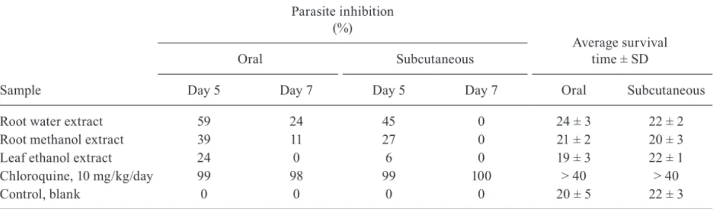
In vivo antiplasmodial activity - Water extracts of the roots of T. grandiflora were the most active samples in vivo in P. berghei-infected mice in the Peters 4-day suppressive test. On day 5 after infection, water extract of the roots administered by mouth and subcutaneously was accompanied by the observation of 59% and 45% suppression, respectively, of P. berghei growth vs. un-treated controls (Table II). Methanol extracts of the roots of T. grandiflora administered orally exhibited border-line moderate inhibitory activity (39% suppression of P. berghei growth) on the fifth day after infection in P. ber-ghei-infected mice, but by the seventh day this effect had decreased substantially (Table II). Low in vivo activities (24-27% parasite suppression) were observed on day 5 for the methanol extracts of roots administered by subcutane-ous injection and the ethanol extracts of leaves adminis-tered orally, but by day 7 the suppressive effects of these extracts was null. In general, mean survival times for P. berghei-infected mice which had received T. grandiflora
extracts were equivalent to untreated controls (Table II).
TABLE I
In vitro median inhibition concentrations (IC50 ± standard deviation) of extracts and fractions of Tachia grandiflora
against K1 strain of Plasmodium falciparum
Extract/fraction
P. falciparum IC50 (µg/mL)
Methanol extracts of roots > 50.0 Chloroform fractions of roots 10.5 Ethanol extracts of leaves > 50.0 Chloroform fractions of leaves 35.8 Water extracts of roots > 50.0 Chloroquine diphosphate salt 0.13
Fig. 2: dose-response curve for drug resistant Plasmodium falciparum
W-2 strain in the presence of different concentrations of amplexine (djalonenol, tetrahydro-3-(hydroxymethyl)-4-[1-(hydroxymethyl)-2-propenyl]-2H-pyran-2-one) in a representative experiment. IC50:
DISCUSSION
As the Krettli group found for the extracts of the roots of a Tachia sp. from the Brazilian Amazon (Car-valho et al. 1991, Brandão et al. 1992), we found that the extracts of roots and leaves of T. grandiflora studied herein were inactive (IC50 > 50 µg/mL) in vitro against P. falciparum (Table I). Through liquid-liquid partitioning of polar extracts, it was possible to obtain chloroform fractions of the roots and leaves. For the first time, in vitro antiplasmodial activity of a Tachia sp. against the human malaria parasite P. falciparum has been demon-strated (for these 2 chloroform fractions).
Little is known about the chemical composition of
Tachia spp. Previously, Pio Corrêa (1926) described the isolation of a “bitter yellow glycoside (tachinine), bitter white, transparent crystalline plates (tachic acid) and ro-settes of fine needles of a non-bitter alkaloid (caferanine)” from caferana (originally cited species: T. guianensis). The structures of these substances were not defined. Based on the diagnostic morphological characters of the drawing of caferana in Pio Correa’s book, the species was either
Tachia lancisepala or Tachia siwertii, both scientifically described only in 2005 (Struwe et al. 2005).
From the active root and leaf chloroform fractions of
T. grandiflora the antiplasmodial tetra-oxygenated xan-thone decussatin (1) and the rare seco-iridoid aglycone amplexine (2) were, respectively, isolated (Pohlit et al. 2012). Tetra-oxygenated xanthones like 1 are commonly isolated from gentians and seco-iridoids are also isolated from gentians. However, seco-iridoids generally occur as glycosides (Jensen & Schripsema 2002).
Decussatin (1), has been previously isolated from
Swertia alata C.B. Clarke (Gentianaceae) and has been shown by Karan et al. (2005) to have an in vitro protective effect against malaria parasites. In general, hydroxyxan-thones such as 1 act through a parasite specific mecha-nism of action by blocking haemozoin formation in the P. falciparum digestive vacuole through binding to haeme in soluble haeme-xanthone complexes (Riscoe et al. 2005).
In the present work, amplexine (2) isolated from the leaves of T. grandiflora inhibited the growth of the blood stages of the W-2 strain of P. falciparum in vitro. Am-plexine (2) is the dihydroxy monoterpene aglycone of the
secoiridoid 1-O-β-D-glucopyranosylamplexine that was
first isolated from the leaves of Anthocleista amplexi-caulis Baker (Gentianaceae) (Rasoanaivo et al. 1994) and later from the stems of Anthocleista djalonensis A. Chev. (Onocha et al. 1995). Interestingly, in Madagascar, a leaf decoction of A. amplexicaulis is used as a remedy for malaria (Rasoanaivo et al. 1994). This is the first re-port on the antiplasmodial activity of terpenoid 2. Inter-estingly, terpenes arrest P. falciparum development by inhibiting the biosynthesis of polyprenylated (terpenoid) primary metabolites such as dolichols, ubiquinones and menaquinones during the growth of P. falciparum para-sites within infected red blood cells (Goulart et al. 2004). The presence of antiplasmodial compounds 1 and 2 in
T. grandiflora is interesting given the widespread tradi-tional use of this plant in the treatment of malaria in the Amazon Region.
The Krettli group collected a Tachia sp. (originally cited species: T. guianensis) in the Brazilian Amazon in São Félix do Xingú, state of Pará (Carvalho et al. 1991, Carvalho & Krettli 1991, Brandão et al. 1992, Krettli et al. 2001). Root infusions were prepared according to tradi-tional practice and administered by mouth to P. berghei -infected mice at doses of 100, 500 and 1,000 mg/kg/day in the 4-day suppressive test. The inhibition obtained was ca. 40% vs. controls. Based on the known distributions of
Tachia spp, it is likely that the plant studied by the Krettli group was either T. grandiflora, the very rare T. siwertii
Struwe, Kinkade & Maas, or maybe even Tachia occiden-talis Maguire and Weaver. The in vivo activity of T. gran-diflora in P. berghei-infected mice herein, together with the Krettli et al.’s (2001) results points to the antimalarial potential of caferana (Tachia spp) infusions (Table II).
Extracts and isolated natural products that inhibit
P. falciparum in vitro often may have little or no effect
TABLE II
In vivo suppression of Plasmodium berghei in infected mice and average mouse survival after oral and subcutaneous treatment with 500 mg/kg/day of extracts of Tachia grandiflora in the Peters 4-day suppressive test
Sample
Parasite inhibition (%)
Average survival time ± SD
Oral Subcutaneous
Day 5 Day 7 Day 5 Day 7 Oral Subcutaneous
Root water extract 59 24 45 0 24 ± 3 22 ± 2
Root methanol extract 39 11 27 0 21 ± 2 20 ± 3
Leaf ethanol extract 24 0 6 0 19 ± 3 22 ± 1
Chloroquine, 10 mg/kg/day 99 98 99 100 > 40 > 40
Control, blank 0 0 0 0 20 ± 5 22 ± 3
against P. berghei in vivo. This was observed for the iso-lated natural product 4-nerolidylcatechol that in general was active against P. falciparum strains in vitro, but only provided a suppressive effect on P. berghei in mice at high dose (600 mg/kg/day) (Rocha e Silva et al. 2011). On the other hand, the alkaloids ellipticine, olivacine and cryptolepine are active both in vitro against P. falcipa- rum (IC50 = 0.35-1.4 µM) and in vivo against P. berghei
in infected mice (10-100 mg/kg/day) (Rocha e Silva et al. 2012). The lack of in vitro antimalarial activity observed for polar extracts of T. grandiflora studied herein and the
Tachia sp. studied by Krettli et al. (2001) mean that these extracts contain substances that mask, suppress or dilute the in vitro activity of antimalarial components that are present in these extracts and concentrated during the fractionation step that led to the preparation of the active chloroform fractions. Future work on the chemical com-position of Tachia spp should feature an initial extraction step followed by liquid-liquid partitioning of the extract to generate simple fractions of increasing polarity that should be assayed both for in vivo activity against P. ber-ghei and in vitro activity in a bioguided process.
Despite the affirmation in Pio Corrêa (1926) that cafer-ana plants present toxicity. The extracts and fractions of roots and leaves of T. grandiflora were not toxic to hu-man fibroblastsin vitro (IC50 > 50 µg/mL). Interestingly, Pohlit et al. (2007) found that fractions of leaf extracts of T. grandiflora were cytotoxicin vitro to five tumour cell lines (IC50 = 4.1-25.4 µg/mL). Thus, there is a selec-tive in vitro cytotoxic effect of the leaf extracts and frac-tions against the faster growing tumour cells than against normal (human fibroblast) cells. Selective cytotoxicity and genotoxicity are common features of many drugs, in-cluding antimalarials such as artesunate and artemether. Mota et al. (2011) demonstrated that artesunate is cyto-toxic and genocyto-toxic to human lymphocytes and Alcân-tara et al. (2013) demonstrated that artemether was more cytotoxic to human lymphocytes than to gastric cancer cell line PG100. More studies on the in vitro cytotoxicity, mutagenicity and genotoxicity of T. grandiflora against different normal human cells and in vivo studies in ani-mal models are needed to evaluate the potential pharma-cological applications of T. grandiflora in humans.
ACKNOWLEDGEMENTS
To Prof Dr Antoniana Ursine Krettli, whose ethnobotanic surveys in the Brazilian Amazon and dedication to the experi-mental evaluation of Amazonian antimalarial plants have had a direct bearing on this study and been an inspiration to the authors for more than a decade, to Prof Maria das Graças Lins Brandão (UFMG, Brazil), for information and clarifications regarding publications co-authored by her, and to Tatiane Cris-tina Mesquita da Silva and to Etienne Louis Jacques Quig-nard, for collaboration.
REFERENCES
Ahmed SA, Gogal Jr RM, Walsh JE 1994. A new rapid and simple non-radioactive assay to monitor and determine the proliferation of lymphocytes: an alternative to [3H] thymidine incorporation
assay. J Immunol Methods 15: 211-224.
Alcântara DD, Ribeiro HF, Cardoso PC, Araújo TM, Burbano RR, Guimarães AC, Khayat AS, de Oliveira Bahia M 2013. In vitro
evaluation of the cytotoxic and genotoxic effects of artemether, an antimalarial drug, in a gastric cancer cell line (PG100). J Appl Toxicol33: 151-156.
Andrade-Neto VF, Pohlit AM, Pinto ACS, Silva ECC, Nogueira KL, Melo MRS, Henrique MC, Amorim RCN, Silva LFR, Costa MRF, Nunomura RCS, Nunomura SM, Alecrim WD, Alecrim MGC, Chaves FCM, Vieira PPR 2007. In vitro inhibition of Plasmodium falciparum by substances isolated from Amazonian antimalarial plants. Mem Inst Oswaldo Cruz102: 359-365.
Brandão MGL, Grandi TSM, Rocha EMM, Sawyer DR, Krettli AU 1992. Survey of medicinal plants used as antimalarials in the Amazon. J Ethnopharmacol36: 175-182.
Carvalho LH, Brandão MGL, Santos-Filho D, Lopes JLC, Krettli AU 1991. Antimalarial activity of crude extracts from Brazilian plants studied in vivo in Plasmodium berghei-infected mice and in vitro against Plasmodium falciparum in culture. Braz J Med Biol Res 24: 1113-1123.
Carvalho LH, Krettli AU 1991. Antimalarial chemotherapy with natural products and chemically defined molecules. Mem Inst Oswaldo Cruz 86 (Suppl. II): 181-184.
Dua VK, Ojha VP, Roy R, Joshi BC, Valecha N, Devi CU, Bhatnagar MC, Sharma VP, Subbarao SK 2004. Anti-malarial activity of some xanthones isolated from the roots of Andrographis panicu-lata. J Ethnopharmacol 95: 247-251.
Goulart HR, Kimura EA, Peres VJ, Couto AS, Duarte FAA, Katzin AM 2004. Terpenes arrest parasite development and inhibit bio-synthesis of isoprenoids in Plasmodium falciparum. Antimicrob Agents Chemother 48: 2502-2509.
Jensen SR, Schripsema J 2002. Chemotaxonomy and pharmacology of gentianaceae. In L Struwe, V Albert (eds.), Gentianaceae - Systematics and natural history, Cambridge University Press, Cambridge, p. 573-631.
Karan M, Bhatnagar S, Wangtak P, Vasisht K 2005. Phytochemical and antimalarial studies on Swertia alata Royale. Acta Horticul-turae 675: 139-145.
Krettli AU, Andrade-Neto VF, Brandão MGL, Ferrari WMS 2001. The search for new antimalarial drugs from plants used to treat fever and malaria or plants randomly selected: a Review. Mem Inst Oswaldo Cruz 96: 1033-1042.
Lambros C, Vanderberg JP 1979. Synchronization of Plasmodium fal-ciparum erythrocytic stages in culture. J Parasitol 65: 418-420.
Maguire B, Weaver Jr RE 1975. The neotropical genus Tachia (Genti-anaceae). J Arnold Arbor 56: 103-125.
Milliken W 1997. Plants for malaria plants for fever: medicinal spe-cies in Latin America - a bibliographic survey, The Royal Bo-tanic Gardens, Kew, 121 pp.
Mors WB, Rizzini CT, Pereira NA 2000. Medicinal plants of Brazil, RA de Filipps (ed.), Medicinal plants of the world 6, Reference Publication, Algonac, 501 pp.
Mota TC, Cardoso PC, Gomes LM, Vieira PC, Corrêa RM, Santana PD, Miranda MS, Burbano RM, Bahia MO 2011. In vitro evalu-ation of the genotoxic and cytotoxic effects of artesunate, an an-timalarial drug, in human lymphocytes. Environ Mol Mutagen 52: 590-594.
Onocha AP, Okorie AD, Connolly DJ, Roycroft DS 1995.
Monoter-pene diol, iridoid glucoside, and dibenzo-α-pyrone from Antho-cleista djalononensis. Phytochemistry 40: 1183-1189.
Pio Corrêa M 1926. Dicionário de plantas úteis do Brasil e das exóti-cas cultivadas, Vol. 1, Imprensa Nacional, Rio de Janeiro, 391 pp.
Pohlit AM, Quignard ELJ, Nunomura SM, Tadei WP, Hidalgo AF, Pinto ACS, Santos EVM, Morais SKR, Saraiva RCG, Ming LC, Alecrim AM, Ferraz AB, Pedroso ACS, Diniz EV, Finney EK, Gomes EO, Dias HB, Souza KS, Oliveira LCP, Don LC, Queiroz MMA, Henrique MC, Santos M, Lacerda Jr OS, Pinto PS, Silva SG, Graça YR 2004. Screening of plants found in Amazonas state, Brazil, for activity against Aedes aegypti larvae. Acta Ama-zonica 34: 97-105.
Pohlit AM, Santos EVM, Silva TCM, Morais SKR, Nunomura SM, Struwe L 2012. A rare secoiridoid monoterpene and a xanthone from Tachia grandiflora Maguire & Weaver. Biochem Syst Ecol 44: 267-269.
Pohlit AM, Tigre RF, Cavalcanti BC, Moraes MO, Costa-Lotufo LV, Moraes MEA, Santos EVM, Morais SKR, Nunomura SM, Pessoa CO 2007. Cytotoxic fractions from the leaves of Tachia grandi-flora. Pharm Biol 45: 429-433.
Quignard ELJ, Pohlit AM, Nunomura SM, Pinto ACS, Santos EVM, Morais SKR, Alecrim AM, Pedroso ACS, Cyrino BRB, Melo CS, Finney EK, Gomes EO, Souza KS, Oliveira LCP, Don LC, Silva LFR, Queiroz MMA, Henrique MC, Santos M, Pinto PS, Silva SG 2003. Screening of plants found in Amazonas state for lethal-ity towards brine shrimp. Acta Amazonica 33: 93-104.
Rasoanaivo P, Nicoletti M, Multari G, Palazzino G, Galeffi C 1994. Secoiridoids and related monoterpenes of Anthocleista amplexi-caulis. Fitoterapia 65: 38-43.
Rieckmann KH, Sax LJ, Campbell GH, Mrema JE 1978. Drug sen-sitivity of Plasmodium falciparum. An in vitro microtechinique.
Lancet 1: 22-23.
Riscoe M, Kelly JX, Winter R 2005. Xanthones as antimalarial agents: discovery, mode of action and optimization. Curr Med Chem 12: 2539-2549.
Rocha e Silva LFR, Montoia A, Amorim RCN, Melo MRS, Henrique MC, Nunomura SM, Costa MRF, Andrade-Neto VF, Costa DS, Dantas GR, Lavrado J, Moreira R, Paulo A, Pinto AC, Tadei WP, Zacardi RS, Eberlin MN, Pohlit AM 2012. Comparative in vitro
and in vivo antimalarial activity of the indole alkaloids ellipti-cine, olivaellipti-cine, cryptolepine and a synthetic cryptolepine analog.
Phytomedicine 20: 71-76.
Rocha e Silva LFR, Pinto ACS, Pohlit AM, Quignard ELJ, Vieira PPR, Tadei WP, Chaves FCM, Samonek JFV, Lima AJL, Costa MRF, Alecrim MGC, Andrade-Neto VF 2011. In vivo and in vitro antimalarial activity of 4-nerolidylcatechol. Phytother Res 25: 1181-1188.
Schmidt TJ, Khalid SA, Romanha AJ, Alves TMA, Biavatti MW, Brun R, Costa FB, de Castro SL, Ferreira VF, de Lacerda MVG, Lago JHG, Leon LL, Lopes NP, Amorim RCN, Niehues M, Ogungbe IV, Pohlit AM, Scotti MT, Setzer WN, Soeiro MNC, Steindel M, Tempone AG 2012a. The potential of secondary metabolites from plants as drugs or leads against protozoan neglected diseases - Part I. Curr Med Chem 19: 2128-2175.
Schmidt TJ, Khalid SA, Romanha AJ, Alves TMA, Biavatti MW, Brun R, Costa FB, de Castro SL, Ferreira VF, de Lacerda MVG, Lago JHG, Leon LL, Lopes NP, Amorim RCN, Niehues M, Ogungbe IV, Pohlit AM, Scotti MT, Setzer WN, Soeiro MNC, Steindel M, Tempone AG 2012b. The potential of secondary metabolites from plants as drugs or leads against protozoan neglected diseases - Part II. Curr Med Chem19: 2176-2228.
Schultes RE, Raffauf RF 1990. The healing forest: medicinal and toxic plants of the northwest Amazonia, Dioscorides Press, Or-egon, 500 pp.
Silva AF, Sampaio PTB, Pohlit AM 2006. Propagação de Tachia gran-diflora por estaquia de galhos. Rev Fitos (ALANAC) 2: 89-93.
Struwe L, Kinkade MP, Maas PJM 2005. Two new Brazilian species of Tachia (Gentianaceae: Helieae). Blumea 50: 457-462.
Tchamo DN, Dijoux-Franca MG, Mariotte AM, Tsamo E, Daskiewicz JB, Bayet C, Barron D, Conseil G, Di Pietro A 2000. Prenylated xanthones as potential P-glycoprotein modulators. Bioorg Med Chem Lett 10: 1343-1345.
WHO - World Health Organization 2001. In vitro micro-test (Mark III) for the assessment of the response of Plasmodium falciparum
![Fig. 1: decussatin (1-hydroxy-3,7,8-trimethoxyxanthone) (1) and amplex- amplex-ine (djalonenol, tetrahydro-3-(hydroxymethyl)-4-[1-(hydroxymethyl)-2 -propenyl]-2H-pyran-2-one) (2) isolated from Tachia grandiflora.](https://thumb-eu.123doks.com/thumbv2/123dok_br/15787589.645486/2.892.443.789.895.1043/decussatin-trimethoxyxanthone-djalonenol-tetrahydro-hydroxymethyl-hydroxymethyl-propenyl-grandiflora.webp)
![Fig. 2: dose-response curve for drug resistant Plasmodium falciparum W-2 strain in the presence of different concentrations of amplexine (djalonenol, tetrahydro-3-(hydroxymethyl)-4-[1-(hydroxymethyl)-2-propenyl]-2H-pyran-2-one) in a represe](https://thumb-eu.123doks.com/thumbv2/123dok_br/15787589.645486/4.892.83.423.938.1103/resistant-plasmodium-falciparum-concentrations-djalonenol-tetrahydro-hydroxymethyl-hydroxymethyl.webp)