Antimalarial drugs disrupt ion homeostasis in malarial parasites
Marcos L Gazarini, Carlos AO Sigolo, Regina P Markus, Andrew P Thomas*,
Célia RS Garcia/
+Departamento de Fisiologia, Instituto de Biociências, Universidade de São Paulo, Rua do Matão, travessa 14, no 321, 05508-900 São Paulo, SP, Brasil *Department of Pharmacology and Physiology, University of Medicine and Dentistry of New Jersey,
Newark, New Jersey, US
Plasmodium chabaudi malaria parasite organelles are major elements for ion homeostasis and cellular sig-naling and also target for antimalarial drugs. By using confocal imaging of intraerythrocytic parasites we demonstrated that the dye acridine orange (AO) is accumulated into P. chabaudi subcellular compartments. The AO could be released from the parasite organelles by collapsing the pH gradient with the K+/H+ ionophore nigericin (20 µM), or by inhibiting the H+-pump with bafilomycin (4 µM). Similarly, in isolated parasites loaded with calcium indicator Fluo 3-AM, bafilomycin caused calcium mobilization of the acidic calcium pool that could also be release with nigericin. Interestingly after complete release of the acidic compartments, addition of thapsigargin at 10 µM was still effective in releasing parasite intracellular calcium stores in parasites at trophozoite stage. The addition of antimalarial drugs chloroquine and artemisinin resulted in AO release from acidic compartments and also affected maintenance of calcium in ER store by using different drug concentrations.
Key words: malaria - Plasmodium chabaudi - calcium - chloroquine - artemisinin - acidic compartment
During the course of its intraerythocytic life, Plas-modium causes well documented biochemical and mor-phological changes in both pathogen and host cell (Howard 1982, Coppel 1986, Garcia et al. 1997, Kirk 2001). Acidic compartments are organelles present in
Plasmodium and several trypanosomatids (Docampo & Moreno 2001). For Plasmodium, it has been suggested that these organelles are the food vacuole in which he-moglobin is digested and antimalarial drugs such as chlo-roquine are accumulated (Yayon et al. 1984). Chloro-quine is though to act by preventing the monomeric hemin from forming the inert and insoluble polymer hemozoin which is derived from digestion of hemoglobin in the acidic vacuole (Slater & Ceram 1992). Defining the sites of chloroquine action in Plasmodium,as well as deter-mining how it is related to proton activity in the acidic compartmentis of major importance in the physiology of these parasites.It has been reported that the antima-larial artemisinin act on PfATP6 (Ca2+ ATPase) in P. falciparum, thus affecting Plasmodium Ca2+ homeosta-sis (Uhlemann et al. 2005).
Regulation of internal pH is important to parasite survival as this sets the environment for intracellular enzymes to function. It has been reported that P. fal-ciparum and P. chabaudi extrude H+ to the extracellu-lar medium by a Na+ independent mechanism (Saliba &
Kirk 1999). Other mechanisms for pH regulation de-scribed in malaria parasites may involve the V-H+ -AT-Pase and the V-H+-PPase in acidocalcisomes (Mar-chesini et al. 2000, McIntosh & Vaidya 2002, Saliba et al. 2003, Biagini et al. 2003). We have reported the ex-istence of Ca2+ pools in the acidic compartment of ro-dent as well as human malaria parasites as a possible mechanism for the regulation of calcium concentration in these parasites (Garcia et al. 1998, Passos & Garcia 1998). Calcium homeostasis in malaria parasites has been extensively studied by several authors (Tanabe et al. 1982, Krungkrai & Yuthavong 1983, Scheibel et al. 1987, Wasserman et al. 1990, Adovelande et al. 1993, Garcia et al. 1997, 1998, Gazarini et al. 2003, Gazarini & Garcia 2004, Beraldo et al, 2005, Budu et al. 2007). Interestingly, in P. chabaudi, the second messenger IP3, mobilizes Ca2+ not only from the classical ER-like pool but also from an acidic compartment, implying that these parasites utilize a calcium-mediated cell signaling mechanism similar to higher organisms (Passos & Garcia 1998). Nevertheless, we still have no clear pic-ture of signaling events and the role of the acidic com-partment in malaria parasite signaling, or its significance in the therapeutic actions of antimalarial drugs such as chloroquine (Yayon et al. 1984, Dzekunov et al. 2000). Ca2+ and pH changes are crucial for parasite enzyme activity such as proteases (Na et al. 2004, Farias et al. 2005). In this context, understanding the mechanisms uti-lized by Plasmodium for the maintenance of its ion ho-meostasis is crucial. In the present work, we have stud-ied the physiological mechanisms by which H+ and Ca2+ activity are related in the acidic compartments of P. chabaudi, and how these are affected by the antimalarials chloroquine and artemisinin. Unraveling the mechanisms by which these organelles contribute to cell function may be important for developing alternative strategies for therapeutic approaches to malaria.
Financial support: Fapesp, CNPq
+Corresponding author: cgarcia@usp.br
MATERIALS AND METHODS
P. chabaudi parasites - Synchronous parasitemia by
P. chabaudi (clone AJ) was maintained in female mice (Balb/c) by transfer of infection. Parasitemia was about 50%. After collecting the blood, cells were washed with PBS (137 mM NaCl, 2.7 mM KCl, 4.3 mM Na2HPO4, 1.4 mM NaH2PO4 and 1 mM CaCl2) and leucocytes and plate-lets were removed by a powdered cellulose column. Para-sitemia was determined from Giemsa-stained thin films.
Parasite isolation - The infected RBCs were then washed twice by centrifugation at 1500 g for 7 min in PBS. Infected RBC (107 ml-1) was lysed in PBS with 60 µg.ml-1 saponin. RBC membranes were removed by cen-trifugation (9000 g for 10 min at 4oC), the parasites were washed twice in MOPS buffer (116 mM NaCl, 5.4 mM KCl, 0.8 mM MgSO4, 5.5 mM D-glucose, 50 mM MOPS, and 2 mM CaCl2, pH 7.2) and dispensed in the same buffer at room temperature (Passos & Garcia 1998).
Confocal microscopy - In vitro parasite-infected RBCs (106 cells ml-1) were loaded with acridine orange (AO) (5 µM) for 5 min at room temperature and washed once with MOPS buffer as described above. Cells were resuspended in the same buffer and placed in a micros-copy chamber. The data acquisition was performed with Zeiss confocal microscope (LSM 510) using the LSM 510 software, version 2.5; excitation 488 nm (Argon laser) and emission collected with a bandpass filter of 505-550 nm (Fluo3-AM). Calcium measurements were performed with isolated parasites incubated with Fluo-3 AM (5 µM) for 50 min at 37oC and placed in microscopy cham-bers at room temperature. The configurations for image acquisition were the EX 488 nm and Em > 560 nm for AO measurements. Software-based analysis allowed fluores-cence imaging in the whole field of view or in a selected cell as a function of time. This was accomplished by defin-ing areas of interest on a given image frame and requestdefin-ing the software to construct a graphical representation of in-tensity against time.
Experiments were carried out with at least three dif-ferent cell preparations. Traces represent typical single cell responses unless indicated otherwise.
Spectrofluorimetric measurement - Isolated para-sites (107 cells ml-1) were incubated with MOPS buffer in 2 ml cuvette. The measurements were performed in a Shimadzu RF-5301 PC spectrofluorimeter at 37oC and dyes fluorescence (Fluo-3 AM and AO) was measured continuously after the drug addition. Parasites were in-cubated with calcium indicator Fluo-3 AM (5µM) for 50 min at 37oC in MOPS buffer containing 2 mM CaCl2 and probenecid (40 µM) or AO (5 µM) incubated for 5 min at 37oC in the same buffer. The dyes parameters are excitation 490 nm and emission collected at 530 nm for Fluo-3 AM and AO fluorescence measurement was ob-tained with Ex-495 nm/Em-590 nm.
Extracellular H+ levels - The effect of chloroquine on H+ mobilization in isolated parasites was recorded using a Cytosensor microphysiometer (Mol Devices, US), a silicon-based biosensor system. This instrument continuously monitors the extracellular pH surrounding
cells in culture, and reports cell activation by measuring the increases in extracellular acidification rate occurring in response to agonist stimulation (McConnell et al. 1992). The isolated parasites was resuspended in DMEM (GIBCO) plus 30% agarose to improve parasites adherence onto the polycarbonate membrane of the transwell capsules (3 µm pore size) at a density of 2.5 × 10⁵ cells.
RESULTS
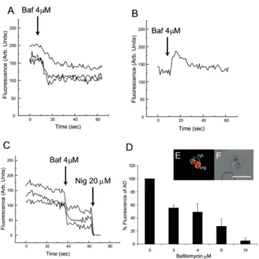
We have investigated the dynamics of solute exchange in the acidic compartments of isolated and intact blood-stage malaria parasite, P. chabaudi. To study intracellu-lar pH changes, we loaded infected red blood cells at trophozoite stage (P. chabaudi) with AO. Dye mobiliza-tion through compartments was measured by selecting areas of interest (cytosol and acid compartment) by con-focal imaging (Fig. 1). Addition of H+-ATPase inhibitor bafilomycin (4 mM) to the cells led to alkalinization of acidic compartments as indicated by the partial release of accumulated AO (Fig. 1A), and simultaneously in-creases the cytosol fluorescence (Fig. 1B). The remain-ing AO could be released by addition of the K+/H+ iono-phore nigericin to the cells (20 µM), which totally re-leased the compartmentalized AO fluorescence (Fig. 1C). Dose-dependent addition of bafilomycin promotes the depletion of AO stores. A remarkable (over 90%) re-lease of AO fluorescence occurs with addition of bafilomycin at 10 µM (Fig. 1D).
The effect of the antimalarial chloroquine on AO ac-cumulation in subcellular compartments (Fig. 2A) in in-fected erythrocyte (Fig. 2B) shows that chloroquine (80
µM) promotes extrusion of AO from the acidic com-partments, presumably as a result of alkalinization. The subsequent addition of H+-ATPase inhibitor bafilomycin (4 µM) caused complete depletion of the residual AO from the acidic compartments (Fig. 2C). Taken together, the data of Figs 1 and 2 suggest that P. chabaudi para-sites display a bafilomycin-sensitive acidic compartment that can be affected in a dose-dependent manner and is also a target for chloroquine.
By using spectrofluorimeter measurements and iso-lated parasites (107 cells) at trophozoite stage previously incubated with AO (5 µM) for 5 min/37oC we showned that AO could be released in a dose-dependent manner by addition of artemisinin and chloroquine (Fig. 3).
We have previously demonstrated that the H+-pump inhibitor 7-chloro-4-nitrobenz-2-oxa-1,3-diazole (NBD-Cl) caused release of Ca2+ in Fluo-3 loaded P. falciparum
on Ca2+ release (Fig. 4C). Interestingly, neither bafi-lomycin (Fig. 4D) or nigericin (Fig. 4E) was able to de-plete ER Ca2+ pool, as a subsequent addition of thapsigargin (Thg) promoted a second calcium rise.
To investigate the effect of the antimalarials drugs chloroquine and artemisinin in calcium homeostasis we
carried out experiments with isolated parasites loaded with calcium indicator Fluo-3 AM (5 µM). Fig 5 shows spectrofluorimetric measurements of calcium release by Thg (10 µM) with parasites previously incubated (15 min at 37oC) with different concentrations of chloro-quine and artemisinin (0-80 µM) in a high calcium
ex-Fig. 1: effect of the H+ pump inhibitor bafilomycin on Plasmodium chabaudi within the intact RBC. Infected RBC was loaded with AO (5 µM) as described in the Methods Section. A: effect of bafilomycin (Baf) (4 µM) on organelles selected areas stained with AO in single cell; B: cytosolic area with acidification promoted by bafilomycin; C: Bafilomycin and nigericin (Nig) effect on subcellular areas; D: dose-response effect of bafilomycin on AO release from intracellular stores; E: fluorescence imaging with selected areas (cytosol and organelles); F: phase contrast. *(D) bar plot represent % of control fluorescence (arbitrary units) remaining from ten cells in three different trials. Scale bar of 10 µm.
Fig. 2: action of the antimalarial chloroquin and bafilomycin on acidic compartments in Plasmodium chabaudi. Infected RBC loaded with 5 µM AO; A: fluorescence image; B: phase contrast; C: changes in AO fluores-cence (arbitrary units) in selected subcellular compartments promoted by chloroquine (Clq) (80 µM). Scale bar of 10 µm.
Fig. 3: effect of antimalarial drugs chloroquine and artemisinin on acri-dine orange mobilization from acidic compartment. Isolated parasites loaded with 5 µM AO for 5 min. Different concentrations of drugs (Art-artemisinin and Clq-chloroquine) were added and fluorescence measure-ment was performed in spectrofluorimeter cuvette. Fluorescence intensi-ties (arbitrary units - AU) represent at least three different cell preparations.
Fluorescence Increase (A.U)
antimalarial concentration (µM)
40 35
30
25
20
15 45
5
0 10
40 75
30
25 50
15 5
1 10
tracellular medium (2 mM). The results show that previ-ous parasite incubation with chloroquine or artemisinin leads to a dose-dependent reduction of total amount of calcium release by thapsigargin.
When parasites are exposed to chloroquine, the cy-tosolic pH decrease as a result of extrusion of H+ from acidic compartments and the parasites (P. falciparum) try to restore the cytosolic pH level using plasma mem-brane mechanisms (Saliba & Kirk 1999, Marchesini et al. 2000, Saliba et al. 2003). By using microphysiometry we showed that addition of chloroquine to P. chabaudi
at the trophozoite stage led to a dose-dependent increase in the extracellular acidification rate (Fig. 6). The mean 50% maximal effective concentration (EC50) for three experiments was 16.29 ± 0.4 µM (n = 3) of chloroquine. This data reveal an activation of plasma membrane mecha-nism of H+ extrusion under chloroquine treatment.
DISCUSSION
The antimalarial drug chloroquine is thought to ac-cumulate in acidic compartments, although its mecha-nisms of action are controversial (Slater & Cerami 1992, Dorn et al. 1995, Waller et al. 2003). The resistance to chloroquine observed in P. falciparum is dependent to mutations in specific transporter (PfCRT), by using het-erologous expression Reeves et al. (2006) observed a increase of lysosomes acidification in mammalian cells expressing PfCRT, providing new information about acidification process and chloroquine resistance.
Fig. 4: H+ pump inhibitor-bafilomycin induces Ca2+ release in intact
Plas-modium chabaudi parasites. P. chabaudi parasites were loaded with the
calcium indicator Fluo-3 AM (5 µM) as described in the Methods Section and cytosolic fluorescence intensities measured in confocal microscopy were normalized as (F1-maximal fluorescence after drug addition/F0-fluo-rescence before drug addition). A: dose-response effect of bafilomycin (Baf) on cytosolic calcium fluorescence (mean data of fluorescence ratio remaining from ten cells in three different trials); B: addition of bafilomycin (increase of 1.3 ± 0.1; n = 6) and nigericin (Nig) (20 µM); C: addition of nigericin (1.42 ± 0.1; n = 6) and bafilomycin (4 µM); D: addition of bafilomycin (1.5 ± 0.2; n = 6) and thapsigargin (Thg) (10 µM); E: addition of nigericin (1.65 ± 0.3; n = 6) and thapsigargin (10 µM).
Ca2+ mobolization by Thg (10µM)
Acidification rate (% of base line)
Log[Clq]µM
30
20
10
1.5
1.0 2.0
[Ca
2+
]cyt
(
µ
M)
Concentration (µM) of pre-incubated antimalarial
1.75
1.50
1.25
1.00
0.75
0.25
0.00 0.50
80 40 20 5
0 10
Art Clq
Fig. 6 : concentration-response curve for chloroquine-induced acidifica-tion rate in Plasmodium chabaudi. Exposure to chloroquine for 7 min with concentrations between 10 µM and 60 µM. Concentration-response curve was calculated as the difference from baseline and the peak. EC50 calculated from this curve is 16.29 mM (n = 3).
Fig. 5: effect of antimalarials drugs on calcium maintenance on endoplas-mic reticulum in Plasmodium chabaudi. Isolated parasites (107 cells ml-1) were loaded with calcium indicator Fluo-3 AM (5 µM) for 50 min at
We have previously shown that the acidic pool also functions as a Ca2+ store in permeabilized malaria para-sites and that chloroquine causes Ca2+ release from this store (Passos & Garcia 1998). In the present study, in-tact parasites were labeled with the calcium indicator, Fluo-3 AM to measure parasite cytosolic Ca2+ mobili-zation, and we also investigated the acidic pools in intact parasites within the RBC, using confocal microscopy.
The data in the present work indicate the existence of mechanisms for H+ homeostasis in P. chabaudi para-sites. The acidic vacuole in malaria parasite is the site of hemoglobin digestion and it is believed of drug action. In the rodent malaria parasite P. chabaudi the second messenger IP3 is known to induced-Ca2+ mobilization from this compartment (Passos & Garcia 1998). These findings are of major significance, because they raise the possibility that chloroquine and artemisinin action in malaria parasites may involve alterations in ion ho-meostasis. This is especially important in view of our work showing that IP3-dependent Ca2+ signaling is involved in the progression of the malarial parasite cell cycle (Hotta et al. 2000, Gazarini et al. 2003, Beraldo et al. 2005).
By using confocal microscopy and analyzing P. chabaudi loaded with AO within the intact RBC, we showed a H+ mobilization, corresponding to a fluores-cence decrease when V-type H+-ATPase was inhibited by bafilomycin (Fig. 1). Simultaneously, parasite cyto-sol fluorescence was transiently increased as the AO was released, thus indicating that cellular mechanisms were operating to restore cytosolic pH (Fig. 1B). Bafilomycin completely discharge the AO pool at 10 µM (Fig. 1D) while Ca2+ release reach the maximal values at addition of 4 µM of H+ pump inhibitor (Fig. 4A).
The interaction of antimalarial artemisinin in P. falciparum was reported to occur through inhibition of
PfATP6, a SERCA-type ATPase (Uhlemann et al. 2005). Our data shows the ability of both antimalarial in modi-fying the calcium and proton dynamics in P. chabaudi
internal stores (Fig. 5).
We also addressed the metabolic response evoked by chloroquine with cytosensor microphysiometer (Fig. 6). These results showed that chloroquine stimulated a H+ extrusion response with an EC50 of 16 µM in P. chabaudi, using plasma membrane mechanisms to re-cover the intracellular pH (Saliba & Kirk 1999).
Our data provide new information of acidic organelle physiology, the high concentration of bafilomycin re-quired for complete reversion fluorescence from AO, suggests a low density of H+ pumps or susceptibility to the drug. However chloroquine action promoted a more extensively effect on parasite ion homeostasis than the specific H+ pumps inhibitor bafilomycin. The micromo-lar concentration of antimamicromo-larial drugs required in our assay may represent the cell physiology differences be-tween the Plasmodium species and we are search for extension of organelles capacity in ion maintenance.
The fact that Ca2+ antagonists block malaria parasite development is well known. In addition, reversal of chlo-roquine resistance in vitro by several Ca2+ antagonists such as verapamil has also been extensively reported (Deloron et al. 1991, Adovelande et al. 1998).
We have demonstrated that host melatonin activates parasite calcium signaling via the second messenger IP3, which appears to be important in the proliferation and maturation of the intraerythrocytic malaria parasites (Hotta et al. 2000). More recently, we also verify cal-cium mobilization with AFMK, a product of melatonin degradation in Plasmodium-infected erythrocytes (Budu et al. 2006). Finally, although the molecular mechanism of chloroquine and artemisinin action is not fully under-stood, its affect on calcium and proton activity indicates the importance in elucidate calcium homeostasis and signaling mechanisms on malarial parasites.
ACKNOWLEDGEMENTS
To Fapesp for funding CRSG and RPM. MLG received fel-lowship from Fapesp and CAS from CNPq.
REFERENCES
Adovelande J, Deleze J, Schrevel J 1998. Synergy between two calcium channel blockers, verapamil and fantofarone (SR33557), in reversing chloroquine resistance in Plasmo-dium falciparum. Biochem Pharmacol55: 433-440. Beraldo FH, Almeida FM, da Silva AM, Garcia CR 2005. Cyclic
AMP and calcium interplay as second messengers in mela-tonin-dependent regulation of Plasmodium falciparum cell cycle. J Cell Biol170: 551-557.
Biagini GA, Bray PG, Spiller DG, White MR, Ward SA. 2003. The digestive food vacuole of the malaria parasite is a dynamic intracellular Ca2+ store. J Biol Chem278: 27910-27915.
Budu A, Peres R, Bueno VB, Catalani LH, Garcia CRS. 2007. N1-acetyl-N2-formyl-5-methoxykynuramine modulates the cell cycle of malaria parasites. J Pineal Research (in press). Coppel RL, Culvenor JG, Bianco AE, Crewther PE, Stahl HD, Brown GV, Anders RF, Kemp DJ 1986. Variable antigen as-sociated with the surface of erythrocytes infected with ma-ture stages of Plasmodium falciparum. Mol Biochem Parasitol 20: 265-277.
Deloron P, Basco LK, Dubois B, Gaudin C, Clavier F, Le Bras J, Verdier F. 1991. In vitro and in vivo potentiation of chloro-quine against malaria parasites by an enantiomer of amlodipine. Antimicrob Agents Chemother35: 1338-1342. Docampo R, Moreno SN. 2001. The acidocalcisome. Mol
Biochem Parasitol114: 151-159.
Dorn A, Stoffel R, Matile H, Bubendorf A, Ridley RG. 1995. Malarial haemozoin/beta-haematin supports haem polymer-ization in the absence of protein. Nature 374: 269-271. Dzekunov SM, Ursos LM, Roepe PD 2000. Digestive vacuolar pH
of intact intraerythrocytic P. falciparum either sensitive or re-sistant to chloroquine. Mol Biochem Parasitol 110: 107-124. Farias SL, Gazarini ML, Melo RL, Hirata IY, Juliano MA, Juliano L,
Garcia CR. 2005. Cysteine-protease activity elicited by Ca2+ stimulus in Plasmodium. Mol Biochem Parasitol 141: 71-79. Garcia CR, Ann SE, Tavares ES, Dluzewski AR, Mason WT, Paiva FB. 1998. Acidic calcium pools in intraerythrocytic malaria parasites. Eur J Cell Biol 76: 133-138.
mitochon-drion senses cytosolic Ca2+ fluctuations. Biochem Biophys Res Commun 321: 138-144.
Gazarini ML, Thomas AP, Pozzan T, Garcia CR. 2003. Calcium signaling in a low calcium environment: how the intracellular malaria parasite solves the problem. J Cell Biol 161: 103-110. Hayward R, Saliba KJ, Kirk K 2006. The pH of the digestive vacuole of Plasmodium falciparum is not associated with chloroquine resistance. J Cell Sci 119: 1016-1025. Hotta CT, Gazarini ML, Beraldo FH, Varotti FP, Lopes C, Markus
RP, Pozzan T, Garcia, C.R. 2000. Calcium-dependent modu-lation by melatonin of the circadian rhythm in malarial para-sites. Nat Cell Biol 2: 466-468.
Howard RJ. 1982. Alterations in the surface membrane of red blood cells during malaria. Immunol Rev 61: 67-107. Kirk K 2001. Membrane transport in the malaria-infected
eryth-rocyte. Physiol Rev 81: 495-537.
Krungkrai J, Yuthavong Y. 1983. Enhanced Ca2+ uptake by mouse erythrocytes in malarial (Plasmodium berghei) infection. Mol Biochem Parasitol7: 227-235.
Marchesini N, Luo S, Rodrigues CO, Moreno SN, Docampo R 2000. Acidocalcisomes and a vacuolar H+-pyrophosphatase in malaria parasites. Biochem J 347: 243-253.
McConnell HM, Owicki JC, Parce JW, Miller DL, Baxter GT, Wada HG, Pitchford S 1992. The cytosensor microphy-siometer: biological applications of silicon technology.
Science257: 1906-1912.
McIntosh MT, Vaidya AB. 2002. Vacuolar type H+ pumping pyrophosphatases of parasitic protozoa. Int J Parasitol32: 1-14.
Na BK, Shenai BR, Sijwali PS, Choe Y, Pandey KC, Singh A, Craik CS, Rosenthal PJ. 2004. Identification and biochemical charac-terization of vivapains, cysteine proteases of the malaria para-site Plasmodium vivax. Biochem J378: 529-538.
Passos AP, Garcia CR. 1998. Inositol 1,4,5-trisphosphate induced Ca2+ release from chloroquine-sensitive and -insensitive in-tracellular stores in the intraerythrocytic stage of the malaria parasite P. chabaudi. Biochem Biophys Res Commun245: 155-160.
Reeves DC, Liebelt DA, Lakshmanan V, Roepe PD, Fidock DA, Akabas MH. 2006. Chloroquine-resistant isoforms of the
Plasmodium falciparum chloroquine resistance transporter acidify lysosomal pH in HEK293 cells more than chloroquine-sensitive isoforms. Mol Biochem Parasitol150: 288-299. Saliba KJ, Allen RJ, Zissis S, Bray PG, Ward SA, Kirk K. 2003.
Acidification of the malaria parasite’s digestive vacuole by a H+-ATPase and a H+-pyrophosphatase. J Biol Chem278: 5605-5612.
Saliba KJ, Kirk K. 1999. pH regulation in the intracellular ma-laria parasite, Plasmodium falciparum. H(+) extrusion via a v-type H(+)-ATPase. J Biol Chem274: 33213-33219. Scheibel LW, Colombani PM, Hess AD, Aikawa M, Atkinson
CT, Milhous, WK 1987. Calcium and calmodulin antagonists inhibit human malaria parasites (Plasmodium falciparum): implications for drug design. Proc Natl Acad Sci USA84: 7310-7314.
Slater AF, Cerami A. 1992. Inhibition by chloroquine of a novel haem polymerase enzyme activity in malaria trophozoites.
Nature355: 167-169.
Tanabe K, Mikkelsen RB, Wallach DF. 1982. Calcium transport of Plasmodium chabaudi-infected erythrocytes. J Cell Biol 93: 680-684.
Uhlemann AC, Cameron A, Eckstein-Ludwig U, Fischbarg J, Iserovich P, Zuniga FA, East M, Lee A, Brady L, Haynes RK, Krishna S. 2005. A single amino acid residue can deter-mine the sensitivity of SERCAs to artemisinins. Nat Struct Mol Biol12: 628-629.
Waller KL, Muhle RA, Ursos LM, Horrocks P, Verdier-Pinard D, Sidhu AB, Fujioka H, Roepe PD, Fidock DA. 2003. Chlo-roquine resistance modulated in vitro by expression levels of the Plasmodium falciparum chloroquine resistance trans-porter. J Biol Chem278: 33593-33601.
Wasserman M, Vernot JP, Mendoza PM. 1990. Role of calcium and erythrocyte cytoskeleton phosphorylation in the invasion of Plasmodium falciparum. Parasitol Res76: 681-688. Yayon A, Cabantchik ZI, Ginsburg H. 1984. Identification of the