19 19 19 19 19 Mem Inst Oswaldo Cruz, Rio de Janeiro, Vol. 92, Suppl.II: 19-32, 1997
Immunological System and
Schistosoma mansoni
:
Co-evolutionary Immunobiology. What is the Eosinophil
Role in Parasite-host Relationship?
Henrique L Lenzi/
+, Ronaldo G Pacheco
*, Marcelo Pelajo-Machado/
++,
Mônica S Panasco, Waldemiro S Romanha, Jane A Lenzi
Departamento de Patologia, Instituto Oswaldo Cruz, Av. Brasil 4365, 21045-900 Rio de Janeiro, RJ, Brasil *Clínica Médica “B”, Departamento de Medicina Geral, Hospital Universitário Gaffrée e Guinle, Universidade
do Rio de Janeiro, Rua Mariz e Barros 775, 20270-004 Rio de Janeiro, RJ, Brasil
Schistosomes, ancestors and recent species, have pervaded many hosts and several phylogenetic levels of immunity, causing an evolutionary pressure to eosinophil lineage expression and response.
Schistosoma mansoni adult worms have capitalized on the apparent adversity of living within the me-senteric veins, using the dispersion of eggs and antigens to other tissues besides intestines to set a systemic activation of several haematopoietic lineages, specially eosinophils and monocytes/macroph-ages. This activation occurs in bone marrow, spleen, liver, lymph nodes, omental and mesenteric milky spots (activation of the old or primordial and recent or new lymphomyeloid tissue), increasing and making easy the migration of eosinophils, monocytes and other cells to the intestinal periovular granu-lomas. The exudative perigranulomatous stage of the periovular reaction, which present hystolitic char-acteristics, is then exploited by the parasites, to release the eggs into the intestinal lumen. The authors hypothesize here that eosinophils, which have a long phylogenic story, could participate in the parasite - host co-evolution, specially with S. mansoni, operating together with monocytes/ macrophages, upon parasite transmission.
Key words: eosinophils - phylogeny - Schistosoma mansoni - egg release - granuloma - co-evolution
The immune system and Schistosoma mansoni
co-evolved for millions of years. Therefore, there has been plenty of time for the development of complex and intimate interactions between the two. Most recent research, either in paleontology or in molecular biology, tends to demonstrate that not only hominids but also modern humans originated from Africa. This means that the contact between humans and schistosomes started long ago in Af-rica, perhaps in the early emergence of the human lineage. In Asia, this contact could only have oc-curred later, either during the migration of Homo erectus (around 1.2 millions years ago?), or
dur-ing that of Homo sapiens sapiens (around 80.000
years ago?) (Combes 1990a).
HISTORY OF SCHISTOSOMA MANSONI (PHYLOG-ENY)
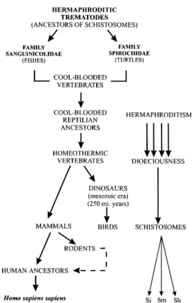
Ancestors of schistosomes were probably her-maphroditic trematodes that lived in the blood ves-sels of cold-blooded vertebrates. Members of the
+ Corresponding author. Fax: +55-21-590.3495 ++CAPES fellowship
Received 3 September 1997 Accepted 30 September 1997
family Sanguinicolidae parasitized fish, and the family Spirochidae inhabit the circulatory system of freshwater and marine turtles worldwide (Platt 1992), and they are generally considered similar to the ancestors of schistosomes. It seems likely that the transition from hermaphroditism to dioeciousness (male and female genitals do not occur in the same individual) in blood flukes ac-companied the acquisition of homeothermy by their host, such as the mammals and birds (Basch 1990). These homeothermic vertebrates have probably evolved from cold-blooded reptilian ancestors. Evolutionary selection toward the schistosome pattern was probably driven by profound physi-ological adaptations in dinosaurs or in derivative transitional forms to birds and mammals, as these animals radiated rapidly and broadly during the
Mesozoic era (Basch 1990). Schistosoma species
of humans and larger mammals have separated each other fairly recently, while Schistosomatium of
Prob-20 20 20 20
20 Schistosome - Host Co-evolution • HL Lenzi et al.
ably, the rodents were important in this lateral trans-ference. This hypothesis is reinforced by the kin-ship, attested by the use of the same vector species and by the success of experimental hybridization between S. mansoni and S. rhodaini from Kenya
(Combes 1990b) (Fig. 1).
and freshwater sponges have acquired the struc-tural prerequisites for an immune system, neces-sary for the establishment of individuality (Van de Vyver 1970, Hildeman et al. 1979, Müller et al. 1979). It is estimated that the oldest multicellular animals, the sponges, diverged from the other ani-mals approximately 800 millions years ago (Müller et al. 1994). Studies indicate that morphoregulatory molecules, cell adhesion molecules, as well as homeodomain proteins have developed during the transition period in the evolution from the Protist to the Animalia (Müller et al. 1994).
Multicellular animals above the sponge level can be conveniently divided into two groups:
Diploblastic and Triploblastic. The diploblastic
animals comprise the phylum Cnidara, or Co-elenterata (hydras, jellyfish, sea anemones and corals). In contrast to sponges, cnidarians possess a gut cavity lined by endoderm, as do most other animals (= coelenteron, or gastrovascular cavity). Their two cell layers are the ectoderm (epidermis) and the endoderm (gastrodermis) lining the gas-trovascular cavity. Between these two layers there is an extracellular layer called mesoglea. The me-soglea ranges from a thin, non-cellular basal lamina, as in hydras and many other hydrozoans, to a thick, fibrous, jelly-like, connective tissue with or without mesenchymal cells (Ruppert & Barnes 1994).
The triploblastic animals have a third cellular layer, the mesoderm, between the ectoderm and endoderm. The triploblastic are bilateral animals and have well-developed, mesodermally derived tissues and organs, which create regulated extra-cellular compartments (evolution of
com-partmentation). The coelenteron in the cnidarians could not evolve into a regionally specialized gut. In large bilateral animals, the multifunctional co-elenteron lining was replaced by two new epithe-lia that delineate a total of three new compartments: the gut cavity and its specialized lining, which
func-tion primarily in digesfunc-tion and absorpfunc-tion; the co-elom and its lining mesothelium for hydrostatic
support, circulation, reproduction and excretion; and a specialization of the connective tissue called the blood-vascular system, which is important in circulation (Ruppert & Barnes 1994). Some bilaterians have an unpartitioned coelom that is continuous throughout the body, in which the co-elomic fluid reaches all tissues and is the sole cir-culatory system. In most bilateral animals, how-ever, the coelom is divided by septa and mesenter-ies, and because of them, the coelomic fluid can only circulate locally. For whole-body transport, these animals have evolved a blood-vascular sys-tem, which consists of fluid-filled channels in the connective tissue (blood vessels) (Ruppert & Fig. 1: schematic view of the history of the schistosomes and
the switch of their host during the evolutional process (Sj =
Schistosoma japonicum; Sm = S. mansoni; Sh = S. haematobium).
HISTORY OF THE HAEMATOLYMPHOPOIETIC SYS-TEM (PHYLOGENY OF THE IMMUNE SYSSYS-TEM)
Recent results gathered while sequencing genes from sponges (Porifera) indicate that the kingdom of Animalia is a monophyletic group of multicel-lular organisms (arising or descendent from a single cell type) (Morris 1993, Müller et al. 1994).
21 21 21 21 21 Mem Inst Oswaldo Cruz, Rio de Janeiro, Vol. 92, Suppl. II, 1997
Barnes 1994). In the great majority of multicellu-lar animals the following fluids are present: tissue fluid, coelomic fluid and blood. Lymph is derived from blood plasma modified in its passage through the tissues.
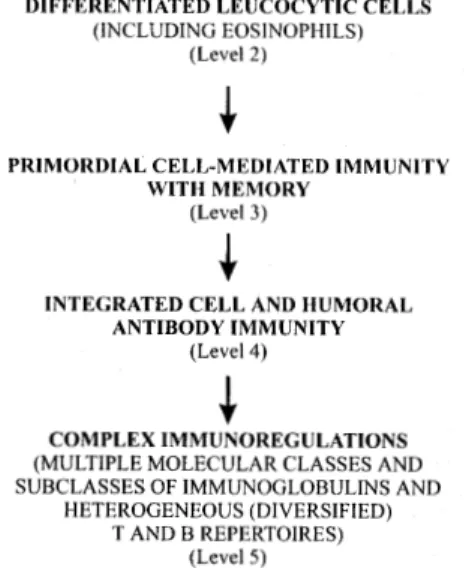
Certain major sequential steps in the phylog-eny of immunologic reactivity have been suggested by Tam et al. (1976). Cell or species-specific ag-gregation (level 1) could be observed in plants, sponges and protozoans. Specific immunorecog-nition/ immunoimcompatibility (level 2) was first evident at the invertebrate level of coelenterates, while cell-mediated immunity with at least short-term memory (level 3) has been detected initially in advanced invertebrates, notably annelids, echi-noderms and possibly, mollusks. Immunoincom-patibility as regularly shown by allogenic contact reactions appears primitive and has surely persisted as an effective surveillance mechanism through-out metazoan phylogeny. Progressively more dif-ferentiated leucocytic cells probably assumed the second-level function as adaptative specialization continued during phylogeny, and were detected in cnidarians (coelenterates), tunicates (adult urochor-dates) and vertebrates. Primordial cell-mediated immunity with memory was regarded as a third-level associated with cooperation of granulocytes, macrophages and T lymphocytes in allograft-type reactions. This function became well developed in primitive fishes associated increasingly with longer-lived memory. Integrated cell and humoral antibody immunity (level 4) may have first evolved in advanced bony fishes. At this vertebrate level, helper T cells and B cells capable of producing two or more molecular classes of antibodies were demonstrable. If the thymus is indeed the source of both T and B lymphocytes in fishes and am-phibians, evolution of the bursa of Fabricius as a separate source of B cells may be merely a special adaptation of the reptilian-avian branch of the phylogenic tree (Tam et al. 1976). Complex immunoregulation in birds and mammals, involv-ing multiple classes and subclasses of immunoglo-bulins and heterogeneous T and B repertoires, con-stituted the level 5 of immunologic organization (Fig. 3).
The divergence of ancestral protostomes and deuterostomes occurred 500 to 600 million years ago, a time in which all of the extant variants of animal body forms appeared. The “Big Bang” of gene duplication occurred after tunicates (from tunicates to chordates). In the deuterostomes, elas-mobranchs were the most primitive species in which genes specifying MHC products have al-ready been identified (Bartl et al. 1994, Bartl & Weissman 1994).
Immunoglobulins and recombination activat-ing genes were also detected for the first time in elasmobranchs and in lower deuterostomes. It has recently been hypothesized that the B cells of sharks most probably resemble the CD5+ cells of man, which produce polyspecific IgM often show-ing autoantibody activity and that their T cells re-semble γ/δ T cells rather than a/b TCR-bearing helper cells (Marchalonis & Schluter 1994, Bartl et al. 1994, Horton & Ratcliffe 1996).
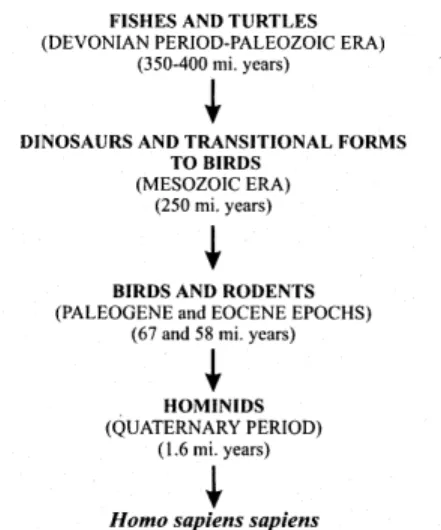
WHAT KIND OF IMMUNE SYSTEM DID THE SCHIS-TOSOMES MEET DURING THEIR PHYLOGENY?
The schistosomes and their ancestors probably presented the following sequential animals as co-evolutive hosts: 1st.: fishes and turtles; 2nd.: dino-saurs and derivative forms transitional to birds; 3rd.: birds and rodents; 4th.: hominids. Therefore, the schistosomes cohabited with animals originated since the Devonian period of the Paleozoic era, occurring from 350 to 400 millions years ago and characterized by the dominance of fishes and the advent of amphibians and ammonites. Later on, the schistosomes lived with animals from the Per-mian, Triassic, Jurassic and Cretaceous eras. Ro-dentia and hominids appeared in Paleogene (Eocene epoch) and Quaternary Periods, respec-tively (Fig. 2).
Fig. 2: schistosomes and co-evolutive hosts according to geo-logical scale of time.
22 22 22 22
22 Schistosome - Host Co-evolution • HL Lenzi et al.
The parasites accumulate slow and asynchro-nously in hepatic vessels, where they can be found from day 4 to day 23, as young and/ or adult speci-mens (Faust et al. 1934, Coelho 1970). The accu-mulation of parasites in mouse liver appears to be complete by 21 days post-infection (Wilson 1987). Wilson and Coulson (1986) estimated that 97 % of those schistosomula which reach the liver do so via the splanchnic capillaries and hepatic portal system. Most of them fail to negotiate the sinuso-ids to reach the hepatic vein, instead they trans-form into blood-feeding worms and begin to grow, ultimately pairing and migrating upstream to the mesenteric venules for oviposition (Wilson 1990). Trapping of schistosomula in the liver is not com-pletely efficient, and in a previously uninfected mouse, an estimated 14 - 30% may return to the lungs (Wilson et al. 1986).
In mice, intrahepatic paired adult worms ap-pear for the first time, on day 20 of infection (Lenzi 1991), the schistosomae begin to migrate to me-senteric vessels from the third week onward (Pinto & Almeida 1948) and the occurrence of eggs lay-ing is detected between 30 and 34 days after infec-tion (Brener 1956, Prata 1957).
The initiation of parasite growth after arrival in the liver may be under endogenous control by ecdysteroids (Nirde et al. 1983) and these hormones may in addition moderate the process of tegument membrane turnover (Torpier et al. 1982).
Once schistosomula have transformed to adult worms, they tend to live inside mesenteric venous vessels, although some of them, specially the fe-males, may undertake further intravascular migra-tion, as we have often observed in Swiss webster mice, from 40 to 160 days after infection (Lenzi 1991).
Advantages of the final habitat and egg dis-persion - The habitat of most adult S. mansoni
within the mesenteric veins of the host caused three biological consequences: (1) long and cozy life within the nutrient-rich mesenteric veins, produc-ing a prodigious total number of eggs (Bash 1990); (2) easy access of the eggs to the intestinal vessels and (3) dispersal of the eggs and parasitic soluble products to extraintestinal organs (mainly to the liver), provoking systemic reactions (Lenzi et al. 1987, 1995).
Extraintestinal consequences -
Schistosomia-sis is essentially an intravascular infection, and schistosomes live in direct contact with the me-senteric or portal vein endothelium. The adult worms produce large numbers of intravascular eggs daily and continuously secrete and excrete numer-ous inert, toxic or antigenic substances that can interfere directly with the contiguous endothelium surface (Lenzi et al. 1988) or can provoke simul-Fig. 3: major steps in phylogeny of immunologic reactivity
ex-perienced by schistosomes (see Tam et al. 1976).
Paleoparasitology should try to answer the question if the schistosomes have enjoyed a long relationship with their hosts extending into the early Mesozoic prior to the breakup of Pangaea (conti-nental drift). Using Brooks and McLennan (1993) analysis on parasite evolutionary biology, we can say that, at the moment, we have very little infor-mation about the difference, if any, in the relation-ship between the degree of pathology, the age of the parasite-host relationship and the type of host switch. For example, the assertion that high patho-genicity is an evidence of recent and still imper-fect parasite-host relationships (Hegner 1926) needs to be confirmed.
INTERACTION BETWEEN SCHISTOSOMES AND RE-CENT HOSTS
Migratory pathways - Human and
experimen-tal infections with S. mansoni are characterized by
23 23 23 23 23 Mem Inst Oswaldo Cruz, Rio de Janeiro, Vol. 92, Suppl. II, 1997
taneous and parallel activation of eosinophil meta-plasia in the liver and eosinopoiesis in the bone marrow and stimulation of extramedullary hemato-poiesis in lymph nodes and omental and mesen-teric milky spots (Byram et al. 1978, Borojevic et al. 1981, Weinberg et al. 1992, Lenzi et al. 1987b, 1990, 1995).
Activation of the old or primordial (immune system) lymphomyeloid tissue - Coelom was a key
evolutionary innovation that has occurred in the body plan of the major animal phyla (Raven & Johnson 1996, Erwin et al. 1997). A major advan-tage of the coelomate body plan was that it allowed contact between mesoderm and endoderm, facili-tating the occurrence of primary induction pro-cesses during development (Raven & Johnson 1996). For example, contact between mesoderm and endoderm permitted localized portions of the digestive tract to develop into complex, highly spe-cialized regions like the stomach. The presence of a coelom also allowed the digestive tract, by its coiling or folding within the coelom, to be longer than the animal itself. Such an arrangement lim-ited the animal’s exposure to predators due to the capacity to hide large amount of food during the digestive process (Raven & Johnson 1996).
The intraembryonic coelom in humans and ver-tebrate animals is divided into pericardial, pleural and peritoneal cavities. In these three cavities, spe-cially in the omentum of the peritoneal cavity and in pericardiodiaphragmatic membrane, there are the milky spots (MS) which were described by Recklinghausen in 1863 in young rabbits (Recklinghausen 1863). We proposed that milky spots (individual structures), considered as a whole, constitute an organized coelom-associated lymphomyelopoietic tissue (CALT), which repre-sents the main immune tissue in the peritoneal cav-ity (Lenzi et al. 1996). Like the thymus, CALT appears to be a mixed lymphoid organ, with sec-ondary (Dux et al. 1986, Mandache et al. 1987) and/ or primary lymphoid organ functions, being an important site of monocyte/ macrophage (Beelen et al. 1980, Wisffels et al. 1992) and B1 cell (CD5+/ IgM+) (Solvason et al. 1992) generation, and of plasma cell maturation, not dependent on germi-nal center (Weinberg et al. 1992, Lenzi et al. 1996). The peritoneum (probably the milky spots) might either serve as a preferential site for extrathymic T cell differentiation - perhaps a non-conventional T cell lineage - or might provide a microenvironment that favors the expansion and/ or accumulation of a particular T cell repertoire (Kroemer et al. 1993). As the schistosome adult worms live inside the mesenteric veins, which are connected with MS vasculature, their products can easily reach the milky spots. In fact, when activated by
schistoso-mal infection, the milky spots showed, in mice, pronounced lymphocytosis, plasmocytogenesis (IgM > IgG > IgA > IgG2a > IgG1) and myelomonocytosis. The lymphocytes were mainly of the B1 type (double positive CD5/ IgM), with smaller number of T cells [TCR αβ (+), TCR γδ
(+), CD3 (+), CD5 (+)] and conventional B2 cells [B220 (+), CD23 (+)]. The infection also caused an increase of myeloid/ erythroid proliferation and differentiation, mainly at 50 and 90 days after in-fection, with expression of monocyte/ macroph-age, eosinophil, neutrophil, megakaryocyte and erythroid lineages. Some milky spots were rich in mucosal and connective tissue mast cells (Lenzi et al. 1992, 1996).
Milky spots, during S. mansoni infection,
pre-sented different histologic patterns that were modu-lated, turning from lymphomonocytic to lymphoplasmocytic, showing an intermediate stage rich in eosinophils.
CALT system is probably equivalent to very primitive lymphomyeloid tissues that develop dur-ing evolution of vertebrates (Horton & Ratcliffe 1996). It is an appropriate environment for myelomonocytic and lymphoid lineages, specially B1 cells (CD5 B cells). These cells are highly con-nected, multispecific and over-represent 3’ VH gene families (Mitchison 1992).
Activation of recent or new lymphomyeloid tis-sue - Our observations, together with data from
the literature, point to the following conclusive results: During the murine schistosomal infection there are three distinct evolutional phases: (a) low or non-productive (before 30-35 days of infection); (b) acute-productive (between 35 and 70-90 days) and (c) chronic-productive (after 70-90 days of infection).
24 24 24 24
24 Schistosome - Host Co-evolution • HL Lenzi et al.
decreasing eosinophil and IgE levels, and enhanced macrophage function in chronically infected mice. Only a sequential study during different phases of infection, documenting the cytokine production
in situ (immunocytochemical and PCR/ in situ
hy-bridization, using tissue sections) in the respond-ing lymphoid organs and in the granulomas, could yield new insights of profound importance on TH1 or TH2 responses in schistosomiasis. For a better criticism on this subject we recommend the papers of Kelso (1995) and Zhang and Tarleton (1996).
Exploitation of the host responses by S. mansoni to continue its life cycle - S. mansoni have
capital-ized the primordial and recent systemic responses of the host caused by the dispersal of eggs and their products, using the new formed cells to compose the periovular reaction, and consequently to con-tinue the life cycle. This phenomenon was quali-fied by Damian (1987) as an example of immune exploitation by the parasite, making possible its propagation to new host. Torres and Pinto (1945), analyzing the lesions in male armadillo (Euphractus sexcinctus), experimentally infected
by S. mansoni, suggested for the first time in the
literature that the egg release to the feces is due to the inflammatory process surrounding the eggs. They pointed out that the main factors affecting the liberation of S. mansoni eggs, as observed in
the armadillo model, are the following: (1) the ex-trusion of eggs from the capillaries and their tran-sient fixation in the mucous coat; (2) the forma-tion of a cellular infiltrate around the extruded ova; (3) the histolysis of this cellular infiltrate as well as of the surrounding tissue; (4) disintegration of the walls of the adjoining Lieberkühn’s glands as the histolysis increases, and consecutive transfer of the eggs to the Lieberkühn’s crypt and (5) their further elimination in conjunction with the intesti-nal juice secreted by the glands.
Doenhoff et al. (1978, 1981) and Dunne et al. (1983) provided the first evidence that the passage of S. mansoni eggs through the intestinal wall of
experimentally infected mice, until they are ex-creted in the feces, depends on immunological mechanisms. Damian (1987), like Torres and Pinto (1945) also proposed that the granuloma is the agent of egg translocation to the intestinal lumen, due to the mobility of T and B lymphocytes, plasma cells, eosinophils, neutrophils, fibroblasts, mac-rophages and multinucleated giant cells. In fact, Lenzi et al. (1987a, 1991), analyzing intestinal se-rial sections of mice, observed that the eggs re-leased to the feces were always wrapped in inflam-matory cells, specially eosinophils and monocytes. This observation was confirmed in three different models: Swiss webster (Rodentia, Muridae),
Calomys callosus (Rodentia, Cricetidae) and
Nectomys squamipes (Rodentia, Cricetidae), and
allow us to conclude that the egg excretion is de-pendent of the exudative-pre-granulomatous stage
to schistosome eggs. This stage has a lytic charac-ter, which prepares the space by destruction of the parenchyma to the establishment and organization of the granulomatous stage. In the intestine, this stage destroy, focally, the usual intestinal extracel-lular matrix (ECM) components in the chorion, form a cellular wave in front of the eggs (unidirec-tional preovular wave), which corrodes the epithe-lia basement membrane, causing destruction or detachment of the superposed epithelial cells, thereby opening channels for the passage of the eggs to the intestinal lumen (Lenzi et al. 1987a, 1991). The histolytic effects are probably derived from eosinophils and/or monocytes collagenases, elastase and non-specific proteases. When the eosi-nophils are blocked, for instance by anti-IL-5 (Sher et al. 1990), the local histolysis can be done by monocytes alone or by monocytes and neutrophils. The eosinophils and monocytes appear to be chemi-cally attracted by some intraluminal or epithelial factor(s) and the cells of the preovular waves are highly CD11a (+), CD11b (+), CD18(+), CD44(+) and ICAM (-), and apparently move by haptotaxy on a gradient of laminin and tenascin in the upper part of villosities. ICAM-1 was detected only in lamina propria mucosae and not in the preovular wave and in the epithelial cells (Lenzi, unpublished data). When we treated S. mansoni-infected Swiss
webster mice with Dexametasone (0.75 mg/ kg I.M., 72/ 72 hr) from the day 16 to 45, 55 and 70 after infection, the ratio between eggs in the feces/ eggs in the tissues decreased up to 60% due to re-duction in the excretion and increase in egg reten-tion in the tissues by drug effects on periovular reaction (Lenzi 1991).
A comparison of neutral protease (collagenase, elastase) levels within the granulomas and granu-loma secretion showed that the large granugranu-lomas (or pre-granulomas ?) of acutely infected mice contained and secreted more enzymes than their smaller immunomodulated counterparts (Truden & Boros 1985).
More recently, Ngaiza and Doenhoff (1990) and Ngaiza et al. (1993) have shown that, at least in some models, the anchoring of egg to endothelial cells may be mediated by platelets or their release products, since egg excretion is markedly sup-pressed when S. mansoni-infected mice are
25 25 25 25 25 Mem Inst Oswaldo Cruz, Rio de Janeiro, Vol. 92, Suppl. II, 1997
neutrophils and platelets (?)] destroy fibers of the extracellular matrix of the intestinal chorion and epithelial basal membrane, creating an afibrillar, easily penetrable environment for the eggs, thus allowing them to be passively and mechanically ejected to the feces by intestinal peristalsis. The intestinal mastocytosis that occur during the S. mansoni infection (Lenzi et al. 1987b) probably
increase the intestinal smooth muscle contractil-ity, amplifying the peristalsis (Finkelman et al. 1997). The chance of the eggs to be excreted is entirely probabilistic and it depends on the confluence of momentary factors: embolization to the mucosal layer and exudative pre-granuloma-tous reaction (focal histolysis). Afterward, the pe-riovular reaction evolves to granulomatous stage, with fiber production and internal cohesive orga-nization that retain the eggs in the tissue (Lenzi et al. 1991). Interestingly, Santos et al. (1992) have shown that schistosomal periovular granulomas in the intestines are smaller and contain less collagen than those in the liver. Due to saturation of the mucosal vessels during the infection, occurs a gradative deepening of the granulomas in the in-testinal wall, decreasing the chance of the eggs to be excreted to the feces, favoring their dropping into the peritoneal cavity (Melro & Mariano 1987).
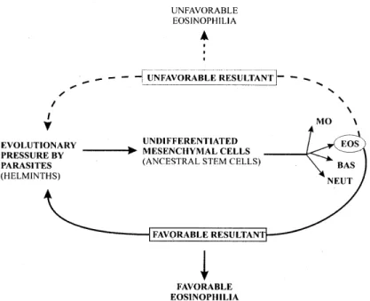
PHYLOGENY OF EOSINOPHILS
The long evolutionary story of Schistosoma
ancestors, dealing with several different species of animals, raises an important question: Why do the schistosome hosts usually present eosinophilia during the infection? Is the evolutionary pressure to eosinophil lineage expression a favorable or unfavorable condition to the parasite? Unfortu-nately, the “military paradigm” applied to the
in-terpretation of the eosinophil function (cytotoxic/ killer cell) has blurred and obstructed a deeper understanding of the eosinophil physiology.
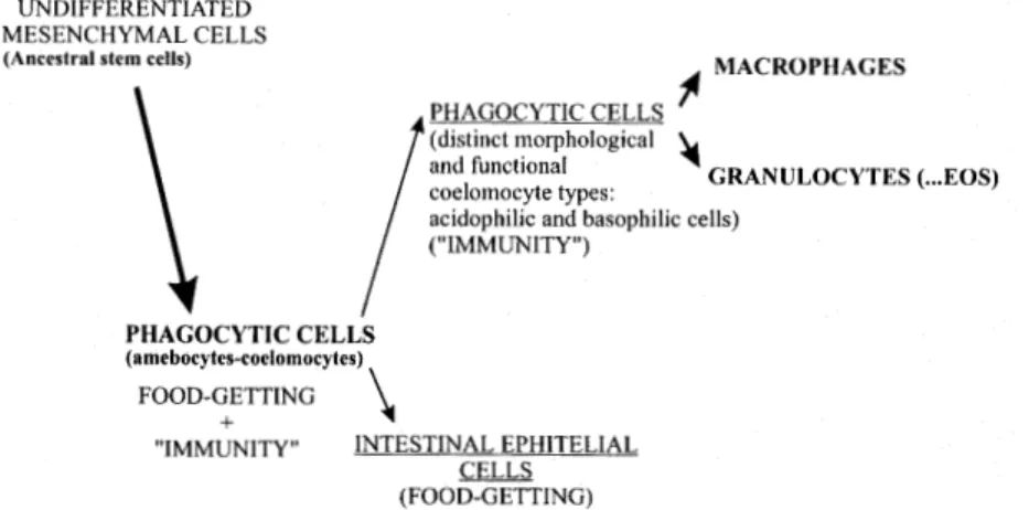
Ancestral cell of the lymphohematopoietic sys-tem existed among primitive invertebrates. This cell probably recognized foreign material or antigens, and responded by phagocytosis. Phagocytic cells have been conserved throughout the phylogenetic scale as macrophages and granulocytes; probably mast cells are also related to this ancestral cell. Thus, the phagocytic cell and its function is com-mon to and pervades all the phylogenetic levels of immunity (= first evolutionary step) (Cooper 1982). When coelomate invertebrates appeared, diverse leukocyte types developed, some of which are con-sidered to be precursors of lymphocytes (Wright & Cooper 1976) (Fig. 4).
Patterson and Landolt (1979) have reported neutrophilic as well as eosinophilic granules in amebocytes of the sea anemone Anthopleura elegantissima (Coelenterata). There have been
ul-trastructural investigations of the blood cells of nemerteans, which are less primitive acoelomates than platyhelminths. Light microscopy observa-tions (Ohuye 1942, Vernet & Gontchanoff 1975, 1976), however, have revealed four or five types of leukocytes, including eosinophilic granulocytes with reniform eccentric nuclei.
In the japanese horseshoe crab Tachypleus tridentatus (artropoda, phylum Chelicerata), the
granular amebocytes have been subdivided by Shishikura and Sekiguchi (1979) into two morpho-logically distinct types, based on the structure of the granular inclusions. The first type, “Type A”, accounts for approximately 10 % of the cell popu-lation, while “Type B” accounts for 90 % of the cells and enclose large, faintly and smaller,
26 26 26 26
26 Schistosome - Host Co-evolution • HL Lenzi et al.
nophilic granules. Deevey (1941) found that the hyaline leukocytes (equivalent to plasmatocytes?) and eosinophils (granular cells?) of Haitian taran-tula, Phormictopus cancerides (arachnid), ingest
a variety of dyes.
Ascidian (protochordata) blood cells are clas-sified into several categories, as follows: (1) un-differentiated cells (hemoblasts or lymphocytes); (2) leukocytes (hyaline or granular); (3) vacuolated cells (signet ring, compartment, or morula); (4) various pigmented cells and (5) nephrocytes. The granules of granular leukocytes are homogeneous, strongly electron-dense, and with conventional blood stains, may be acidophilic, basophilic or neutrophilic (Wright & Ermak 1982). Eosinophils have been described in the blood and tissues of a number of different fishes, including lower verte-brates such as the elasmobranchs (nurse shark (Gingilymostoma cirratum) (Hyder et al. 1983),
torpedoes (Torpedo marmorata risso and Torpedo acellata rafinisque) (Grimaldi et al. 1983), carps
(Smith et al. 1970), stripped bass (Morone saxatilis)
(Bodammer 1986), tench (Kelenyi & Nemeth 1969), loach, (Misgurnus anguillicaudatus)
(Ishizeki et al. 1984) and some teleost species (Davies & Haynes 1975, Bielek 1981). However, eosinophils are absent in hagfishes (Agnathan cy-clostomes) (Linthicum 1975). Curtis et al. (1979)
describe the bone marrow of Plethodon glutinosus
(amphibia) as containing large numbers of devel-oping neutrophils and eosinophils, lymphocytes of various sizes, plasmablasts, and plasma cells. Eosi-nophils are also present in frogs (Kelenyi & Nemeth 1969, Meseguer et al. 1985).
On the evolutionary scale, reptiles are pivotal since they are the progenitors of both avian and mammalian classes. The most prominent and well developed reptilian lymphoid organs are the thymus and spleen. In the early phases of the splenic devel-opment, before the organ becomes lymphopoietic, it contains a large number of granulocytes. In the turtle, the eosinophils are restricted to the subcap-sular region while basophils are scattered through-out the parenchyma. In later development, the sub-capsular eosinophils disappear and the spleen be-comes primarily lymphopoietic (Muthukkaruppan et al. 1982). Eosinophils of turtles and lizards do not contain a crystalloid internum (Kelenyi & Nemeth 1969). A primitive reptile, the tuatara
Splenedon punctatus, has eosinophils without
crys-talloids (Desser & Weller 1979). The American al-ligator (Alligator mississippiensis) presents blood
eosinophils positive for alkaline phosphatase and with some phagocytic capacity for bacteria in vitro
(Mateo et al. 1984). Eosinophils are found in nu-merous mammals other than man, such as buffalos, camels, cattle, goats, sheep, swine (artiodactyla);
cats, dogs, ferrets, mink, raccoons (carnivora); rab-bits (lagomorpha); kangaroos and opossum (mar-supials); horses (perissodactyla); primates and rodentia (familiae Muridae, Cricetidae and Caviidae) (Spry 1988).
The use of molecular probes for the unique constituents in eosinophils will be of great interest to determine the philogeny of eosinophils, mainly in invertebrates. We should also point out that there is considerable variation in the structure and bio-chemical composition of the eosinophils of differ-ent species (Spry 1988).
DISCUSSION AND CONCLUSION
Schistosomes, ancestors and recent species, have pervaded many hosts and several phyloge-netic levels of immunity. In recent hosts, S. mansoni
interacts with and activates the primordial lymphohematopoietic tissue (CALT) and the new immune system, reproducing a phylogenic “memory” (Lenzi et al. 1996). Taking advantages of the egg dispersion, it exploits the host cellular and immune responses to exteriorize its eggs (Damian 1987), maintaining the external concen-tration of eggs above the threshold level below which transmission cannot occur. We hypothesize here that the eosinophils could participate in the parasite host co-evolution, specially with S. mansoni, operating, together with monocytes/
macrophages and platelets in parasite transmission.
S. mansoni infection uses two patterns of
trans-mission: one relatively rapid with high number of eggs and more intense systemic and periovular re-action (eosinophils and monocytes) and other with slow transmission (chronic phase) and down-regu-lated granulomas and fall in the eosinophil and monocyte levels. S. mansoni adult worm have
capi-talized on the apparent adversity of living within the mesenteric vein, using the dispersion of eggs and antigens to other organs besides intestines to set a systemic activation of several hematopoietic lineages, specially eosinophils and monocytes/ macrophages. This activation occurs in bone mar-row, spleen, liver, lymph nodes, omental and me-senteric milky spots (CALT) (Lenzi et al. 1995), increasing the offer and making easy the migra-tion of eosinophils, monocytes and other cells to the intestinal periovular reaction.
ob-27 27 27 27 27 Mem Inst Oswaldo Cruz, Rio de Janeiro, Vol. 92, Suppl. II, 1997
servations exemplify some aspects of the intriguing complexity of the parasite-host relationship estab-lished during very long co-evolution (Fig. 5).
Bloch (1984) called attention to an apparent discrepancy in the extent of the attack on schistosomules by granulocytes between the in vivo
observations and in vitro studies. The later
experi-ments have repeatedly demonstrated that granulo-cytes and other cells involve a major number of schistosomules, adhering to them and killing them. While cell adherence to schistosomules was ob-served in vivo, the number of schistosomules
at-tacked was small. According to Bloch (1984), the reason for the discrepancy may be that in vivo the
parasites are metabolically more active and alter their surface membranes more rapidly, so that cell adherence is not so effective as in in vitro
environ-ment. Lozzi et al. (1996) showed that S. mansoni
primary and re-infection induced an influx of eosi-nophils to regional lymph nodes. However, only in the reinfected animals, were the eosinophils able to adhere to the larval surface, damaged larval be-ing found inside eosinophilic infiltrates. These authors questioned the significance of this adhe-sion, since it could either signify a role in larval death or a phenomenon secondary to larval degen-erative changes.
Why helminths have been responsible for evo-lutionary pressure to eosinophil response in most of their hosts? Is it any particular type of adaptative immune response (Mitchell 1991) that favors the host and the parasite? (Fig. 5).
The eosinophil is an ubiquitous cell, able to hide its purposes for more than 100 years (thousands or millions of years?). What is its role in the normal physiology? To answer this question it will be fun-damental to do phylogenetic and in vivo studies,
together with molecular biology approaches. In this context, McNagny et al. (1996) detected that avian retrovirus-transformed eosinophils and their pre-cursors expresse a 100 KD cell surface glycopro-tein, which presents homology to human melanotransferrin. By analogy to saxiphilin, melanotransferrin may have evolved to bind and inactivate toxic substances present in intestine or generated during kidney filtration or eosinophil maturation. Further experiments are required to elucidate the molecule’s true function (McNagny et al. 1996).
Finally, studies performed in shistosomiasis (and other parasitic diseases) have brought numerous information on the various facets of eosinophil func-tions and led to a better knowledge of eosinophil physiopathology, applicable to other diseases involv-ing eosinophils and specially allergic diseases (Capron & Capron 1992). In fact, infections by para-sites can be useful models to study eosinophil physi-ology and physiopathphysi-ology, because the parasites may function as natural eosinophil stimulants or depressors. According to Samter (1980), eosinophils continue to be “nominated but not elected cells”. Actually the function of eosinophils is complex, varied and still unknown, but can be artificially schematized as in Fig. 6.
28 28 28 28
28 Schistosome - Host Co-evolution • HL Lenzi et al.
ACKNOWLEDGEMENTS
To Luzia Fatima Gonçalves Caputo and Adelaide Lopes de Amorim for their expert technical assistance.
REFERENCES
Ackerman SJ, Weller PF, Nicholson-Weller A 1987. Activation of complement by human eosinophil granule major basic protein. Am Rev Resp Dis 135 (Suppl): A337.
Adrouny A, Seraydarian A, Levine AM, Hungerford GF, Carmel R 1984. Cyclic eosinophilic leucocytosis in eosinophilic leukemia with observations on transcobalamin I and eosinophils. Cancer 54: 1374-1378.
Andrade ZA, Reis MG 1984. Estudo sobre o papel dos eosinófilos na destruição dos esquistossômulos do Schistosoma mansoni in vivo. Mem Inst Oswaldo Cruz 79: 371-373.
Bartl S, Baltimore D, Weissman IL 1994. Molecular evolution of the vertebrate immune system. Proc Nat Acad Sci 91: 10769-10770.
Bartl S, Weissman IL 1994. Isolation and characteriza-tion of major histocompatibility complex class II β genes from the nurse shark.Proc Nat Acad Sci 91: 262-266.
Bash PF 1990. Why do schistosomes have separate sexes? Parasitol Today 6: 160-163.
Beelen RHJ, Fluitsma DM, Hoefsmit ECM 1980. Peroxidatic activity of mononuclear phagocytes de-veloping in milky spots. J Reticuloendothelial Soc 28: 601-609.
Bielek E 1981. Developmental stages and localization of peroxidatic activity in the leucocytes of three te-leost species (Cyprimus carpio L.; Tinca tinca L.; Salmo gairdneri Richardson). Cell Tissue Res 220: 163-180.
Bloch EH 1984. In vivo microscopy of schistosomiasis. IV. The dynamics of the host-parasite responses to Schistosoma mansoni in the hypodermal tissues as observed in transparent chambers in two susceptible hosts during primary and challenge infections. Am J Trop Med Hyg 33: 899-910.
Bodammer JE 1986. Ultrastructural observations on peritoneal exudate cells from the striped bass. Vet Immunol Immunopathol 12: 127-140.
Borojevic R 1966. Étude experimentale de la différentiation des cellules de l’éponge au cours de son dévelopment. Develop Biol 14: 130-153. Borojevic R 1992. Experimental murine schisosomiasis
mansoni: establishment of the chronic phase of the disease. Mem Inst Oswaldo Cruz 87 (Suppl. IV): 171-174.
Borojevic R, Stocker S, Grimaud JA 1981. Hepatic eosi-nophil granulocytopoiesis in murine experimental schistosomiasis mansoni. Br J Exp Pathol 62: 480-489.
Brener Z 1956. Observações sobre a infecção do camundongo pelo Schistosoma mansoni. Rev Bras Malariol D Trop 8: 565-575.
Brooks D, McLennan DA 1993. Parascript. Parasites and the language of evolution. Smithsonian Institu-tion Press, Washington, 429 pp.
Byram JE, Imohiosen EAE, von Lichtenberg F 1978. Tissue eosinophil proliferation and maturation in shistosome-infected mice and hamsters. Am J Trop Med Hyg 27: 267-270.
Capron M, Capron A 1992. Effector functions of eosi-nophils in schistosomiasis. Mem Inst Oswaldo Cruz 87 (Suppl.IV): 167-170.
Chensue SW, Terebuh PD, Warmington KS, Hershey SD, Evanoff HL, Kunkel SL, Higashi GI 1992. Role of IL-4 and IFN-γ in Schistosoma mansoni egg-induced granuloma formation. Orchestration, relative
con-Fig. 6: many functional hypothetical capabilities of eosinophils, which are probably integrated during eosinophil participation in vivo processes, such as inflammation and wound healing (Gansler 1956, Pepper & Lindsay 1960, Tchernitchin et al. 1967, Gleich
29 29 29 29 29 Mem Inst Oswaldo Cruz, Rio de Janeiro, Vol. 92, Suppl. II, 1997
tribution, and relationship to macrophage function. J Immunol 148: 900-906.
Chensue SW, Warmington KS, Ruth J, Lincoln PM, Kunkel SL 1994. Cross-regulatory role of interferon-gamma (IFN-γ), IL-4 and IL-10 in schistosome egg granuloma formation: in vivo regulation of the activ-ity and inflammation. Clin Exp Immunol 98: 395-400. Coelho MV 1970. O parasito - Schistosoma mansoni, p. 1-12. In AS Cunha, Esquistossomose mansoni. Editora da Universidade de São Paulo, São Paulo. Combes C 1990a. Where do human schistosomes come
from? An evolutionary approach. Trend in Ecol Evol 5: 334-337.
Combes C. 1990b. D’où viennent les parasites de l’homme? Ann Parasitol Hum Comp 65 (Suppl. I): 59-64.
Cooper EL 1982. Invertebrate defense systems - An overview, p. 1-35. In N Cohen, MM Sigel (eds). The Reticuloendothelial system - A comprehensive treatise. Vol 3 - Phylogeny and ontogeny. Plenum Press, New York.
Curtis SK, Cowden RR, Nagel JW 1979. Ultrastructure of the bone marrow of the salamander Plethodon glutinosus (Caudata: Plethrodontidae). J Morphol 159: 151.
Dahl R, Venge P 1979. Enhancement of urokinase-in-duced plasminogen activation by the cationic pro-tein of human eosinophil granulocytes. Thromb Res 14: 599-608.
Damian RT 1987. The exploitation of host immune re-sponses by parasites. J Parasitol 73: 3-13. Davies HG, Haynes ME 1975. Light and electron
mi-croscope observations on certain leukocytes in te-leost fish. J Cell Sci 17: 263.
Davis WB, Fells GA, Sun XH, Gadek JE, Venet A, Crys-tal RG 1984. Eosinophil-mediated injury to lung parenchymal cell and interstitial matrix. J Clin In-vest 74: 269-278.
Deevey GB 1941. The blood cells of the Haitian taran-tula and their relation to the moulting cycle. J Morphol 68: 457.
Desser SS, Weller I 1979. Ultrastructural observation on the granular leucocytes of the tuatara Sphenodon punctatus (Gray). Tissue Cell 11: 703-715. Doenhoff MJ, Musallam R, Bain J, McGregor A 1978.
Studies on the host-parasite relationship in Schisto-soma mansoni-infected mice. The immunological dependence of parasite egg excretion. Immunology 35: 771-778.
Doenhoff MJ, Pearson S, Dunne DW, Bickle Q, Lucas S, Bain J, Musallam R, Hassounah O 1981. Immu-nological control of hepatotoxicity and parasite egg excretion in Schistosoma mansoni infections: stage specificity of the reactivity of immune serum in T-cell deprived mice. Trans R Soc Trop Med Hyg 75: 41-53.
Dunne DW, Hassounah O, Musallam M, Lucas S, Pepys MB, Baltz M, Doenhoff MJ 1983. Mechanisms of Schistosoma mansoni egg excretion: parasitological observations in immunossuppressed mice reconsti-tuted with immune serum. Parasite Immunol 5: 47-60.
Dux K, Rouse RV, Kyewski B 1986. Composition of the lymphoid cell populations from omental milky spots during the immune response in C57Bl/Ka mice. Eur J Immunol 16: 1029-1032.
Erwin D, Valentine J, Jablonski D 1997. The origin of animal body plans. Am Scientist 85: 126-137. Faust EC, Jones CA, Hoffman WA 1934. Estudios sobre
la esquistosomiasis mansoni en Puerto Rico. III. Estudio biologico. 2. La etapa mamífera del ciclo vital. The Puerto Rico Publ Health Trop Med 10: 197-254.
Finkelman FD, Shea-Donohue T, Goldhill J, Sullivan CA, Morris SC, Madde KB, Gause WC, Urban Jr JF 1997. Cytokine regulation of host defense against parasitic gastrointestinal nematodes: Lessons from studies with rodent models. Ann Rev Immunol 15: 505-533.
Gansler H 1956. Elektronenmikroskopishe untorsuchungen am uterusmuskel der ratte unter follikehormonwirkring. Virchous Arch 329: 235-244. Gleich GJ, Loegering DA, Kueppers F, Bajaj SP, Mann KG 1974. Physicochemical and biological proper-ties of the major basic protein from guinea pig eosi-nophil granules. J Exp Med 140: 313-332. Grimaldi MC, Dippolito S, Pica A, Della Corte F 1983.
Cytochemical identification of the leukocytes of tor-pedoes (Torpedo marmorata Risso and Torpedo ocellata Rafinisque). Basic Appl Histochem 27: 311-317.
Grzych JM, Pearce E., Cheever A, Caulada ZA, Caspar P, Heiny S, Lewis F, Sher A 1991. Egg deposition is the major stimulus for the production of TH2 cytokines in murine schistosomiasis mansoni. J Immunol 146: 1322-1327.
Hegner RW 1926. The biology of host-parasite relation-ship among protozoa living in man. Quat Rev Biol 1: 393-418.
Henderson GS, Lu X, McCurley TL, Colley DG 1992. In vivo molecular analysis of lymphokine involved in the murine immune response during Schistosoma mansoni infection. II. Quantification of IL-4 mRNA, IFN-γ mRNA and IL-2 mRNA levels in the granu-lomatous livers, mesenteric lymph nodes and spleens during the course of modulation. J Immunol 147: 2261-2269.
Hibbs MS, Mainardi CL, Kang AH 1982. Type-specific collagen degradation by eosinophils. Biochem J 207: 621-624.
Hildeman WH, Johnston IS, Jokiel P 1979. Immunocom-petence in the lowest Metazoan Phylum: transplanta-tion immunity in sponges. Science 204: 420-422. Horton J, Ratcliffe N 1996. Evolution of immunity, p.
15.1-15.22. In I Roitt, J Bronstoff, D Male (eds). Immunology, 4th ed., Mosby, London.
Hyder SL, Cayer ML, Pettey CL 1983. Cell types in peripheral blood of the nurse shark: an approach to structure and function. Tissue Cell 15: 437-455. Ishizeki K, Nawa T, Tachibana T, Sakakura Y, Iida S
30 30 30 30
30 Schistosome - Host Co-evolution • HL Lenzi et al.
CD4+ T-cell function in host-parasite models. Chem Immunol 63: 51-65.
Kelenyi G, Nemeth A 1969. Comparative histochemis-try and electron microscopy of the eosinophil leu-kocytes of vertebrates. I. A study of avian, reptile, amphibian and fish leukocytes. Acta Biol Acad Sci Hung 20: 405-422.
Kelso A 1995. TH1 and TH2 subsets: paradigms lost? Immunol Today 16: 374-379.
Kroegel C, Virchow JC, Luttmann W, Walker C, Warner JA 1994. Pulmonary immune cells in health and dis-ease: the eosinophil leucocyte (Part I). Eur Respir J 7: 519-543.
Kroemer G, Cuende E, Martinez AC 1993. Compart-mentalization of the peripheral immune system. Adv Immunol 53: 157-216.
Lenzi, HL 1991. A dinâmica da resposta hematológica e celular na esquistossomose mansônica murina, com ênfase nas séries eosinofílica e mastocitária. PhD Thesis. Faculdade de Medicina, Universidade Federal de Minas Gerais, Belo Horizonte, 580 pp. Lenzi HL, Lenzi JA 1990. Comparative distribution of
eosinophils in bone marrow, blood and peritoneal cavity in murine schistosomiasis. Braz J Med Biol Res 23: 989-994.
Lenzi HL, Lenzi JA, Rosman FC, Pelajo-Machado M, Mota EM, Panasco MS, Oliveira DN 1995. Ex-tramedullary hematopoiesis in murine schistosomia-sis mansoni. Mem Inst Oswaldo Cruz 90: 169-177. Lenzi HL, Oliveira DN, Borojevic R, Lenzi JA 1992. Milky spots reaction to schistosomal mansoni infec-tion. Mem Inst Oswaldo Cruz 87 (Suppl V): 111-116.
Lenzi HL, Oliveira DN, Pelajo-Machado M, Borojevic R, Lenzi JA 1996. Coelom-associated lymphomyeloid tissue (milky spots): site of lym-phoid and myelomonocytic cell generation. Braz J Med Biol Res 29: 19-24.
Lenzi HL, Lenzi JA, Sobral ACL 1987a. Eosinophils favor the passage of eggs to the intestinal lumen in schistosomiasis. Braz J Med Biol Res 20: 433-435. Lenzi HL, Sobral ACL, Lenzi JA 1987b. In vivo kinet-ics of eosinophils and mast cells in experimental schistosomiasis. Mem Inst Oswaldo Cruz 82 (Suppl. IV): 67-76.
Lenzi HL, Sobral ACL, Lenzi JA 1988. Participation of endothelial cells in murine schistosomiasis. Braz J Med Biol Res 21: 999-1003.
Lenzi HL, Lenzi JA, Kerr IB, Antunes SLG, Mota EM, Oliveira DN 1991. Extracellular matrix in parasitic and infectious diseases. Mem Inst Oswaldo Cruz 86 (Suppl. III): 77-90.
Linthicum DS 1975. Ultrastructure of Hagfish blood leu-kocytes. Adv Exp Med Biol 64: 241-250.
Lozzi SP, Machado CRS, Gerken SE, Mota-Santos TA 1996. Involvement of regional lymphnodes after penetration of Schistosoma mansoni cercariae in naive and infected mice. Mem Inst Oswaldo Cruz 91: 491-498.
Luque EH, Montes GS 1989. Progesterone promotes a massive infiltration of the rat uterine cervix by the eosinophilic polymorphonuclear leukocytes. Anat
Rec 223: 257-265.
Mandache E, Moldveanu E, Negoescu A 1987. Lymphatic follicle-like structure in the stimulated omental milky spots. Morphologie et Embriologie 33: 285-289. Marchalonis JJ, Schluter SF 1994. Development of an
immune system. Ann New York Acad Sci 712: 1-11. Mateo MR, Roberts ED, Enright FM 1984. Morphologic, cytochemical, and functional studies of peripheral blood cells of young healthy American alligators (Alligator mississippiensis). Am J Res 45: 1046-1053. McNagny KM, Rossi F, Smith G, Graf T 1996. The eosi-nophil-specific cell surface antigen, EOS 47, is a chicken homologue of the oncofetal antigen melanotransferrin. Blood 87: 1343-1352.
Melro MCBF, Mariano M 1987. Extra-tissular Schisto-soma mansoni egg granulomata in the peritoneal cavity of mice. Mem Inst Oswaldo Cruz 82 (Suppl. IV): 245-252.
Meseguer J, Lozano MT, Agulleiro B 1985. Ultrastruc-ture of renal granulopoietic tissue of the Rana ridibunda tadpole. J Submicrosc Cytol 17: 391-401. Mitchell GF 1991. Co-evolution of parasites and adap-tive immune responses. Parasitol Today 7: A2-A5. Mitchison NA 1992. Regulation of the immune response, p. 226-275. In JO’D McGee, PG Isaacson, NA Wright (eds), Oxford Textbook of Pathology, Oxford University Press, Oxford.
Morris PJ 1993. The developmental role of the extracel-lular matrix suggests a monophyletic origin of the kingdom animalia. Evolution 47: 152-165. Müller WEG, Müller IM, Gamulin V 1994. On the
mono-phyletic evolution of metazoa. Braz J Med Biol Res 27: 2083-2096.
Müller WEG, Zahn RK, Rijavec M, Britvic S, Kurelec B, Müller I 1979. Aggregation of sponge cells. The Aggregation factor as a tool to estabilish species. Biochem Syst Ecol 7: 49-55.
Muthukkaruppan VR, Borysenko M, El Ridi R 1982. RES structure and function of the reptilia, p. 461-507. In N Cohen, MM Siegel (eds). The reticu-loendothelial system. A comprehensive treatise Vol. 3, Phylogeny and ontogeny. Plenum Press, New York.
Ngaiza JR, Doenhoff MJ 1990. Blood platelets and schis-tosome egg excretion. Proc Soc Exp Biol Med 193: 73-79.
Ngaiza JR, Doenhoff MJ, Jaffe EA 1993. Schistosoma mansoni egg attachment to culture human umbilical vein endothelial cells: An in vitro model of an early step of parasite egg excretion. J Inf Dis 168: 1576-1580.
Nirde P, Torpier G, DeReggi ML, Capron A 1983. Ecdys-one and 20 hydroxyecdysEcdys-one: new hormEcdys-ones for the humam parasite Schistosoma mansoni. FEBS letters 151: 223-227.
Ohuye T 1942. On the blood corpuscles and the hemopoises of nemertean Lineus fuscoviridis and of a sipunculid Dendrostoma minor. Sci Rep Tohoku Imp Univ 17: 187.
31 31 31 31 31 Mem Inst Oswaldo Cruz, Rio de Janeiro, Vol. 92, Suppl. II, 1997
Patterson MJ, Landolt ML 1979. Cellular reactions to injury in the anthozoan Anthopleura elegantissima. J Invert Pathol 33: 189.
Pepper H, Lindsay S 1960. Levels of eosinophils, plate-lets, leukocytes and 17-hydroxycorticosteroids dur-ing normal menstrual cycle. Proc Soc Exp Biol Med 104: 145-147.
Pinto C, Almeida AF 1948. Schstosomiasis mansoni no Brasil. Capítulo IV. Penetração e disseminação do trematóide no organismo dos mamíferos receptíveis. Mecanismos de penetração dos schistosômulos na pele. Monografias do Instituto Oswaldo Cruz 5: 47-57.
Platt TR 1992. A phylogenetic and biogeographic analy-sis of the genera of Spirorchinae (Digenea: Spirorchidae) parasitic in freshwater turtles. J Parasitol 78: 616-629.
Prata A 1957. Biopsia retal na esquistossomose mansoni. Bases e aplicações no diagnóstico e tratamento. Serviço de Educação Sanitária, Rio de Janeiro 198 pp.
Raven PH, Johnson GB 1996. Biology, 4th ed., Wm C Brown Publishers, Dubuque, IA., 1310 pp. Recklinghausen F von 1863. Uber eiter und
bindesgewebs-korjerchen. Virchows Arch Pathol Anat 28: 157-166.
Reis MG, Andrade ZA 1987. Functional significance of periovular granuloma in schistosomiasis. Braz J Med Biol Res 20: 55-62.
Ruppert EE, Barnes RD 1994. Invertebrate Zoology, 6th ed. Saunders College Publishing, London, 1051 pp. Salvini-Plawen L 1978. On the origin and evolution of the lower metazoa. Zeitschrift für Zoologische Systematik und Evolutionforschung 16: 40-88. Samter M 1980. Eosinophils-nominated but not elected.
N Engl J Med 303: 1175-1176.
Santos RO, Barbosa Jr AA, Andrade ZA 1992. Dynamic of fibrosis production and resorption in intestinal schistosomiasis of mice. Mem Inst Oswaldo Cruz 87: 25-31.
Sher A, Coffman RL, Hieny S, Scott P, Cheever AW 1990. Interleukin 5 is required for the blood and tis-sue eosinophilia but not granuloma formation in-duced by infection with Schistosoma mansoni. Proc Natl Acad Sci USA 87: 61-65.
Shishikura F, Sekiguchi K 1979. Comparative studies on hemocytes and coagulogens on Asian and Ameri-can horseshoe crabs, p. 185-201. In E Cohen Bio-medical applications of horseshoe crabs (Limulidae), Liss, New York.
Smith AM, Wivel NA, Potter M 1970. Plasmacytopoiesis in the pronephros of the carps. Anat Rec 167: 351.
Solvason N, Chen X, Shu F, Kearney JF 1992. The fetal omentum in mice and humam. A site enriched for precursors of CD5 B cells early in development. Ann New York Acad Sciences 651: 10-20.
Spry CJF 1988. Eosinophils. A comprehensive review and guide to the scientific and medical literature. Oxford University Press, Oxford, 484 pp.
Tam MR, Reddy AL, Karp RD, Hildemann WH 1976. Phylogeny of cellular immunity among vertebrates,
p. 98-119. In JJ Marchalonis, Comparative Immu-nology. Blackwell Scientific Publications, Oxford. Tchernitchin A 1967. Autoradiographic study of
(6,7-3H) oestradiol - 17 β incorporation into rat uterus. Steroids 10: 661-668.
Torpier G, Hirn M, Nirde P, Dereggi M, Capron A 1982. Detection of ecdysteroids in the human trematode Schistosoma mansoni.Parasitology 84: 123-130. Torres CM, Pinto C 1945. Lesões produzidas pelo
Schis-tosoma mansoni no tatu (Euphractus sexcinctus), mecanismos de eliminação dos ovos e sensibilidade da espécie animal nas infecções experimentais. Mem Inst Oswaldo Cruz 43: 301-348.
Truden JL, Boros DL 1985. Collagenase, elastase and nonspecific protease production by vigorous on immunomodulated liver granulomas on acid granu-loma macrophages/ eosinophils of S. mansoni- in-fected mice. Am J Pathol 121:166-175.
Van de Vyver G 1970. La non confluence intraspécifique chez les spongiares et la notion d’individu. Ann Embryol Morphol 3: 251-262.
Venge P, Dahl R, Hällgren R 1979. Enhancement of F XII-dependent reactions by eosinophil cationic pro-tein. Thromb Res 14: 641-649.
Vernet G, Gontcharoff M 1975. Étude auto-radiographique de l’incorporation de l’acide 4-aminolércetinique 3H et 55Fe dans les éléments fig-ures du sang de Lineus lacteus Montagu (Hétéronémertes).CR Acad Sci Ser D 280: 1413. Vernet G, Gontcharoff M 1976. Cytological Study of
the blood corpuscles of Lineus lacteus (Rhynchocoela, Lineidae). Cytobios 17: 137. Weiler JM, Gleich GJ 1988. Eosinophil granule major
basic protein regulates generation of classical and alternative amplification pathway C3 convertases in vitro. J Immunol 140: 1605-1610.
Weinberg DF, Baldo-Correa AE, Lenzi HL, Borojevic R 1992. Schsitosoma mansoni: peritoneal plasmatocytogenesis and polypoid transformation of mesenteric milky spots in infected mice. Exp Parasitol 74: 408-416.
Weinstock JV, Blum AM 1990a. Release of substance P by granuloma eosinophils in response to secreta-gogues in murine schistosomiasis mansoni. Cell Immunol 125: 380-385.
Weinstock JV, Blum AM 1990b. Detection of vasoac-tive intestinal peptide and localization of its mRNA within granulomas of murine schistosomiasis. Cell Immunol 125: 291-300.
Weinstock JV, Blum AM, Walder J, Walder R 1988. Eosinophils from granulomes in murine schistoso-miasis mansoni produce substance P. J Immunol 141: 961-966.
Weller PF, Rand TH, Finberg RW 1991. Human eosino-phil function as HLA-DR dependent, MHC-re-stricted antigen-presenting cells. FASEB J 5: A640. Wijffels JFAM, Henrickx RJBM, Steenbergen JJE, Eestermans IL, Beelen RHJ 1992. Milky spots in the mouse omentum may play an important role in the origin of peritoneal macrophages. Res Immunol 143: 401-409.
develop-32 32 32 32
32 Schistosome - Host Co-evolution • HL Lenzi et al.
ment and migration in the mammalian host, p. 115-146. In D Rollinson, AJG Simpson (eds). The biol-ogy of schistosomes: from genes to latrines. Aca-demic Press, New York.
Wilson RA 1990. Leaky livers, portal shunting and im-munity to schistosomes. Parasitol Today 6: 354-358. Wilson RA, Coulson PS 1986. Schistosoma mansoni: dynamics of migration through the vascular system of the mouse. Parasitology 92: 83-100.
Wilson RA, Coulson PS, Dixon B 1986. Migration of the schistosomula of Schistosoma mansoni in mice vaccinated with radiation-attenuated cercariae, and normal mice: an attempt to identify the site and tim-ing of parasite death. Parasitology 92:101-116. Wright RK, Cooper EL 1976. Phylogeny of Thymus and
Bone Marrow-bursa Cells. Elsevier, North-Holland, Amsterdam, 325 pp.
Wright RK, Ermak TH 1982. Cellular defense systems of the protochordata, p. 283-319. In N Cohen, MM Sigel (eds). The reticuloendothelial system. A com-prehensive treatise. Plenum Press, New York. Zhang L, Tarleton RL 1996. Characterization of cytokine
production in murine Trypanosoma cruzi infection by in situ immunocytochemistry: lack of associa-tion between susceptibility and type 2 cytokine pro-duction. Eur J Immunol 26: 102-109.