mTOR Inhibition Attenuates Dextran Sulfate
Sodium-Induced Colitis by Suppressing T Cell
Proliferation and Balancing TH1/TH17/Treg
Profile
Shurong Hu
1☯, Mengmeng Chen
1☯, Yilin Wang
2, Zhengting Wang
1, Yaofei Pei
3,
Rong Fan
1, Xiqiang Liu
4, Lei Wang
1, Jie Zhou
1, Sichang Zheng
1, Tianyu Zhang
1,
Yun Lin
1, Maochen Zhang
1, Ran Tao
4¤b*
, Jie Zhong
1¤a*
1Department of Gastroenterology, Ruijin hospital, Shanghai Jiaotong University School of Medicine, Shanghai, PR China,2Department of Surgery, Cancer hospital, Fudan University, Shanghai, PR China, 3Department of Surgery, Ruijin hospital, Shanghai Jiaotong University School of Medicine, Shanghai, PR China,4Department of Hepatobiliary-Pancreatic Surgery, Zhejiang Provincial People’s Hospital, Hangzhou, Zhejiang Province, PR China
☯These authors contributed equally to this work.
¤a Current address: Department of Gastroenterology, Ruijin Hospital, Shanghai Jiaotong University School of Medicine, PR China
¤b Current Address: Department of Hepatobiliary-Pancreatic Surgery, Zhejiang Provincial Peoples’Hospital (ZJPPH), Hangzhou, Zhejiang Province, PR China
*[email protected](JZ);[email protected](RT)
Abstract
It has been established that mammalian target of Rapamycin (mTOR) inhibitors have
anti-inflammatory effects in models of experimental colitis. However, the underlying mechanism
is largely unknown. In this research, we investigate the anti-inflammatory effects of
AZD8055, a potent mTOR inhibitor, on T cell response in dextran sulfate sodium
(DSS)-induced colitis in mice, a commonly used animal model of inflammatory bowel diseases
(IBD). Severity of colitis is evaluated by changing of body weight, bloody stool, fecal
consis-tency, histology evaluation and cytokine expression. We find that AZD8055 treatment
atten-uates DSS-induced body weight loss, colon length shortening and pathological damage of
the colon. And AZD8055 treatment decreases colonic expression of genes encoding the
pro-inflammatory cytokines interferon-γ, interleukin (IL)-17A, IL-1β,IL-6 and tumor necrosis
factor(TNF)-a and increases colonic expression of anti-inflammatory cytokines IL-10. We
show that AZD8055 treatment decreases the percentages of CD4+ T cells and CD8+
T cells in spleen, lymph nodes and peripheral blood of mice. We also find that AZD8055
treatment significantly reduces the number of T helper 1(TH1) cells and TH17 cells and
increases regulatory T (Treg) cells in the lamina propria and mesenteric lymph nodes.
Fur-thermore, we demonstrates that AZD8055 suppresses the proliferation of CD4+ and CD8+
T cells and the differentiation of TH1/TH17 cells and expands Treg cells in vitro. The results
suggest that, in experimental colitis, AZD8055 exerts anti-inflammatory effect by regulating
T helper cell polarization and proliferation.
a11111
OPEN ACCESS
Citation:Hu S, Chen M, Wang Y, Wang Z, Pei Y, Fan R, et al. (2016) mTOR Inhibition Attenuates Dextran Sulfate Sodium-Induced Colitis by Suppressing T Cell Proliferation and Balancing TH1/TH17/Treg Profile. PLoS ONE 11(4): e0154564. doi:10.1371/journal. pone.0154564
Editor:Hossam M Ashour, Wayne State University, UNITED STATES
Received:January 4, 2016
Accepted:April 17, 2016
Published:April 29, 2016
Copyright:© 2016 Hu et al. This is an open access article distributed under the terms of theCreative Commons Attribution License, which permits unrestricted use, distribution, and reproduction in any medium, provided the original author and source are credited.
Data Availability Statement:All relevant data are within the paper.
Funding:This study was supported by a Grant-in-Aid for Scientific Research (15ZR1426400, 14XJ10014) from Shanghai Science and Technology Committee for the Promotion of Science in China.
Competing Interests:The authors have declared that no competing interests exist.
Introduction
Inflammatory bowel diseases (IBD) which consist of Crohn
’
s disease (CD) and ulcerative
coli-tis (UC), are chronic heterogeneous intestinal disorders, which remain clinically challenging
[1,
2]. Currently, the drugs for IBD patients are limited. The precise etiology of IBD remains
unknown, although it is generally accepted that it result from an overactive immune response
to commensal bacteria within the gut in genetically predisposed individuals [3].
Helper T cells have a significant role in IBD pathogenesis [4]. TH1, TH2, TH17 and
regula-tory T cells (Tregs) form an important quarter of helper T cells [5,
6]. Studies have been shown
that TH1, TH2, and TH17 cells were essential for defenses against excessive entry of
microor-ganisms [7,
8]. Intestinal immune homeostasis depends on the regulation and balance of these
T cell subgroups. It has been shown that deregulated overexpansion and activation of effector
cells in relation to regulatory T cells can lead to intestinal inflammation like IBD [9,
10].
The T cell transfer induced colitis has been used to study T cell response in IBD. In this study,
CD4+CD45RBhi T cells are transferred into immune-deficient mice. Since this model depends
on genetically compromised mice and an unbalance of naïve and Treg cells which is not seen in
wild type mice, it does not reflect the immunological courses of the development of pathogenic
T cells in healthy animals [11
–
13]. On the other hand, DSS-induced colitis model is a classic and
stable model of murine colitis, which can be used in all backgrounds of mice. Many drugs used
in IBD patients are also available for this model [14
–
16]. Previous studies have shown that
DSS-induced colitis is often not considered as a good model for T cell involvement, since it is chemical
damage model which can be induced without the help of T cells. However, recent studies show
that T cells especially pro-inflammatory, antigen-specific CD4+ T cells accumulate at the site of
inflammation, and do progress during DSS-induced colitis model, suggesting that DSS model
can be used to study T cell development during intestinal inflammation [17
–
19].
Mammalian target of Rapamycin (mTOR) is a protein kinase that regulates cell survival, cell
growth, cell proliferation and autophagy. Besides its crucial role in tumorigenesis, recent
stud-ies show that mTOR participates in adjusting adaptive immune response and modulating
CD4+ or CD8+ T cell polarization, as well as increasing the percentage of Treg cells [20
–
22].
Farkas et al showed that Rapamycin, an mTOR inhibitor, reduced leukocyte migration as
effec-tively as immunosuppressant cyclosporine A (CsA) in DSS-induced murine colitis [23].
Matsuda et al found that Everolimus, a Rapamycin analog, prevented colitis in
interleukin-10(IL-10)
–
/
–
model by decreasing the percentage of CD4+ T cells in the colonic mucosa and
reducing IFN-γ
production [24].
mTOR functions in two multi-protein complexes, mTORC1 and mTORC2. mTORC1 is
suppressed by Rapamycin, but Rapamycin can
’
t block mTOR activity completely due to its
inability to influence mTORC2 directly [25,26]. On the other hand, ATP-competitive mTOR
inhibitor AZD8055 targets the ATP site and inhibits any mTOR-containing complex [27].
AZD8055 not only inhibits phosphorylation of the mTORC1 substrates p70S6K and 4E-BP1,
but also phosphorylation of the mTORC2 substrates AKT and downstream protein [28]. In
spite of the emerging role of RAPA-resistant mTOR in immune cell function, the effect of
AZD8055 on T cells has not been fully studied. In this study, we investigate the effect of
AZD8055 in DSS-induced colitis. We find that AZD8055 attenuates DSS-induced colitis by
inhibiting T-cell proliferation and balancing TH1/TH17/Treg profile.
Materials and Methods
Ethics Statement
All experimental procedures were performed in accordance with the criteria issued by the
Chi-nese ethics committee for animal studies, formulated by the Ministry of Science and Technology
Interferon gamma; TNF-α, tumor necrosis factorof the People
’
s Republic of China. The animal protocols were examined and approved by the
Ethical Committee on Animal Experiments at Shanghai Ruijin Hospital. All endeavors were
made to alleviative suffering.
Mice
6
–
8 weeks old male C57BL/6 mice were gained from Shanghai Laboratory Animal Center
(SLAC) and housed under specific pathogen-free environment in the Research Center for
Experimental Medicine of Shanghai Ruijin Hospital. All animal studies were authorized and
handled according to the guidelines of the Ethics Committee of Ruijin Hospital.
Reagents
AZD8055 was purchased from Selleck (China). It was dissolved in dimethyl sulfoxide (DMSO)
and prepared as 10mM stock solutions and stored at -20°C for in vitro studies. For in vivo
administration, AZD8055 solution was diluted in sterile emulsifiers.
Induction of Dextran Sodium Sulfate (DSS) Colitis and assessment of
acute colitis
Mice were randomly divided into three groups, Wild type (WT), DSS, DSS+AZD8055 (n = 8 in
each group). For acute colitis, mice were subjected to 4% (w/v) DSS (MW 36, 000
–
50, 000, MP
Biomedical) in their drinking water for 7 (day0
–
6) days. Mice were monitored daily by change
of body weight, gross rectal bleeding, and stool consistency. The disease activity index score
(DAI) was described previously (Table 1) [29]. Mice were treated daily intra-peritoneally (i.p.)
with 10mg/kg AZD8055 or emulsifier as control from day 0 to day 6 and were sacrificed on
day 7.
Histological examination
The colons were embedded in paraffin and stained with hematoxylin-eosin (H&E). To evaluate
the histological inflammation, we adopted the histological damage score: edema; crypt loss;
erosion/ulceration and mono- and poly-morphonuclear cells infiltration. The degree of colon
inflammation scored from 0 to 20 [30]. The analysis was performed by two investigators who
were blinded to the experiment.
Preparation of lamina propria (LP) lymphocytes
LP lymphocytes were isolated according to previously described study [31]. Briefly, the
intes-tines were opened longitudinally, washed with PBS to remove stool, and shaken in Hank's
Bal-anced Salt Solution (HBSS) supplemented with 4% FBS and 5mM EDTA (Sigma-Aldrich) at
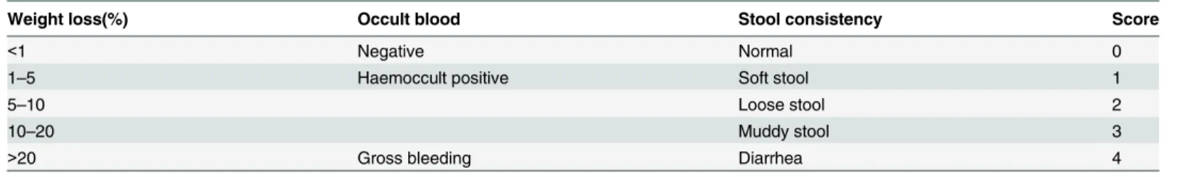
Table 1. The disease activity index score (DAI).
Weight loss(%) Occult blood Stool consistency Score
<1 Negative Normal 0
1–5 Haemoccult positive Soft stool 1
5–10 Loose stool 2
10–20 Muddy stool 3
>20 Gross bleeding Diarrhea 4
Five grades of weight loss and stool consistency and three grades of occult blood.
37°C for 25 min. This procedure was to remove epithelial cells and intraepithelial lymphocytes.
The colon fragments were then incubated with RPMI 1640 supplemented with 4% FBS and
1mg/ml collagenase type IV (R&D) and Dnase I (Sigma-Aldrich) at 37°C for 25 min with
stir-ring. The digested tissues were pooled together and separated on a 40/80% discontinuous
Per-coll gradient (GE Healthcare). The gradient separation was centrifuged at 2500rpm for 25 min
at room temperature. Lamina propria mononuclear cells were collected at the interface of the
Percoll gradient, washed, and suspended in RPMI 1640 containing 4% FBS.
Establishment of T cell Proliferation and Differentiation in vitro
Splenocytes and lymph nodes were isolated and prepared as mononuclear cells from mice.
CD4
+T cells and CD8
+T cells were respectively negatively selected using the CD4
+T cell
Isola-tion kit (Miltenyi) and CD8
+T cell Isolation kit (Miltenyi). In order to analysis T cell
prolifera-tion, CD4
+T cells and CD8
+T cells were labeled with carboxyfluorescein succinimidyl ester
(CFSE; Invitrogen). Cells were stimulated with mouse antibody CD3/CD28 (Invitrogen) at
2ug/ml for 3 days in the presence of AZD8055 at the indicated concentrations and were
evalu-ated by flow cytometry. In order to analysis T cell differentiation into TH1, TH17 or Treg cells,
naive T cells were sorted by CD4
+CD62L
+T cell Isolation kit II (Miltenyi). Naive T cells were
labeled with CFSE and stimulated with anti-CD3 (2
μ
g/mL), and anti-CD28 (2
μ
g/mL)
antibod-ies under TH0(40 U/mL mouse IL-2), TH1 (40
μ
g/mL anti-mouse IL-4, 40 ng/mL mouse IL-12),
TH17 (50 ng/mL mouse IL-6, 2.5 ng/mL mouse TGF-
β
, 10
μ
g/mL anti-mouse IFN-
γ
, 20 ng/mL
mouse IL-23, 10
μ
g/mL anti-mouse IL-4) or Treg conditions(2 ng/mL mouse TGF-β) for 4 days
in the presence of AZD8055 at specified concentrations and were evaluated by flow cytometry.
Flow cytometry
To quantify the percentage of CD4 and CD8 positive cells, mononuclear cells from spleen,
mesenteric lymph node and lamina propria were washed and incubated with anti-CD3-APC,
anti-CD4-FITC, anti-CD8-PE (ebioscience, San Diego, CA). To determine the percentage of
TH1 and TH17 cells, mononuclear cells were stimulated with PMA (50ng/mL, sigma) and
Ionomycin (1
μ
g/mL, Tocris) and BFA (1:1000, eBioscience) for 6 hours. Cells were then
washed and stained with anti-CD4-FITC. Following CD4 staining, cells were blocked, fixed,
permeabilized and stained with anti-IFN-γ-APC or anti-IL-17-PE-Cy7. To detect the
percent-age of Treg cells, mononuclear cells were stained with anti-CD4-FITC and anti-CD25-APC
and then fixed/permeabilized and incubated with anti-Foxp3-PE. All labeled cells were
detected using FCM on the FACScan Flow Analyzer. The results were evaluated with FlowJo7.6
software [32,
33].
Quantitative reverse-transcription polymerase chain reaction analysis
Total RNA from colonic samples were extracted by TRIzol
1Reagent (Invitrogen Life
Technol-ogies) and were reverse transcribed using oligo (dT) primers (Takara) in accordance with the
manufacturer's instructions. Real-time polymerase chain reaction (PCR) was performed on
QuantiTect SYBR Green PCR Kit. The expressions of target gene were analyzed by their ratios
to the house keeping gene hypoxanthine-guanine phosphoribosyl transferase (HPRT). The
sequence of target genes primer sets were listed in
Table 2.
Statistical Analysis
Results
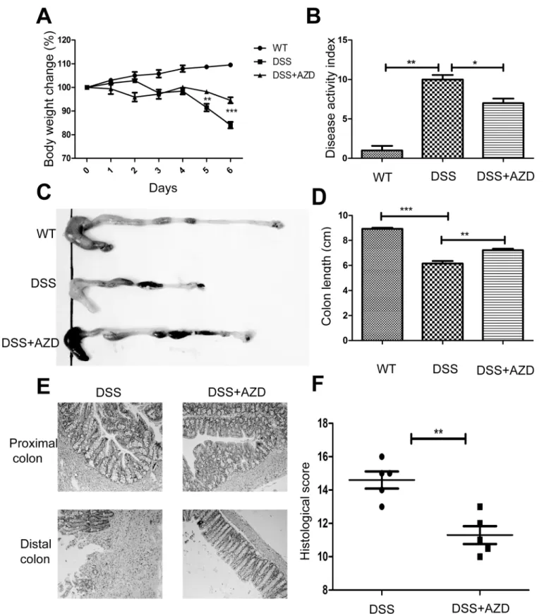
1. AZD8055 attenuates the development of intestinal inflammation
In assessing the role of AZD8055 in the development of colitis, we used 4% DSS to induce
coli-tis. It is known that DSS can induce severe inflammation in mice characterized by persistent
weight loss, rectal bleeding and diarrhea. As shown in
Fig 1A, DSS-treated mice exhibited
pro-found body weight loss, whereas AZD8055 attenuated the loss of body weight. By daily
moni-toring of clinical manifestations such as weight loss, diarrhea, and rectal bleeding, we scored
disease activity index (DAI) according to the standards in
Table 1. We observed that AZD8055
decreased DAI score and prevented colitis (Fig 1B). DSS-induced colon shortening, a marker
of intestinal inflammation, was also ameliorated by AZD8055 (Fig 1C and 1D). The severity of
intestinal inflammation and ulceration were further assessed by histological study using
Hae-matoxylin & eosin (H&E) staining. DSS-induced pathological damage includes epithelial crypt
loss, gross ulceration following massive infiltration of monocytic cells into the mucosa, as well
as edema and congestion of the submucosa. As shown in
Fig 1E and 1F, AZD8055-treated
mice showed less inflammatory cells infiltration and smaller ulceration and greater mucosal
integrity. Thus, we concluded that AZD8055 treatment could relieve DSS-induced colitis.
Results were from three independent experiments.
2. AZD8055 reduces pro-inflammatory mediator production in colon
The hallmark of DSS-induced colitis is the increased production of pro-inflammatory
cyto-kines in the colon. The colon was isolated and real-time PCR was used to assess the expression
profiles of pro-inflammatory cytokines. As shown in
Fig 2, the mRNA levels of
pro-inflamma-tory cytokines, such as IFN-γ, TNF-α, IL-1β, IL-17A and IL-6 in the colon increased in the
presence of DSS administration, whereas AZD8055 treatment decreased the mRNA levels of
these cytokines. And the anti-inflammatory cytokine IL-10 increased under AZD8055
treatment.
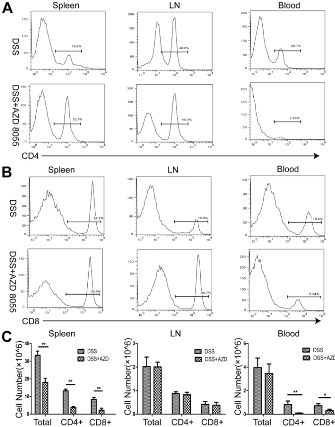
3. AZD8055 decreases the proportion of CD4
+T cells and CD8+ T cells
in vivo
Aberrant immune response is responsible for chronic inflammatory diseases. CD4
+and CD8
+T cells played important role in inflammatory bowel disease. We isolated and prepared
mono-nuclear cells of spleen, peripheral lymph nodes and peripheral blood from DSS-treated and
DSS+AZD8055 treated mice. The results showed that, compared with DSS treatment, DSS
+AZD8055 induced reduction in the number and proportion of CD4+ and CD8+ T cells,
espe-cially in the spleen and peripheral blood (Fig 3).
Table 2. Sequence of primer pairs used in real-time quantitative PCR.
Gene name Forward primer(5’!3’) Reverse primer(5’!3’)
TNF-α CTCTTCAAGGGACAAGGCTG CTCTTCAAGGGACAAGGCTG
IL-1β TTCAGGCAGGCAGTATCA GTCACACACCAGCAGGTTA
IL-6 CCAATGCTCTCCTAACAGA TGTCCACAAACTGATATGC
IFN-γ CAGCAACAACATAAGCGTC CTCAAACTTGGCAATACTC
IL-17A CCTTCACTTTCAGGGTCGAG CAGTTTGGGACCCCTTTACA
IL-10 AGGGCACCCAGTCTGAGAACA CGGCCTTGCTCTTGTTTTCAC
HPRT TCAACGGGGGACATAAAAGT TGCATTGTTTTACCAGTGTCAA
Fig 1. AZD8055 attenuates the development of intestinal inflammation.When the mice developed clinical signs of disease, they were sacrificed. (A) During the course of colitis, body weights were recorded and calculated as percentage of the initial weight at day 1 (n = 8 for AZD, n = 8 for control). (B) Disease activity index (DAI) of each group. (C) Gross morphology of each group. (D) The length of colon of each group. (E) Histological observations of colon sections with H&E staining (Original magnifications, 200×magnification). (F) Histological score of each group. All data represented the mean±SEM. Statistical significance was assessed by t-test.*P<0.05,**P<0.01.
Fig 2. AZD8055 treatment suppresses the expression of pro-inflammatory mediators. (A-F)The expression profiles of IFN-γ, TNF-α, IL-1β, IL-6, IL-17A and IL-10 were determined in the colon of emulsifier or AZD8055-treated mice by real-time PCR. Data was normalized to the expression of HPRT mRNA (n = 8/group). All data represented the mean±SEM. Statistical significance was assessed by t-test.*P<0.05,**P<0.01.
Fig 3. AZD8055 treatment leads to a decrease in the percentage of CD4+ T cells and CD8+ T cells in vivo.(A) The percentage of CD4+T cells in the spleen, lymph nodes and peripheral blood of mice treated with AZD8055 or emulsifier. (B) The percentage of CD8+T cells in the spleen, lymph nodes and peripheral blood of mice treated with AZD8055 or emulsifier. (C) The cell number of CD4+ T cells and CD8+ T cells in the spleen, lymph nodes and peripheral blood of mice treated with AZD8055 or emulsifier. All data represented the mean±SEM. Statistical significance was assessed by t-test.*P<0.05,**P<0.01.
4. AZD8055 suppresses the proliferation of CD4
+T cells and CD8+ T
cells in vitro
As described above, AZD8055 decreased the percentage of CD4
+and CD8
+T cells in spleen,
lymph nodes and peripheral blood. To further confirm the anti-inflammatory effect of
AZD8055 in vitro, we investigated if AZD8055 inhibited the proliferation of CD4
+and CD8
+T cells. CFSE-labeled CD4
+and CD8
+T cells were activated with anti-CD3/CD28 beads in the
presence or absence of increasing concentrations of AZD8055 (10
–
50nM). The proliferation of
CD4
+and CD8+ T cells were analyzed by flow cytometry. As shown in
Fig 4A and 4B, we saw
that the proliferation of CD4
+and CD8
+T cells were inhibited by AZD8055 in a
dose-depen-dent manner.
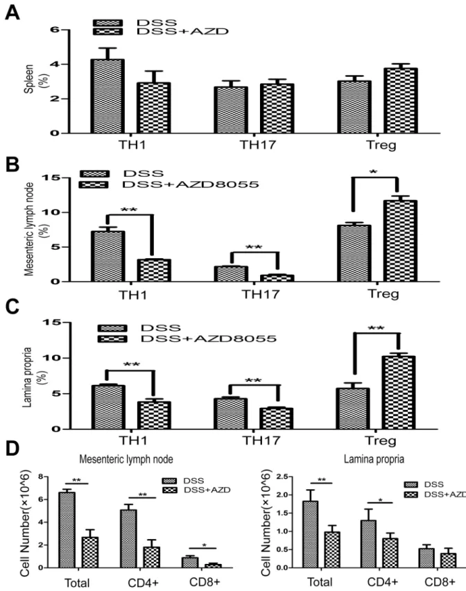
5. AZD8055 balances TH1/TH17/Treg profile in vivo
Through the activation of separate signaling pathways, naive T cells differentiate into helper
T (TH) cells, termed TH1, TH2 and TH17, and induce Treg cells. Dysregulation of TH cells
results in IBD. To test the effect of AZD8055 on the proportion of different T helper cell
sub-sets in spleen, mesenteric lymph nodes and lamina propria, we isolated mononuclear cells of
mesenteric lymph nodes and lamina propria from DSS-treated mice as well as DSS+AZD8055
treated mice. We found that, although there were no differences in splenic TH1, TH17 and
Treg between the control and AZD8055 treated mice (Fig 5A), AZD8055 treatment reduced
the proportion of TH1 cells (CD4+IFN-
γ
+) and TH17 cells (CD4+IL-17+) and increased the
proportion of Treg cells (CD4+FOXP3+) in mesenteric lymph nodes and lamina propria
(Fig 5B and 5C). In addition, the cell number of CD4+ T cells and CD8+ T cells in the presence
of AZD8055 were also decreased in the mesenteric lymph nodes and lamina propria (Fig 5D).
Therefore, AZD8055 ameliorated colitis might through balancing TH1/TH17/Treg cell profile
to maintain normal immune homeostasis.
6. AZD8055 inhibits the proliferation and differentiation of TH1 and TH17
cells in vitro
Next, we determined if AZD8055 could restrain T cell expansion and differentiation in vitro.
CFSE-labeled naive CD4
+T cells were isolated and irritated under TH0, TH1 and TH17
condi-tions for 4 days in the presence of AZD8055 at different concentration (0nM, 20nM, 50nM).
We observed a dose-dependent inhibition of TH1 proliferation and differentiation by
AZD8055. While there was a significant inhibitory effect of AZD8055 on TH1 cell
differentia-tion at a concentradifferentia-tion of 50nM (Fig 6A and 6C). And AZD8055 exhibited the inhibidifferentia-tion on
TH17 cell differentiation at a concentration of 20nM (Fig 6B and 6C).
7. AZD8055 enhances the proliferation and differentiation of Treg cells in
vitro
Fig 4. AZD8055 inhibits the proliferation of CD4+T cells and CD8+T cells in vitro.CD4+T cells and CD8+T cells isolated from spleen and mesenteric lymph node of mice were stimulated with anti-CD3/CD28 and labeled with CFSE in the presence of AZD8055 (10–50nM) for two or three days. (A) The proliferation of CD4+T cells in the presence of AZD8055 were examined by flow cytometry. (B) The proliferation of CD8+T cells in the presence of AZD8055 were examined by flow cytometry. Results were from three independent experiments.
Fig 5. AZD8055 inhibits TH1 and TH17 cell polarization and expands Treg cell polarization in vivo.Lymphocytes were isolated from the spleen, mesenteric lymph nodes and lamina propria and analyzed by flow cytometry on day7. (A) The percentage of TH1 cells (CD4+IFN-γ+), TH17 cells (CD4+IL-17+) and Treg cells (CD4+FOXP3+) in spleen treated with AZD8055 or emulsifier. (B) The percentage of TH1 cells, TH17 cells and Treg cells in mesenteric lymph nodes treated with AZD8055 or emulsifier. (C) The percentage of TH1 cells, TH17 cells and Treg cells in lamina propria treated with AZD8055 or emulsifier. (D) The cell number of CD4 + T cells and CD8+ T cells in the mesenteric lymph nodes and lamina propria treated with AZD8055 or emulsifier. The data were expressed as mean±SEM. Statistical significance was assessed by t-test.*P<0.05,**P<0.01.
Discussion
In this study, we found that ATP-competitive dual mTOR inhibitor AZD8055 ameliorated
DSS-induced colitis by increasing the functional activity of Treg cells and suppressing TH1 and
TH17 cell response in vivo. Previous studies have showed that infusion SCID mice with naive
T cells without Treg cells induces hyperactivity to intestinal commensal bacteria, while infusing
whole T cells prevents inflammation [34
–
36]. Another study also observed that the depletion
of Treg cells in mice deteriorated the disease [37]. In the presence of Interleukin-23(IL-23) or
Interleukin-6 (IL-6), naive T cells differentiate into TH17 cells [38,
39]. The efficacy of
balanc-ing Treg/TH17 cells has an important role in the clinical and experiment model [40
–
42].
Toci-lizumab, a humanized monoclonal antibody against the membrane and the soluble IL-6, and
Ustekinumab, a human monoclonal antibody that blocks IL-12 and IL-23, have been used to
treat IBD patients [43,
44]. Hui Yin et al have found that sirolimus (Rapamycin analogue)
ame-liorated TNBS-induced colitis by promoting differentiation of Treg cells and inhibiting the
generation of TH17 cells [45]. In our study, we found that AZD8055 had the dose-dependent
effect on inhibition of TH17 cells differentiation, and increase of Treg cells proliferation and
differentiation in vitro. Furthermore, we also found that AZD8055 treatment induced the
increase of Treg cells and decrease of TH17 cells in the lamina propria and mesenteric lymph
nodes in mice. Our results suggested that AZD8055 was involved in the regulation of balancing
of Treg/TH17 cells both in vivo and in vitro.
Delgoffe et al reported that mTOR-deficient T cells were unable to differentiate into TH1,
TH2, or TH17 effector cells while they displayed normal activation and IL-2 production upon
initial stimulation [20]. The incapacity of differentiation was related to STAT activation
reduc-ing as well as failure of the up-regulation of lineage specific transcription factors. However,
under normal conditions of activation, T cells lacking mTOR were capable of differentiating
into Foxp3+regulatory T cells. The results indicated that mTOR pathway was critically
involved in the differentiation of CD4+ effector T cells [46,
47]. The first-generation mTOR
inhibitors Rapamycin and its analogues such as Sirolimus and Everolimus employed an
alloste-ric mechanism to block mTORC1 output. Meanwhile, second generation mTOR inhibitors
such as AZD8055 and PP242 competitively targeted the ATP binding site to hinder kinase
activity of both TORC1 and TORC2 [48]. In our study, we found that AZD8055 could suppress
the differentiation and proliferation of TH1 and TH17 cells and increase Treg cells both in vivo
and in vitro.
Recent studies have found that specific B cell subsets, regulatory B cells (Bregs), are potent
immune response regulators and are of crucial importance in a variety of mouse models of
immune-mediated disorders like IBD, systemic lupus erythematous(SLE) and rheumatoid
arthritis (RA) and immune thrombocytopenia patients(ITP) [49
–
53]. Flores-Borja et al.
inves-tigated that healthy Breg cells especially CD19+CD24hiCD38hi B cells could limit the
differen-tiation of naïve T cells into TH1 and TH17 cells and convert effector CD4+ T cells into Treg
cells by producing a large amount of IL-10 [54].
Ashour HM
et al. studied that expansion of
Breg cells were necessary for the induction of T cell tolerance elicited through the anterior
chamber of the eye [55]. Therefore, Bregs may play an important role in the induction of T cell
under TH1 condition (Lower panels). (B) TH17 cells were stimulated with PMA + Ionomycin and stained for CD4 and intracellular expression of IL-17 in the presence of AZD8055. Dot-plots showed the expression of CD4+ IL-17 + TH17 cells under TH0 condition (Upper panels). Scatterplot displayed the differentiation of TH17 cells under TH17 condition (Lower panels). (C) Results were from three independent experiments. All results showed the mean±SEM. Statistical significance was determined by student’s t-test.*P<0.05, **P<0.01,***P<0.005.
Fig 7. AZD8055 expands the proliferation and differentiation of Treg cells in vitro.Naive CD4+T cells were isolated from spleen and lymph nodes of mice, CFSE-labeled, and activated in the presence of AZD8055 (0nM, 20nM) under TH0 and Treg conditions for 4 days. (A) Treg cells were permeabilized and stained for CD4 and intracellular expression of Foxp3 in the presence of AZD8055 (0nM, 20nM). Dot plots showed the differentiation of CD4+ Foxp3+ Treg cells under TH0 and Treg conditions (Upper panels). Scatterplot displayed the proliferation of Treg cells under TH0 and Treg conditions treated with AZD8055 (Lower panels). (B) Results were from three independent experiments. All results showed the mean±SEM. Statistical significance was evaluated by student’s t-test.*P<0.05, **P<0.01,***P<0.005.
tolerance and in maintaining the key balance between TH1/TH17/Treg populations [56]. In
addition, it has also been shown that Granulocyte macrophage colony stimulating factor
(GM-CSF) can differentiates precursor cells into tolerogenic DCs that can expand Treg cell
numbers and function [57,58]. These Treg cells can suppress effector T cells through secretion
of IL-10 [59]. GM-CSF has a critical role in regulating the immune response and maintaining
immunological tolerance by inducing specialized cell types from precursors or by affecting
phenotypes of mature cell populations [60
–
62]. The protective effect of GM-CSF has been
investigated in IBD and Type 1 diabetes (T1D) [63
–
66]. Adoptive transfer of GM-CSF could
directly expand Treg cells which was shown in diabetes in mouse models [66,
67]. Thus
GM-CSF may exert a far-reaching influence on the state of immune tolerance referring to a
wide array of autoimmune diseases.
It has been shown that Rapamycin inhibites induced chronic colitis by decreasing leukocyte
migration. However, the effect of Rapamycin on T cells regulation has not been investigated.
Since Rapamycin also inhibits mTORC2 activity after the prolonged treatment, it would be
interesting to compare the efficiency of Rapamycin with AZD8055 in DSS-induced colitis. In
our study, we showed that ATP-competitive dual mTOR inhibitor AZD8055 exhibited potent
immunosuppressive properties and was used therapeutically in countering autoimmunity and
preventing allograft rejection. In future studies, we will use mTORC1 or mTORC2 knockout
mice to study the effect of mTOR complexes on DSS-induced mice. We will also investigate the
effect of mTOR inhibition on the immune cells from human biopsy in IBD patients and
explore possible mechanism.
Although AZD8055 has been used for its anti-tumor activity, its application in the
autoim-mune diseases still lacks comprehensive understanding [68
–
71]. The immune regulation of
AZD8055 is involved in a variety of immune cells and cytokines. Our study showed that
AZD8055 attenuated DSS-induced colitis and also explored its possible mechanisms in
immune regulation. This study indicates that ATP-competitive mTOR inhibitor may offer a
promising alternative pharmaceutical strategy to manage IBD.
Acknowledgments
The authors would like to thank Professor Yu Li (Shanghai Jiaotong University Medical
School) for critical comments and help with the manuscript.
Author Contributions
Conceived and designed the experiments: J. Zhong RT. Performed the experiments: SRH
MMC YLW. Analyzed the data: ZTW YFP RF XQL. Contributed reagents/materials/analysis
tools: LW J. Zhou SCZ TYZ YL MCZ. Wrote the paper: SRH MMC.
References
1. Kaser A, Zeissig S, Blumberg RS. Inflammatory bowel disease. Annu Rev Immunol. 2010; 28: 573–621. doi:10.1146/annurev-immunol-030409-101225PMID:20192811
2. Sartor RB. Mechanisms of disease: pathogenesis of Crohn's disease and ulcerative colitis. Nat Clin Pract Gastroenterol Hepatol. 2006; 3: 390–407. PMID:16819502
3. Brown SJ, Mayer L. The immune response in inflammatory bowel disease. Am J Gastroenterol. 2007; 102: 2058–2069. PMID:17561966
4. Zenewicz LA, Antov A, Flavell RA. CD4 T-cell differentiation and inflammatory bowel disease. Trends Mol Med. 2009; 15: 199–207. doi:10.1016/j.molmed.2009.03.002PMID:19362058
6. Makita S, Kanai T, Oshima S, Uraushihara K, Totsuka T, Sawada T, et al. CD4+CD25bright T cells in human intestinal lamina propria as regulatory cells. J Immunol. 2004; 173: 3119–3130. PMID:
15322172
7. Izcue A, Coombes JL, Powrie F. Regulatory T cells suppress systemic and mucosal immune activation to control intestinal inflammation. Immunol Rev. 2006; 212: 256–271. PMID:16903919
8. Annunziato F, Cosmi L, Santarlasci V, Maggi L, Liotta F, Mazzinghi B, et al. Phenotypic and functional features of human Th17 cells. J Exp Med. 2007; 204: 1849–1861. PMID:17635957
9. Yen D, Cheung J, Scheerens H, Poulet F, McClanahan T, McKenzie B, et al. IL-23 is essential for T cell-mediated colitis and promotes inflammation via IL-17 and IL-6. J Clin Invest. 2006; 116: 1310–1316. PMID:16670770
10. Abraham C, Cho JH. Inflammatory bowel disease. N Engl J Med. 2009; 361: 2066–2078. doi:10.1056/ NEJMra0804647PMID:19923578
11. Wu X, Dou Y, Yang Y, Bian D, Luo J, Tong B, et al. Arctigenin exerts anti-colitis efficacy through inhibit-ing the differentiation of Th1 and Th17 cells via an mTORC1-dependent pathway. Biochem Pharmacol. 2015; 96: 323–336. doi:10.1016/j.bcp.2015.06.008PMID:26074264
12. Takedatsu H, Michelsen KS, Wei B, Landers CJ, Thomas LS, Dhall D, et al. TL1A (TNFSF15) Regu-lates the Development of Chronic Colitis by Modulating Both T-Helper 1 and T-Helper 17 Activation. Gastroenterology. 2008; 135: 552–567. doi:10.1053/j.gastro.2008.04.037PMID:18598698
13. Heiseke AF, Faul AC, Lehr HA, Förster I, Schmid RM, Krug AB, et al. CCL17 Promotes Intestinal Inflammation in Mice and Counteracts Regulatory T Cell–Mediated Protection From Colitis. Gastroen-terology. 2012; 142: 335–345. doi:10.1053/j.gastro.2011.10.027PMID:22057112
14. Sokol H, Conway KL, Zhang M, Choi M, Morin B, Cao Z, et al. Card9 Mediates Intestinal Epithelial Cell Restitution, T Helper 17 Responses, and Control of Bacterial Infection in Mice. Gastroenterology. 2013; 145: 591–601. doi:10.1053/j.gastro.2013.05.047PMID:23732773
15. Mudter J, Yu J, Zufferey C, Brüstle A, Wirtz S, Weigmann B, et al. IRF4 Regulates IL-17A Promoter Activity and Controls RORct dependent Th17 Colitis In Vivo. Inflamm Bowel Dis. 2011; 17: 1343–1358. doi:10.1002/ibd.21476PMID:21305677
16. Brown JB, Cheresh P, Zhang Z, Ryu H, Managlia E, Barrett TA. P-selectin Glycoprotein Ligand-1 Is Needed for Sequential Recruitment of T-helper 1 (Th1) and Local Generation of Th17 T Cells in Dextran Sodium Sulfate (DSS) Colitis. Inflamm Bowel Dis. 2012; 18: 323–332. doi:10.1002/ibd.21779PMID:
22009715
17. Ustyugova IV, Zhi L, Wu MX. Reciprocal Regulation of the Survival and Apoptosis of Th17 and Th1 Cells in the Colon. Inflamm Bowel Dis. 2012; 18: 333–343. doi:10.1002/ibd.21772PMID:21618360
18. Wurbel MA, McIntire MG, Dwyer P, Fiebiger E. CCL25/CCR9 Interactions Regulate Large Intestinal Inflammation in a Murine Model of Acute Colitis. PLoS One. 2011; 6: e16442. doi:10.1371/journal. pone.0016442PMID:21283540
19. Morgan ME, Zheng B, Koelink PJ, van de Kant HJ, Haazen LC, van Roest M, et al. New perspective on dextran sodium sulfate colitis: antigen-specific T cell development during intestinal inflammation. PLoS One. 2013; 8: e69936. doi:10.1371/journal.pone.0069936PMID:23936123
20. Delgoffe GM, Kole TP, Zheng Y, Zarek PE, Matthews KL, Xiao B, et al. The mTOR kinase differentially regulates effector and regulatory T cell lineage commitment. Immunity. 2009; 30: 832–844. doi:10. 1016/j.immuni.2009.04.014PMID:19538929
21. Shin HJ, Baker J, Leveson-Gower DB, Smith AT, Sega EI, Negrin RS. Rapamycin and IL-2 reduce lethal acute graft-versus-host disease associated with increased expansion of donor type CD4+CD25+ Foxp3+ regulatory T cells. Blood. 2011; 118: 2342–2350. doi:10.1182/blood-2010-10-313684PMID:
21734238
22. Robb RJ, Lineburg KE, Kuns RD, Wilson YA, Raffelt NC, Olver SD, et al. Identification and expansion of highly suppressive CD8(+)FoxP3(+) regulatory T cells after experimental allogeneic bone marrow transplantation. Blood. 2012; 119: 5898–5908. doi:10.1182/blood-2011-12-396119PMID:22538855
23. Farkas S, Hornung M, Sattler C, Guba M, Steinbauer M, Anthuber M, et al. Rapamycin decreases leu-kocyte migration in vivo and effectively reduces experimentally induced chronic colitis. Int J Colorectal Dis. 2006; 21: 747–753. PMID:16228179
24. Matsuda C, Ito T, Song J, Mizushima T, Tamagawa H, Kai Y, et al. Therapeutic effect of a new immuno-suppressive agent, everolimus, on interleukin-10 gene-deficient mice with colitis. Clin Exp Immunol. 2007; 148: 348–359. PMID:17437423
25. Thomson AW, Turnquist HR, Raimondi G. Immunoregulatory functions of mTOR inhibition. Nat Rev Immunol. 2009; 9: 324–337. doi:10.1038/nri2546PMID:19390566
27. Thoreen CC, Kang SA, Chang JW, Liu Q, Zhang J, Gao Y, et al. An ATP-competitive mammalian target of rapamycin inhibitor reveals rapamycin-resistant functions of mTORC1. J Biol Chem. 2009; 284: 8023–8032. doi:10.1074/jbc.M900301200PMID:19150980
28. Chresta CM, Davies BR, Hickson I, Harding T, Cosulich S, Critchlow SE, et al. AZD8055 is a potent, selective, and orally bioavailable ATP-competitive mammalian target of rapamycin kinase inhibitor with in vitro and in vivo antitumor activity. Cancer Res. 2010; 70: 288–298. doi: 10.1158/0008-5472.CAN-09-1751PMID:20028854
29. Cooper HS, Murthy SN, Shah RS, Sedergran DJ. Clinicopathologic study of dextran sulfate sodium experimental murine colitis. Lab Invest. 1993; 69: 238–249. PMID:8350599
30. ten Hove T, van den Blink B, Pronk I, Drillenburg P, Peppelenbosch MP, van Deventer SJ. Dichotomal role of inhibition of p38 MAPK with SB 203580 in experimental colitis. Gut. 2002; 50: 507–512. PMID:
11889071
31. Atarashi K, Nishimura J, Shima T, Umesaki Y, Yamamoto M, Onoue M, et al. ATP drives lamina propria T(H)17 cell differentiation. Nature. 2008; 455: 808–812. doi:10.1038/nature07240PMID:18716618
32. Chen Z, Kim SJ, Chamberlain ND, Pickens SR, Volin MV, Volkov S, et al. The novel role of IL-7 ligation to IL-7 receptor in myeloid cells of rheumatoid arthritis and collagen-induced arthritis. J Immunol. 2013; 190: 5256–66. doi:10.4049/jimmunol.1201675PMID:23606539
33. Bhattacharya P, Fan J, Haddad C, Essani A, Gopisetty A, Elshabrawy HA, et al. A novel pancreaticβ -cell targeting bispecific-antibody (BsAb)can prevent the development of type 1 diabetes in NOD mice. Clin Immunol. 2014; 153: 187–98. doi:10.1016/j.clim.2014.04.014PMID:24792135
34. Martin B, Banz A, Bienvenu B, Cordier C, Dautigny N, Bécourt C, et al. Suppression of CD4+ T lympho-cyte effector functions by CD4+CD25+ cells in vivo. J Immunol. 2004; 172: 3391–3398. PMID:
15004137
35. Karlsson F, Robinson-Jackson SA, Gray L, Zhang S, Grisham MB. Ex vivo generation of regulatory T cells: characterization and therapeutic evaluation in a model of chronic colitis. Methods Mol Biol. 2011; 677: 47–61. doi:10.1007/978-1-60761-869-0_4PMID:20941602
36. Martin B, Auffray C, Delpoux A, Pommier A, Durand A, Charvet C, et al. Highly self-reactive naive CD4 T cells are prone to differentiate into regulatory T cells. Nat Commun. 2013; 4: 2209. doi:10.1038/ ncomms3209PMID:23900386
37. Veltkamp C, Ruhwald R, Giesem T, Autschbach F, Kaden I, Veltkamp R, et al. CD4+CD25+ cell deple-tion from the normal CD4+ T cell pool prevents tolerance toward the intestinal flora and leads to chronic colitis in immunodeficient mice. Inflamm Bowel Dis. 2006; 12: 437–446. PMID:16775487
38. Kitani A, Xu L. Regulatory T cells and the induction of IL-17. Mucosal Immunol. 2008; Suppl 1: : S43–46. doi:10.1038/mi.2008.51PMID:19079228
39. Luckheeram RV, Zhou R, Verma AD, Xia B. CD4+T cells: differentiation and functions. Clin Dev Immu-nol. 2012; 2012: 925135 doi:10.1155/2012/925135PMID:22474485
40. Kleinewietfeld M, Hafler DA. The plasticity of human Treg and Th17 cells and its role in autoimmunity. Semin Immunol. 2013; 25: 305–312. doi:10.1016/j.smim.2013.10.009PMID:24211039
41. O'Garra A, Vieira P. Regulatory T cells and mechanisms of immune system control. Nat Med. 2004; 10: 801–805. PMID:15286781
42. Ogino H, Nakamura K, Ihara E, Akiho H, Takayanagi R. CD4+CD25+ regulatory T cells suppress Th17-responses in an experimental colitis model. Dig Dis Sci. 2011; 56: 376–386. doi:10.1007/ s10620-010-1286-2PMID:20521112
43. Ito H. Treatment of Crohn's disease with anti-IL-6 receptor antibody. J Gastroenterol. 2005; 40 Suppl 16:32–4. PMID:15902961
44. Sandborn WJ, Gasink C, Gao LL, Blank MA, Johanns J, Guzzo C, et al. Ustekinumab induction and maintenance therapy in refractory Crohn's disease. N Engl J Med. 2012; 367: 1519–1528. doi:10. 1056/NEJMoa1203572PMID:23075178
45. Yin H, Li X, Zhang B, Liu T, Yuan B, Ni Q, et al. Sirolimus ameliorates inflammatory responses by switching the regulatory T/T helper type 17 profile in murine colitis. Immunology. 2013; 139: 494–502. doi:10.1111/imm.12096PMID:23480027
46. Mondino A, Mueller DL. mTOR at the crossroads of T cell proliferation and tolerance. Semin Immunol. 2007; 19: 162–172. PMID:17383196
47. Yurchenko E, Shio MT, Huang TC, Da Silva Martins M, Szyf M, Levings MK, et al. Inflammation-driven reprogramming of CD4+ Foxp3+ regulatory T cells into pathogenic Th1/Th17 T effectors is abrogated by mTOR inhibition in vivo. PLoS One. 2012; 7: e35572. doi:10.1371/journal.pone.0035572PMID:
22545118
49. Blair PA, Noreña LY, Flores-Borja F, Rawlings DJ, Isenberg DA, Ehrenstein MR, et al. CD19
+CD24hiCD38hi B cells exhibit regulatory capacity in healthy individuals but are functionally impaired in systemic lupus erythematosus patients. Immunity. 2010; 32: 129–40. doi:10.1016/j.immuni.2009. 11.009PMID:20079667
50. Maseda D, Candando KM, Smith SH, Kalampokis I, Weaver CT, Plevy SE, et al. Peritoneal cavity regu-latory B cells (B10 cells) modulate IFN-γ+CD4+ T cell numbers during colitis development in mice. J Immunol. 2013; 191: 2780–95. doi:10.4049/jimmunol.1300649PMID:23918988
51. Lund FE,Randall TD. Effector and regulatory B cells: modulators of CD4+ T cell immunity. Nat Rev Immunol. 2010; 10: 236–47. doi:10.1038/nri2729PMID:20224569
52. Salinas GF, Braza F, Brouard S, Tak PP, Baeten D. The role of B lymphocytes in the progression from autoimmunity to autoimmune disease. Clin Immunol. 2013; 146: 34–45. doi:10.1016/j.clim.2012.10. 005PMID:23202542
53. Hua F, Ji L, Zhan Y, Li F, Zou S, Chen L,et al. Aberrant frequency of IL-10-producing B cells and its association with Treg/Th17 in adult primary immune thrombocytopenia patients. Biomed Res Int. 2014; 2014:571302. doi:10.1155/2014/571302PMID:25057496
54. Flores-Borja F, Bosma A, Ng D, Reddy V, Ehrenstein MR, Isenberg DA, et al. CD19+CD24hiCD38hi B cells maintain regulatory T cells while limiting TH1 and TH17 differentiation. Sci Transl Med. 2013; 5: 173ra23. doi:10.1126/scitranslmed.3005407PMID:23427243
55. Ashour HM,Niederkorn JY. Expansion of B cells is necessary for the induction of T-cell tolerance elic-ited through the anterior chamber of the eye. Int Arch Allergy Immunol. 2007; 144: 343–6. PMID:
17671393
56. Ashour HM,Seif TM. The role of B cells in the induction of peripheral T cell tolerance. J Leukoc Biol. 2007; 82: 1033–9. PMID:17656652
57. Bhattacharya P, Gopisetty A, Ganesh BB, Sheng JR, Prabhakar BS. GM-CSF induced, bone-marrow-derived dendritic cells can expand natural Tregs and induce adaptive Tregs by different mechanisms. J Leukoc Biol. 2011; 89: 235–49. doi:10.1189/jlb.0310154PMID:21048215
58. Gopisetty A, Bhattacharya P, Haddad C, Bruno JC Jr, Vasu C, Miele L, et al.OX40L/Jagged1 cosignal-ing by GM-CSF-induced bone marrow-derived dendritic cells is required for the expansion of functional regulatory T cells. J Immunol. 2013; 190: 5516–25. doi:10.4049/jimmunol.1202298PMID:23630352
59. Gangi E, Vasu C, Cheatem D, Prabhakar BS. IL-10-producing CD4+CD25+ regulatory T cells play a critical role in granulocyte-macrophage colony stimulating factor-induced suppression of experimental autoimmune thyroiditis. J Immunol. 2005; 174: 7006–13. PMID:15905543
60. Rowin J, Thiruppathi M, Arhebamen E, Sheng J, Prabhakar BS, Meriggioli MN. Granulocyte macro-phage colony stimulating factor treatment of a patient in myasthenic crisis: effects on regulatory T cells. Muscle Nerve. 2012; 46: 449–53. doi:10.1002/mus.23488PMID:22907239
61. Bhattacharya P, Thiruppathi M, Elshabrawy HA, Alharshawi K, Kumar P, Prabhakar BS. GM-CSF: An immune modulatory cytokine that can suppress autoimmunity. Cytokine. 2015; 75: 261–71. doi:10. 1016/j.cyto.2015.05.030PMID:26113402
62. Bhattacharya P, Budnick I, Singh M, Thiruppathi M, Alharshawi K, Elshabrawy H, et al. Dual Role of GM-CSF as a Pro-Inflammatory and a Regulatory Cytokine: Implications for Immune Therapy. J Inter-feron Cytokine Res. 2015; 35: 585–99. doi:10.1089/jir.2014.0149PMID:25803788
63. Bernasconi E, Favre L, Maillard MH, Bachmann D, Pythoud C, Bouzourene H, et al. Granulocyte-macrophage colony-stimulating factor elicits bone marrow-derived cells that promote efficient colonic mucosal healing. Inflamm Bowel Dis. 2010; 16: 428–41. doi:10.1002/ibd.21072PMID:19639560
64. Sainathan SK, Hanna EM, Gong Q, Bishnupuri KS, Luo Q, Colonna M, et al. Granulocyte macrophage colony-stimulating factor ameliorates DSS-induced experimental colitis. Inflamm Bowel Dis. 2008; 14: 88–99. PMID:17932977
65. Gathungu G, Kim MO, Ferguson JP, Sharma Y, Zhang W, Ng SM, et al. Granulocyte-macrophage col-ony-stimulating factor autoantibodies: a marker of aggressive Crohn's disease. Inflamm Bowel Dis. 2013; 19: 1671–80. doi:10.1097/MIB.0b013e318281f506PMID:23749272
66. Gaudreau S, Guindi C, Menard M, Besin G, Dupuis G, Amrani A. Granulocyte macrophage colony-stimulating factor prevents diabetes development in NOD mice by inducing tolerogenic dendritic cells that sustain the suppressive function of CD4+CD25+ regulatory T cells. J Immunol. 2007; 179: 3638–47. PMID:17785799
68. Willems L, Chapuis N, Puissant A, Maciel TT, Green AS, Jacque N, et al. The dual mTORC1 and mTORC2 inhibitor AZD8055 has anti-tumor activity in acute myeloid leukemia. Leukemia. 2012; 26: 1195–202. doi:10.1038/leu.2011.339PMID:22143671
69. Li Q, Song XM, Ji YY, Jiang H, Xu LG. The dual mTORC1 and mTORC2 inhibitor AZD8055 inhibits head and neck squamous cell carcinoma cell growth in vivo and in vitro. Biochem Biophys Res Com-mun. 2013; 440: 701–706. doi:10.1016/j.bbrc.2013.09.130PMID:24103749
70. Sini P, James D, Chresta C, Guichard S. Simultaneous inhibition of mTORC1 and mTORC2 by mTOR kinase inhibitor AZD8055 induces autophagy and cell death in cancer cells. Autophagy. 2010; 6: 553–554. doi:10.4161/auto.6.4.11671PMID:20364113