2
MOLECULAR MECHANISMS UNDERLYING THE ACTION OF
HISTONE DEACETYLASES INHIBITORS (HDACIs) IN OVARIAN
CANCER
Fernanda Maria Gonçalves da Silva Orientadora: Ana Maria Félix de Campos Pinto, Professora Associada Co-orientadora: Jacinta Serpa, Professora Auxiliar
Tese para obtenção do grau de Doutor em Ciências da Vida na Especialidade em Biomedicina
i The result chapters presented in this thesis are manuscripts published or in preparation for subsequent publications. I clarify that I have participated fully in the conception and execution of the experimental work, interpretation of the results and manuscript drafting.
ii
INDEX
ABSTRACT ... vi
RESUMO ... x
FIGURE INDEX ... xiv
TABLE INDEX ... xvi
ACKNOWLEDGMENTS ... xviii
LIST OF PUBLICATIONS ... xx
ABBREVIATIONS ... xxii
CHAPTER 1 ... 1
INTRODUCTION ... 2
1.1Epithelial ovarian cancer (EOC) ... 2
1.1.1 Epidemiology and Risk factors ... 2
1.1.2 Histological Types ... 4
1.1.2.1 Serous carcinoma ... 5
1.1.2.1.1 Low grade serous carcinomas (LGSC) ... 5
1.1.2.1.2 High grade serous carcinomas (HGSC) ... 6
1.1.2.2 Endometrioid carcinomas (EC) ... 7
1.1.2.3 Clear cell carcinomas (CCC) ... 8
1.1.2.4 Mucinous carcinomas (MC) ... 10
1.1.2.5 Other types of ovarian carcinomas ... 11
1.1.3 Clinical presentation ... 11
1.1.4 Treatment of ovarian cancer ... 14
1.2Molecular features of ovarian cancer ... 18
1.2.1 Pathways involved in ovarian cancer ... 19
1.2.1.1 Notch Signaling Pathway... 19
1.2.1.2 Mitogen-activated protein kinases (MAPK) pathway ... 21
1.2.1.3 PI3K/PTEN/AKT pathway ... 22
1.2.2 Genes involved in ovarian cancer ... 25
iii
1.2.2.2 Hepatocyte nuclear factor 1β (HNF1β) ... 27
1.2.2.3 Cyclins and Cyclin-dependent-Kinase inhibitors ... 28
1.3Epigenetics and ovarian cancer ... 29
1.3.1 DNA methylation ... 30
1.3.2 Histone acetylation ... 33
1.3.3 HDAC inhibitors ... 35
1.3.3.1 Mechanisms of action of HDACI ... 36
1.3.3.2 Butyric acid ... 38
1.3.3.3 Vorinostat ... 39
AIMANDTHESISOUTLINE ... 42
CHAPTER 2 ... 44
PROTEIN EXPRESSION PROFILE OF HISTONE DEACETYLASES (HDAC)1,2,3,4,6 AND PHOSPHOHDAC4/5/7 IN OVARIAN CANCER ... 44
ABSTRACT ... 45
INTRODUCTION ... 46
MATERIAL AND METHODS ... 47
RESULTS ... 53
DISCUSSION ... 64
CHAPTER 3 ... 70
THE IN VITRO EFFECT OF EPIGENETICS REGULATORY DRUGS, ISOLATED AND COMBINED WITH CONVENTIONAL CHEMOTHERAPY, IN EPITHELIAL OVARIAN CANCER (EOC) ... 70
ABSTRACT ... 71
INTRODUCTION ... 72
MATERIAL AND METHODS ... 75
RESULTS ... 79
DISCUSSION ... 90
CHAPTER 4 ... 96
iv
ABSTRACT ... 97
INTRODUCTION ... 98
MATERIAL AND METHODS ... 99
RESULTS ... 103
DISCUSSION ... 109
CHAPTER 5 ... 112
FUNCTIONAL REDUNDANCY OF THE NOTCH PATHWAY IN OVARIAN CANCER CELL LINES ... 112
ABSTRACT ... 113
INTRODUCTION ... 114
MATERIAL AND METHODS ... 115
RESULTS ... 117
DISCUSSION ... 121
CHAPTER 6 ... 124
ESTABLISHMENT AND CHARACTERIZATION OF A NOVEL OVARIAN HIGH GRADE SEROUS CARCINOMA CELL LINE -IPO-SOC43 ... 124
ABSTRACT ... 125
INTRODUCTION ... 126
MATERIAL AND METHODS ... 126
RESULTS ... 135
DISCUSSION ... 151
CHAPTER 7 ... 154
GENERAL DISCUSSION ... 156
CONCLUSIONS ... 160
FUTURE PERSPECTIVES ... 162
vi
ABSTRACT
Epithelial ovarian cancer (EOC) is the most lethal gynecological malignancy, despite advances in treatment. The most common histological type, high grade serous carcinoma (HGSC) is usually diagnosed at an advanced stage, and although this type of tumors frequently responds to surgery and platinum-based chemotherapy, they usually recur. Ovarian clear cell carcinoma (CCC) is an unusual histological type, which is known to be intrinsically chemoresistant and is associated with poor prognosis in advanced stages. Hence, the discovery of new therapeutic strategies urges and the fact that histone deacetylases (HDACs) expression is increased in ovarian cancer points out HDAC inhibitors (HDACIs) as an attractive approach.
Our hypothesis is that HDACIs are useful drugs to treat ovarian cancer. So, the main goal was to disclose the molecular mechanisms underlying the action of HDACIs in ovarian cancer.
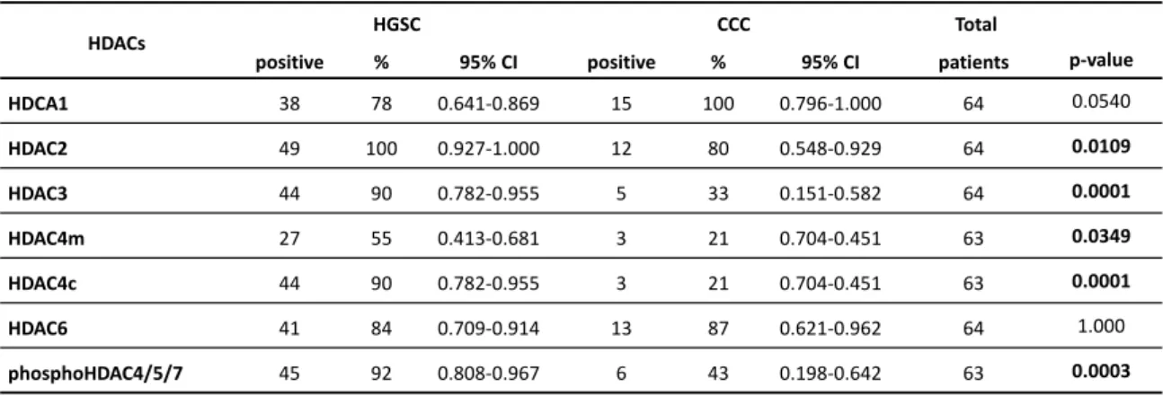
We investigated the expression profile of HDAC, class I (HDAC1, 2, 3) and class II (HDAC4, 6 and phosphoHDAC4/5/7) in ovarian cancer. HDACs protein expression was analyzed by immunohistochemistry on tissue microarrays (TMA) from 64 patients with ovarian cancer (49 cases of HGSC and 15 cases of CCC). Our results showed that HGSC expressed HDAC1, 2, 3, 4, 6 and phosphoHDAC4/5/7 whereas CCC expressed HDAC1, 2 and 6. HDAC2, 3, 4 and pHDAC4/5/7 were associated with HGSC type. Kaplan-Meier curves showed that HDACs expression was not associated with survival. We also evaluated the association of HDACs expression profile with the cell cycle markers and we found that HDAC1 expression was statistically associated with p21 expression in HGSC.
vii line exposed to vorinostat, by the study of immunofluorescence of cleaved caspase 3 and the ratio of BAX/BCL-2 proteins, which increased after vorinostat exposure. Cell migration was decreased by vorinostat in both cancer cell lines. Our results also indicated that the association of different drugs were able to potentiate the effect of standard chemotherapy, mainly in ES2 cell line (CCC), supporting that EOC treatment can benefit from combined chemical and epigenetic therapy.
Considering that CCC is a unique clinical, histopathological and molecular entity within ovarian cancer and having HNF1β as a pivotal pro-survival gene we evaluate the
role of HNF1β in cell cycle arrest and apoptosis in CCC upon vorinostat exposure. Our results showed that vorinostat induced increased levels of HNF1β and at the same time cell cycle arrest and apoptosis in ES2 cells. This effect on HNF1β is associated with increased acetylation load of HNF1β. This study confirms that epigenetic modulation affects not only the conformation of chromatin and gene expression, but also it may alter the function and the turnover of proteins other than histones.
Also, epigenetic modulation of signaling pathways have been reported in HGSC and CCC, including the overexpression of Notch pathway elements and we investigated the modulation of the Notch pathway by vorinostat in ovarian cancer. Using immunofluorescence and quantitative polymerase chain reaction, our results revealed that vorinostat activated the Notch pathway in CCC and HGSC cell lines, through different Notch ligands. The activation of Notch pathway by vorinostat, in CCC and HGSC cell lines, culminated in the increased expression of the same downstream transcription factors, hairy enhancer of split (Hes1) 1 and 5, and Hes-related proteins 1
and 2. Therefore, vorinostat modulates the expression of several downstream targets of the Notch pathway and independent Notch receptors and ligands that are expressed in HGSC and CCC. This upregulation of the Notch pathway may explain why vorinostat therapy fails in ovarian carcinoma treatment, as shown in certain clinical trials.
viii and 3D cell cultures. Characterization studies confirmed that IPO-SOC43 cell line is of EOC origin and maintains morphological and molecular features of the primary tumor. The success of the cell line growth in a 3D systems, allows it to be used in more complex assays than those performed in 2D models. IPO-SOC43 is available for public research and wehope it can contribute to enrich the in vitro models addressing EOC heterogeneity, being useful to investigate EOC and to develop new therapeutic modalities.
In conclusion, the results in vitro proved that there is benefits of combined therapy, using drugs with action of epigenetic modulation and conventional therapy. This thesis also pave the path for future studies on the role of HDACs cancer cells pathophysiology, serving as markers for the design of personalized and specific therapeutic strategies.
x
RESUMO
O carcinoma do ovário é a neoplasia ginecológica mais letal. Vários fatores são identificados como responsáveis pela baixa eficácia no tratamento do carcinoma do ovário. Assim, é postulado que a elevada taxa de mortalidade se deve principalmente, a dificuldades no diagnóstico da doença numa fase inicial e à resistência à terapêutica convencional. Deste modo, a investigação de novas estratégias terapêuticas é essencial e urgente.
A expressão aumentada das desacetilases de histonas (HDAC) identificada em carcinomas do ovário pode indicar que os inibidores de HDACs (HDACIs) possam ser uma alternativa terapêutica. Os HDACIs são uma classe de agentes anti-neoplásicos, que bloqueiam a desacetilação de histonas e outras proteínas, causando paragem do ciclo celular, diferenciação e/ou apoptose das células neoplásicas. Vários HDACIs estão a ser testados em ensaios clínicos, quer como agentes únicos quer em terapias combinadas, apresentando alguns resultados positivos no combate a vários tumores sólidos e hematológicos. Os mecanismos moleculares do efeito anti-tumoral dos HDACIs não estão completamente esclarecidos, nem a avaliação da sensibilidade e/ou resistência das células a fármacos com influência na regulação epigenética.
O cancro do ovário é um conjunto vasto de neoplasias distintas sendo os carcinomas o grupo mais prevalente. Atualmente, não é possível, de modo fiável prever o curso clínico da doença nem a resposta individual à quimioterapia. No entanto, a identificação morfológica do tipo de carcinoma do ovário tem valor prognóstico independente em análise multivariada, pelo que a avaliação de potenciais biomarcadores que possam constituir alvos terapêuticos deverá ser efetuada de modo independente, pois previsivelmente cada tipo histológico deverá ter uma resposta específica ao tratamento.
xi Neste projeto foram estudados apenas dois tipos histológicos de carcinoma do ovário: seroso de alto grau (HGSC), que é o mais frequente e o carcinoma de células claras (CCC) que apesar de pouco frequente é atualmente resistente à terapia convencional.
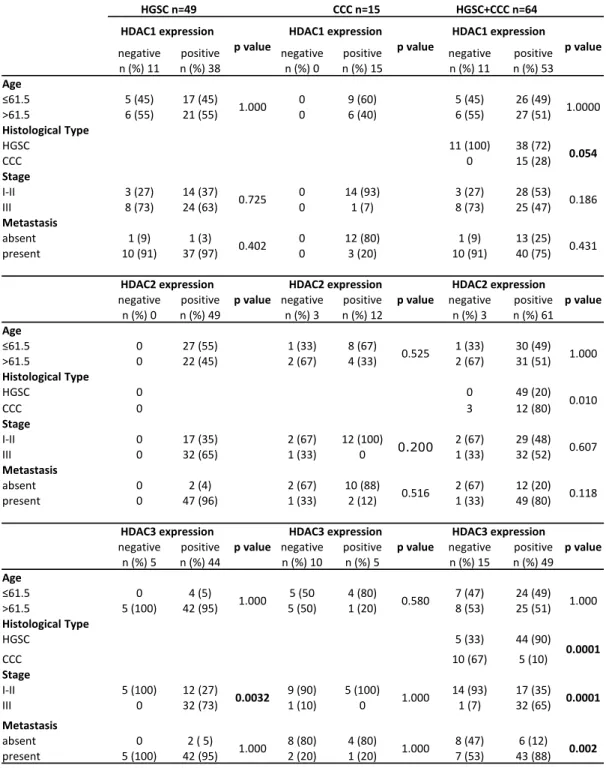
Assim, um dos primeiros objetivos desta tese foi caracterizar o perfil de expressão das HDACs em carcinomas do ovário e validar a relevância de testar HDACIs como agentes de regulação epigenética, no tratamento do carcinoma do ovário. Os resultados em doentes com cancro do ovário mostram que o perfil de expressão das HDACs se associa ao tipo histológico. No HGSC verifica-se uma associação das HDACs 2, 3, 4 e das HDAC4/5/7 fosforiladas, enquanto que no carcinoma de células claras (CCC), as HDAC1, 2 e 6 apresentam níveis elevados de expressão e verifica-se uma diminuição da expressão da HDAC3, HDAC4 e das HDAC4/5/7 fosforiladas. A expressão das HDACs 2, 3, 4 e das HDAC4/5/7 fosforiladas estão associadas ao tipo histológico sendo mais expressas no HGSC. A expressão da HDAC3 está ainda associada a parâmetros clínicos, nomeadamente ao estádio mais avançado e à metastização; bem como a HDAC4c e a HDAC4/5/7 fosforiladas.
Considerando que as HDACs estão sobre-expressas nestes tipos histológicos de carcinomas do ovário, seguidamente, pretendemos avaliar o efeito epigenético dos HDACIs em modelos in vitro de carcinoma de ovário e compreender as alterações decorrentes da exposição aos HDACIs, bem como perceber o efeito destes na dinâmica molecular e viabilidade celular. Para tal, usamos duas linhas celulares, de dois tipos histológicos já avaliados, uma linha de HGSC - OVCAR3 e uma linha celular de CCC - ES2.
xii cumulativo na morte celular, principalmente na linha celular ES2 (CCC). A migração celular também é afetada pelo vorinostat em ambas as linhas celulares de carcinoma de ovário.
Os resultados mostram também que o vorinostat induz aumento do HNF1β e ao mesmo tempo paragem do ciclo celular e apoptose nas células ES2. O aumento da
expressão do HNF1β é acompanhado pelo aumento dos seus níveis da acetilação.
Este estudo confirma que a modulação epigenética afeta a conformação da cromatina e da expressão genética, mas também pode alterar a função e o turnover de outras proteínas além das histonas. Pela primeira vez é observado que a proteína
HNF1β pode ser acetilada, contudo serão necessários mais estudos para validar o
papel da acetilação na função e degradação da proteína HNF1β.
Nesta tese foi avaliado também a modulação de outras vias de sinalização importantes no carcinoma do ovário, como por exemplo a via Notch através da exposição a HDACIs, nomeadamente vorinostat e ácido butírico. E os nossos resultados mostram que o vorinostat ativa a via Notch nas linhas celulares de CCC e HGSC, através de diferentes ligandos da via Notch. No CCC, a ativação da via Notch parece ocorrer através dos ligandos Delta (Dll) 1, 2 e 3, enquanto nos HGSC, os ligandos Dll1, Jagged 1 e 2 são os mais expressos. A ativação da via Notch pelo vorinostat, em ambas linhas celulares, culminou na expressão aumentada dos mesmos genes alvo da via Notch, Hes1 e 5 e Hey 1 e 2.
Assim, o vorinostat modula a expressão de vários fatores de transcrição da via Notch, independentemente do painel de receptores e dos ligandos expressos nas linhas de HGSC e CCC. Esta redundância da via Notch pode explicar porque a terapêutica isolada com vorinostat falha no tratamento do carcinoma do ovário, como observado em vários ensaios clínicos.
xiii Foram efetuados estudos de caracterização desta linha que demonstram que a linha celular IPO-SOC43 é de origem epitelial e que mantém características morfológicas e moleculares do tumor primário. As células mantêm-se em cultura em monocamada (2D) e em agregados em 3D, com taxas de proliferação e viabilidade dentro do esperado para uma linha neoplásica maligna. O sucesso da viabilidade em culturas 3D permite o seu uso em ensaios mais complexos, nomeadamente, na análise da contribuição dos vários componentes do microambiente tumoral na progressão da doença e resposta à terapêutica.
A linha celular IPO-SOC43 está disponível para ser utilizada experimentalmente e esperamos que possa contribuir para enriquecer os modelos in vitro que abordam a heterogeneidade dos EOC e desenvolver novas modalidades terapêuticas.
Em suma, os resultados contribuem para a implementação racional de novas e diferentes abordagens terapêuticas. Os resultados mostram que in vitro há benefício de terapêutica combinada, utilizando fármacos com ação de modelação epigenética e terapia convencional. Esta tese também abre caminho para estudos futuros mais detalhados sobre a função das HDACs na dinâmica celular e não só na regulação epigenética, bem como também na análise de mecanismos subjacentes à ação de proteínas codificadas por genes relevantes em cada tipo histológico de carcinoma do ovário.
Espera-se assim, que a compreensão dos mecanismos subjacentes à ação dos fármacos envolvidos na regulação epigenética possa fornecer orientações para estratégias terapêuticas mais específicas e personalizadas.
xiv
FIGURE INDEX
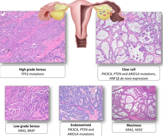
Figure 1. 1 - Representative examples of the five main types of ovarian carcinoma stained with
hematoxylin and eosin (H&E) ... 5
Figure 1. 2 - Ovarian cancer signaling pathways scheme ... 21
Figure 1. 3 – Cytosine metylation ... 31
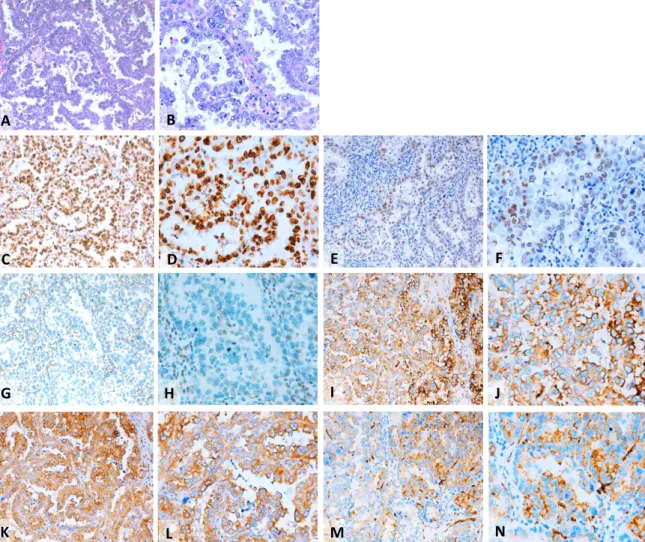
Figure 2. 1- Representative pattern of HDACs protein expression in HGSC in tissue microarray (TMA) sections. ... 54
Figure 2. 2 - Representative pattern of HDACs protein expression in CCC in tissue microarray (TMA) sections ... 55
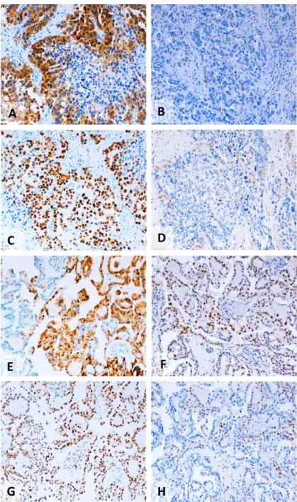
Figure 2. 3 - Representative examples of immunostaining for p16, p21, p53 and cyclin D1 in different histological types of ovarian cancer ... 61
Figure 3. 1 - Mechanism of action of HDAC inhibitors (HDACI) ... 74
Figure 3. 2 - Cell death in ES2 cell line exposed to HDACIs and 5-aza-dC ... 80
Figure 3. 3 - Cell death in OVCAR3 cell line exposed to HDACIs and 5-aza-dC ... 81
Figure 3. 4 - mRNA expression of BCL-2 and BAD in ES2 cell line ... 82
Figure 3. 5 - Expression of BCl-2 and BAX by Western blot ... 83
Figure 3. 6 - Expression of cleaved caspase 3 by immunofluorescence in EOC cell lines... 84
Figure 3. 7 - mRNA expression of BECLIN-1 and ATG-7 in ES2 cell line ... 85
Figure 3. 8 - Cell death in ES2 cell line exposed to vorinostat, 5-aza-dC, carboplatin and/or paclitaxel 86 Figure 3. 9 - Cell death in HGSC-OVCAR3 cell line exposed to vorinostat, 5-aza-dC, carboplatin ... 87
Figure 3. 10 - Cell proliferation curve for ES2 and OVCAR3 cells exposed to HDACIs and 5-aza-dC ... 88
Figure 3. 11 - Wound healing assay in ES2 cells and OVCAR3 cells exposed to vorinostat. ... 89
Figure 4. 1 - Vorinostat affects cell proliferation of clear cell carcinoma (ES2) but not of serous carcinoma (OVCAR3) ... 104
Figure 4. 2 - Long term exposure of Vorinostat induces p21 expression and cell cycle arrest in clear cell carcinoma (ES2) ... 105
xv
Figure 4. 4 - Vorinostat increases cell death in clear cell carcinoma (ES2) ... 107
Figure 4. 5 - Vorinostat exposure increase the levels of HNF1β in clear cell carcinoma (ES2), both in short and long term exposure ... 108
Figure 5.1 – Vorinostat increases the expression of mRNA of Notch receptors, Delta/Jagged ligands and Hey/Hes downstream target genes ... 118
Figure 5. 2 – Vorinostat increases the mRNA expression of Notch receptors, ligands and downstream targets in ovarian serous carcinoma OVCAR3 cell line ... 120
Figure 5. 3 - Schematic representation of Notch pathway in ovarian cancer ... 123
Figure 6. 1 – Morphological characterization of cells from ascitic fluid ... 136
Figure 6. 2 - Characterization of the primary ovarian carcinoma of the patient from whose ascitic fluid IPO-SOC43 was established ... 137
Figure 6. 3 - Detection of cytokeratins and vimentin in IPO-SOC43 cell line by flow cytometry ... 138
Figure 6. 4 - Ovarian biomarkers in IPO-SOC43 cell line ... 139
Figure 6. 5 - Expression of biological markers in 2D cell culturing models in IPO-SOC43 cell line ... 140
Figure 6. 6 - Characterization of IPO-SOC43 cell line in 2D model ... 141
Figure 6. 7 - Proliferation curve of IPO-SOC43 cell line ... 142
Figure 6. 8 - Wound healing assay to determine the migration rate of IPO-SOC43. ... 143
Figure 6. 9 - cCGH profiles of cells from ascitic fluid and IPO-SOC43 cell line ... 144
Figure 6. 10 - Sanger sequencing histogram of TP53 exon 5 ... 147
Figure 6. 11 - Aggregation of IPO-SOC43 with and without ROCK Inhibitor in 3D culture system ... 148
Figure 6. 12 - 3D Culture progression with measurement of aggregates area and growth curve of IPO-SOC43 cells with ROCK Inhibitor ... 149
Figure 6. 13 - Characterization of IPO-SOC43 cell line in 3D model... 150
xvi
TABLE INDEX
Table 1. 1 - TNM and FIGO classifications for Ovarian, Fallopian Tube and Peritoneal Cancer Staging
System ... 15
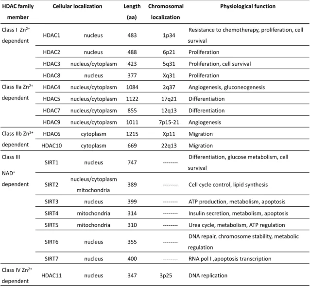
Table 1. 2 - Classes of histone deacetylases; localization in the cell, length, chromosomal localization and function ... 35
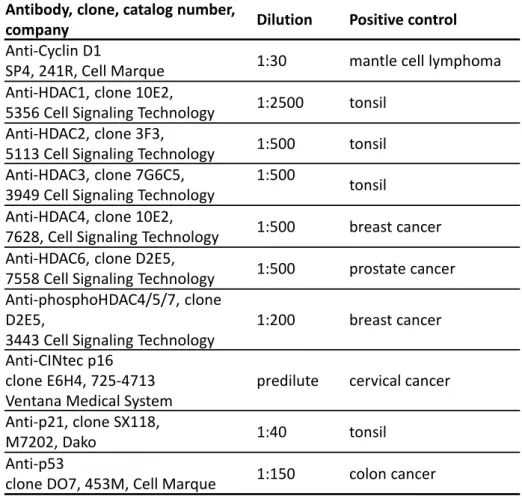
Table 2. 1 - Antibodies used for imunohistochemical study ... 50
Table 2. 2 - Clinical and pathological characteristics of the EOC patients ... 52
Table 2. 3 - Expression of HDACs in EOC commercial lines ... 53
Table 2. 4 - Expression of HDACs in EOC samples and its association with the histological type ... 54
Table 2. 5 - Association of HDACs expression and clinicopathological parameters in EOC ... 57
Table 2. 6 - Multivariate analysis of HDAC3, HDAC4c and phosphoHDAC4/5/7 expression for histological type and metastasis ... 59
Table 2. 7 - Expression of cell cycle markers in EOC commercial cell lines ... 59
Table 2. 8 - Expression of cell cycle markers in EOC samples ... 60
Table 2. 9 - Expression of HDACs expression and cell cycle markers in EOC ... 62
Table 3. 1 - Chemicals used in ovarian cancer cell lines ... 76
Table 6. 1 - Antigens evaluated and antibodies used to characterize IPO-SOC43 cell line ... 130
Table 6. 2 - Primers for PCR of the TP53 gene (Exons 4-9) ... 133
xviii
ACKNOWLEDGMENTS
This PhD thesis would not be possible without a large number of contributions, and for this reason I would like to express my gratitude to my supervisors, Ana Félix, for all the precious mentoring, guidance and motivation and to Jacinta Serpa for guiding me in the field of molecular biology, teaching me professional attitude towards designing, conducting and evaluating laboratory experiments. Both of them generously shared their time and expertise with me, were always open to answer my questions and discuss my ideas and were critical and supportive at the same time. I am deeply indebted to them for helping me.
I am also thankful for all former and present members of the UIPM with a very special thanks to Leonor Remédio, Francisco Caiado, Germana Domingues, Lídia Silva, Sofia Gouveia and Filipa Coelho for all the good work environment, great friendship and helpful discussions.
I would like to thank to the director José Cabeçadas, doctors and all colleagues of the Pathology Department of IPOLFG, especially to Teresa Pereira, Sónia Araújo, Filipa Areia, Cátia Teixeira, Arsénia Manhita and Filipa Antunes for creating a motivating and pleasant working atmosphere. From IPOLFG, I also thank Carmo Martins for her precious help with CGH and to António Guimarães for the valuable clinical data.
I would like to thank to Vitor Espírito Santo and Catarina Brito, from IBET for sharing your knowledge of 3D cultures.
xx
LIST OF PUBLICATIONS
The author declares that has designed and performed research, collected and analyzed data, and wrote the text of the published paper, and the manuscripts in preparation, which are part of this dissertation.
Published original paper:
Silva, F., Félix, A. & Serpa, J. Functional redundancy of the Notch pathway in ovarian cancer cell lines. Oncol. Lett. 12, 2686–2691 (2016).
Published in proceedings (Poster):
Silva, F., Serpa, J., Domingues, G., Silva, G., Almeida, A., Félix, A. Cell death induced by HDACS inhibitors in ovarian cancer cell lines (serous and clear cells carcinomas) – role of NOTCH, TP53 and FN1. BMC Proceedings, 4(Suppl 2):P36 (2010).
Silva, F., Serpa, J., Fernandes, S., Dias, S., Félix, A. HDACS inhibitors effects in Ovarian Clear Cell Carcinoma. SINAL, 6th National Meeting, April, UMinho (2012).
Silva, F., Fernandes, S., Félix, A., Serpa, J., Vorinostat Increases ARID1A splicing variant expression which may regulate HNF1β and cell survival, in Ovarian Clear Cell Carcinoma (CCC). International Journal of Gynecological Cancer, 23 (8) (2013)
xxii
ABBREVIATIONS
ARID1A adenine thymine-rich interactive domain 1A gene AKT akt serine/Threonine kinase
ATCC American Type Culture Collection BAX BCL-2 associated X protein BCL-2 B-cell lymphoma 2
BAD BCL-2 associated agonist of cell death
BRAF v-Raf murine sarcoma viral oncogene homolog B1 BRCA1 breast cancer 1 gene
BRCA2 breast cancer 2 gene CA125 cancer antigen 125 CCNE1 cyclin E1
CDKN1A cyclin dependent kinase inhibitor 1A CTNNB1 catenin beta 1
EOC epithelial ovarian cancer
ER estrogen receptor
FACS fluorescence-activated cell sorting FBS fetal bovine serum
FIGO Féderation Internationale de Gynécologie et d’Obstétrique FITC fluorescein isothiocyanate
FOXM1 forkhead box M1 GSH glutathione peroxidase HAT histone acetyltransferases HDAC histone deacetylases
HDACI histone deacetylases inhibitors HES human hairy and enhancer of split
HEY hairy/enhancer of split related with YRPW motif HER-2 human epidermal growth factor receptor 2
HPRT hypoxanthine-guanine phosphoribosyl transferase
HNF1β hepatocyte nuclear factor 1β
xxiii
LOH heterozygosity
MAPK mitogen-activated protein kinase MLH1 mutL homolog 1
MSH2 mutS homolog 2 MSH6 mutS homolog 6 IHC immunohistochemistry
KRAS kirsten rat sarcoma viral oncogene homolog MAPK mitogen-activated protein kinase
mTOR mammalian target of rapamycin NICD notch intracellular domain
NF1 neurofibromin 1
PAX paired box 2
p16INK4A cyclin-dependent kinase inhibitor 2A
p21 cyclin-dependent kinase inhibitor 1A p53 tumor protein/suppressor p53 PBS phosphate-buffered saline PCR polymerase chain reaction
PFA paraformaldehyde
PIK3CA phosphatidylinositol-4,5-bisphosphate 3-kinase catalytic subunit alpha
PMS2 PMS1 homolog 2, mismatch repair endonuclease PPP2R1A protein phosphatase 2, regulatory subunit A PTEN phosphatase and tensin homolog
ROMA risk of ovarian malignancy algorithm ROS reactive oxygen species
RB1 retinoblastoma transcriptional corepressor 1 RSF1 remodeling and spacing factor 1
SAHA suberoylanilide hydroxamic acid TCGA The Cancer Genome Atlas TMA tissue microarray
TP53 gene encoding tumor protein p53 WHO World Health Organization
2
INTRODUCTION
Cancer is a group of distinct diseases in which cells proliferate out of control and invade other tissues. Cancer cells no longer respond to genes that regulate cell cycle, function, differentiation and death. Tumor cell invasion of surrounding tissues and distant organs is the main cause of morbidity and mortality for most cancer patients1.
The biological process by which normal cells are transformed into malignant cancer cells has been the subject of a large research in the biomedical sciences for many decades1. Cancer is the leading cause of mortality worldwide, being responsible for 8.2
million cancer related deaths in 20122.
1.1 Epithelial ovarian cancer (EOC)
1.1.1 Epidemiology and Risk factors
Epithelial ovarian cancer (EOC) is the most lethal gynecology malignancy3–5 and
the seventh most common in women worldwide. Nearly 240,000 women6 were
estimated to have been diagnosed with ovarian cancer in 2012 with incidence rates varying across the world. Although ovarian cancer accounts for only 4% of all cancers in women7, it has one of the highest death-to-incidence ratios, due to difficulties in
detection, diagnosis and therapy of the disease8. Like most cancers, the risk of ovarian
cancer rises with age: the median age of patients at diagnosis is 60 years, and the average lifetime risk for women in developed countries is around one in 709,10.
Most ovarian carcinomas occur after menopause when the ovaries have little or no physiological role. As a consequence of the absence of abnormal ovarian function associated with major symptoms and the anatomical location (deep in the pelvis) about 70% of the patients are diagnosed with advanced-stage (extra-ovarian) disease and 5-years survival rates are between 20-40%11,12. Although most advanced stage
3
In Portugal, the incidence of ovarian cancer is 490/100.000 (2008) and 5-year survival rate is 40%. Every year, in Portugal, the disease accounts for more than 371 deaths12.
There are several factors that can affect the risk of developing ovarian cancer and therefore ensuring a primary prevention, screening and early detection. One important risk factor is the familial history of ovarian or breast cancer, present in only 10–15% of patients and mainly associated with germline mutations in BRCA1 and
BRCA2 (breast cancer 1, 2 gene) genes14, and also with Lynch syndrome (a group of
hereditary nonpolyposis colorectal cancer), having germline mutations in DNA missmatch repair genes MLH1 (MutL homolog1), MSH2 (MutS homolog 2), MSH6
(MutS homolog 6) and PMS2 (PMS1 homolog 2)15,16. Women with a BRCA1 mutation,
the risk of epithelial ovarian cancer is 39–46%17, and with a BRCA2 mutation is
between 12–20%9,14. Whereas, for women with Lynch syndrome a 3-14% risk of
ovarian cancer is estimated. The management of women with genetic predisposition for ovarian cancer includes surveillance and risk-reducing surgery18.
Moreover, nulliparity, early menarche, late menopause, and increasing age are associated with increased risk, whereas oral contraceptive use, pregnancy, lactation, and tubal ligation are associated with reduced risk19. Several studies have
demonstrated that the use of oral contraceptives for more than five years decreases the risk of ovarian cancer by 20%19. The prophylactic salpingo-oophorectomy may
reduce the risk of ovarian cancer by 80 to 95% and can be recommended for women with high risk of developing the disease20. Survival of ovarian cancer patients has
4
1.1.2 Histological Types
Ovarian tumors mainly arise from three different cell types; epithelial, germ, and sex cord stromal cells21 and several studies have contributed to confirm the
heterogeneous nature of EOC based on their clinicopathological and molecular features and possible putative precursor lesions22–24. EOC represent 90% of ovarian
malignant tumors21 and is a very heterogeneous group of neoplasms that exhibit a
wide range of tumor morphology, clinical manifestations and underlying genetic alterations25.
Current investigations support the general view that serous tumors develop, at least in 50%, from the fallopian tube; endometrioid and clear cell tumors are postulated to arise from endometrial tissue in the ovary (most probably by passing through the fallopian tube) that results in endometriosis and tumors23,24.
According to the latest classification by World Health Organization2 in 2014 and
based on histopathology, immunohistochemistry, and molecular genetic analysis, ovarian epithelial tumors are grouped into seven different types. Low grade tumors include low grade serous carcinoma (LGSC<5%), endometrioid carcinomas (EC; 10%), mucinous carcinomas (MC; 3%), and a subset of clear cell carcinomas (CCC; 10%)26–29.
These tumors develop slowly and in general are confined to the ovary30, genetically
stable, and each histological type has a distinct genetic profile27,29,30. In contrast, high
grade tumors progress rapidly and include high grade serous carcinoma (HGSC), seromucinous tumors (probably), undifferentiated carcinoma and some clear cell carcinomas28 (Figure 1.1). HGSC is the most prevalent type and it is characterized by a
5
Figure 1. 1 - Representative examples of the five main types of ovarian carcinoma stained with hematoxylin and eosin (H&E)
High grade serous carcinoma, clear cell carcinoma, low grade serous carcinoma, endometrioid carcinoma and mucinous carcinoma. Scheme adapted from Jones & Drapkin32 and Karst &
Drapkin33.
1.1.2.1 Serous carcinoma
Serous histology is the most common, representing 70% of EOC34. It is accepted
that LGSC and HGSC are fundamentally different tumor types, and consequently, different diseases2.
1.1.2.1.1 Low grade serous carcinomas (LGSC)
LGSC are rare and account for <5% of all cases of ovarian carcinomas35. They
frequently have a noninvasive serous borderline component (with or without micropapillary pattern) and probably they represent tumor progression of serous borderline tumors beyond microinvasion. Although the presence of small foci of LGSC
Clear cell
PIK3CA, PTEN andARID1A mutations, HNF1βde novo expression High grade Serous
TP53 mutations
Mucinous
KRAS, HER2
Endometrioid
PIK3CA, PTEN and
ARID1A mutations
Low grade Serous
6 in an ovarian borderline tumor is associated with a good prognosis, patients with advanced stage disease fare less favorably. Typically, present as large masses that are confined to one ovary (stage Ia), are indolent, and have good prognosis2,28. They show
a relative genetic stability and usually display a variety of somatic alterations that include mutations in v-Raf murine sarcoma viral oncogene homolog B1 (BRAF) or kirsten rat sarcoma viral oncogene homolog (KRAS) genes, in 33-50% and 27-36%36,
respectively.
Microscopically, LGSC has small papillae of tumor cells exhibiting uniform nuclei within hyalinized stroma, which often contains psammoma bodies. Distinction of LGSC from HGSC relies on cell atypia and mitotic activity. Ki-67 immunoreaction differs between the two tumor types, being the median Ki-67 labeling index lower (2.5%) in LGSC than in HGSC (22.4%)37. The most useful immunomarker is p53 as this tumor type
shows a wild type positivity in the nuclei in contrast with HGSC2.
1.1.2.1.2 High grade serous carcinomas (HGSC)
HGSC are aggressive tumors that are usually diagnosed at an advanced stage, and although they frequently respond to surgery and platinum-based chemotherapy, they eventually recur. Microscopically, HGSC show neoplastic cells displaying papillary and solid growth with slit-like glandular lumens. The tumor cells are usually of intermediate size, with spotted bizarre mononuclear giant cells exhibiting prominent nucleoli26. In contrast to LGSC, these tumors show more than threefold variation in
nuclear size26,38.
In contrast to LGSCs, HGSCs are not associated with serous borderline tumors and typically exhibit TP53 mutations and BRCA alterations39, and most of these tumors
show immunoexpression for p53, BRCA1, WT1 (Wilm’s tumor 1 factor), p16 and have
estrogen receptors (ER) in approximately two-thirds of HGSC cases40.
7
overexpression/amplification of HER2/neu, AKT (AKT serine/ threonine kinase) and loss of heterozygosity (LOH) on chromosomes 7q and 9q are common4. Profiling of mRNA
and miRNA (micro RNA) of 489 Type II tumors, in particular HGSC, performed by The Cancer Genome Atlas (TCGA)31 revealed that almost all samples comprised TP53
mutations (in almost 98% of the cases ) and significant recurring somatic mutations in
NF1 (neurofibromin 1), BRCA1, BRCA2, RB1 (RB transcriptional corepressor 1),
CDK1227–31 (cyclin dependent kinase 12), CCNE1 (cyclin E1), AKT2 (AKT serine/
threonine kinase 2), RSF1 (remodeling and spacing factor 1) and PIK3CA (phosphatidylinositol-4,5-bisphosphate 3-kinase catalytic subunit alpha)31,41. KRAS mutations are rare and BRAF mutations have not been described in HGSC36. In
addition, Notch3 and FoxM1 (forkhead box M1) were found to be involved in HGSC pathophysiology. More recently, the results of the TCGA study were highlighted in another genome-wide report42.
As a result of the chromosomal instability, HGSC are typically aneuploid, with very complex karyotypes. The aCGH (array Comparative Genomic Hybridization) findings of the TCGA study31 reveal gains/amplifications of 1q, 3q 6p 7q, 8q, 12p, 20p
and 20q; and losses of 4p, 4q, 5q, 6q, 8p, 9p, 9q, 11p, 11q ,13q, 14q, 15q, 16p, 16q, 17p, 17q, 18p, 18q, 19p, 19q and 22q some of them already reported in other studies43. Other genetic alterations observed, suggest a co-operative effect, for
example, associations have been found between CCNE1 (cyclin E1) and 12p amplification41, and between MYC (V-Myc Avian Myelocytomatosis viral oncogene
homolog) and 20q amplification by fluorescence in situ hybridization (FISH). Also, positive associations were identified between gains on 19 and 20q, gain of 20q and loss of X, and between the loss of several regions, particularly 17q.
1.1.2.2 Endometrioid carcinomas (EC)
After HGSC, EC and clear cell carcinomas (CCC) are the most frequent types of EOC accounting for approximately 10-15% of all ovarian carcinomas23. The molecular
8 CCC and a direct transition from ovarian atypical endometriosis to EC or CCC, in 15%-32% of cases44.
EC occur most frequently in women of perimenopausal age, and most of them are found at an early stage45. These ovarian tumors are bilateral in 28% of cases and
are associated in 15%–20% of cases with a carcinoma of the endometrium45,46.In the
former presentation, both tumors are endometrioid in type in the majority of cases2.
Up to 42% of cases, evidences of bilateral ovarian or pelvic endometriosis is present. Squamous differentiation inside the tumor occurs in 50% of cases45. Somatic mutations
in CTNNB1 (β-catenin) and PTEN (phosphatase and tensin homolog) genes are the most common genetic alterations identified in ECs, being CTNNB1 mutations associated with a favorable disease outcome47,48. Also PTEN is mutated in 20% of cases
and its inactivation results in the lack of inhibition of PI3K-AKT signaling pathway that inhibits apoptosis and activates proliferation. The finding of LOH at 10q23 and somatic PTEN mutations in endometriotic cysts, adjacent to ECs with similar genetic alterations, provides additional evidence of the precursor role of endometriosis in ovarian carcinogenesis47. An alternative mechanism for the activation of PI3K signaling
in EC is activating mutations of PIK3CA, which encodes the p110 catalytic subunit of PI3K. EC is the most frequently encountered type in patients with hereditary Lynch syndrome49. The reported frequency of microsatellite instability (MI) in ovarian ECs
ranges from 12.5% to 19%50,51.
Recent discovery of ARID1A mutations in ovarian EC as well as in adjacent endometriosis, is considered a particular feature of these tumors52, although it appears
to be a late event in endometrial carcinoma53. EC have expression of vimentin,
cytokeratins (CK7, 97%; CK20, 13%), epithelial membrane antigens (EMA), estrogen and progesterone receptors by immunohistochemical studies54.
1.1.2.3 Clear cell carcinomas (CCC)
CCC is a rare type known to be intrinsically chemoresistant and associate with very poor prognosis in advanced stages55. Tumors are commonly unilateral. As EC, they
9
predominantly in the architectural findings: solid, tubule-cystic and papillary areas mixed in varied proportions. Cytological features are characterized most commonly for polygonal to flattened cells with pale or clear cytoplasm, which is attributed to the accumulation of glycogen and lipids. Densely hyaline basement membrane material expanding the cores of the papillae or deposit between cells is a common feature. Mitoses are less frequent than in other types of EOC55.
Based on genome-wide mutational analysis, the most common mutations identified in CCC include ARID1A mutation in nearly half of the CCC (46%-57%) and loss of BAF250a expression (protein encoded by ARID1A)44. ARID1A gene has been
identified as a novel tumor suppressor44,57 and acts as a chromatin remodeling
modifier, which stimulates cell signaling that can lead to cell cycle arrest and cell death in the event of DNA damage, consequently the dysfunction of ARID1A may result in susceptibility to CCC carcinogenesis through a defect in the repair or replication of damaged DNA58. A recent study indicated that the loss of ARID1A protein expression is
related to short disease-free survival and chemoresistance in CCC59.
Activating mutations of PIK3CA and deletion of PTEN, are found respectively in about 50% and 20% of tumors47, supporting the role of an aberrant PI3K/PTEN
pathway in CCC60. In addition, the overexpression of Notch pathway elements has also
been reported in these tumors61,62. In contrast to various other human tumor types, TP53 and Rb (retinoblastoma protein)mutations are rarely detected in CCC, but these cell cycle regulatory proteins may be involved in the carcinogenesis of CCC through inactivation by ARID1A58 for example. In CCC, no BRCA alterations, chromosomal instability, nor complex karyotypes as seen in HGSC63 have been reported, however
epigenetic modulation of signaling pathways have been described64–66.
Molecular alterations in CCC are mainly unknown and this histological type represent a distinct challenge with a unique and different histology, with de novo expression of HNF1β (hepatocyte nuclear factor1β transcription factor).
HNF1β is involved in the glycogen synthesis67 and regulates several specific
10 iron deposition, and antiapoptosis), annexin 4 (calcium ion binding, epithelial cell differentiation), and UDP Glucuronosyltransferase 1 Family, Polypeptide A Complex (UGT1A1) (detoxifycation)68. It is postulated that HNF1β plays an important role in the
pathogenesis and behavior of CCC69. Promoter hypomethylation of HNF1β, and active
transcription, was previously reported in CCC70,71. Moreover, recently, we showed the
role of HNF1 in the intrinsic chemoresistance exhibited by CCC72.
CCC tumor cells are usually positive for HNF1β (>90%) and are negative for ER
and WT1 in >95% of cases37,40. In addition to its presence in CCC, HNF1β is also
expressed in the majority of tumors with cytoplasmic clearing, including renal CCC, endometrial CCC69 and ovarian and endometrial carcinomas of mixed histology.
1.1.2.4 Mucinous carcinomas (MC)
Mucinous tumors account, approximately, for 3% of EOC. The cells of MC resemble cells of the gastric pylorus, or intestine45,46,73, showing gastrointestinal
differentiation in the majority of these tumors. Primary MC of the ovary are large, unilateral and usually confined to the ovary, without ovarian surface involvement or pseudomyxoma peritonei, while mucinous ovarian metastases are typically small (<10 cm) and bilateral. Malignant mucinous ovarian tumors are often heterogeneous, showing the coexistence of benign-appearing, borderline, noninvasive carcinoma, and invasive components. This morphology suggests a step-wise progression from benign to borderline and from borderline to carcinoma74. The category of mucinous
borderline tumor with intraepithelial carcinoma is used for those tumors that lack obvious stromal invasion but show unequivocally malignant areas75. Mucinous
borderline tumors with intraepithelial carcinoma have a very low risk of recurrence (<5%)76.
The gene expression profile of MC differs from the HGSC, EC and CCC77. KRAS mutations are frequent, (up to 75% of primary MC), and seems to be an
early tumorigenic event78. Using KRAS as a molecular marker, laser capture
microdissection studies have shown the identical KRAS mutation in MC and adjacent mucinous cystadenomas and borderline tumors74 supports the morphological
11
1.1.2.5 Other types of ovarian carcinomas
Seromucinous carcinomas (SMC), Brenner carcinomas (BC) and Undifferenciated carcinomas (UC)
Seromucinous carcinomas (SMC) are composed predominantly of serous and endocervical-type mucinous epithelium. These tumors are quite uncommon and data on the epidemiology is still insufficient2.
Brenner carcinomas (BC) are extremely rare but Brenner benign tumor accounts for 5% of benign ovarian epithelial tumors2. BC histogenesis is not well characterized
nor the genetic and molecular events.
Another uncommon histological type is undifferentiated carcinomas (UC), usually appear as solid masses with extensive necrosis, and a deficient mismatch repair proteins have been found in almost half of the tumors2.
In summary, each histological type of EOC is associated with a distinct profile of clinical, morphological, genetic and molecular alterations, which for the most common low-grade tumors are shared with their respective precursor lesions (borderline tumors and endometriosis) supporting their stepwise progression. In contrast, the high grade tumors, aside from a very high frequency of TP53 mutations and molecular alterations of BRCA1/2, are characterized by marked genetic instability.
1.1.3 Clinical presentation
Ovarian cancer is often described as a ‘silent killer’79 because the symptoms are
non-specific and common to other diseases. Symptoms such as abdominal discomfort, bloating, gas, nausea, and urinary urgency, frequently lead to misdiagnosis80,81, and
contribute for the high percentage of cases that are diagnosed at anadvanced stage. In addition, there is no routine and reliable screening for ovarian cancer82, hence the
12 Approximately 70% of women with ovarian cancer are diagnosed at stage III or IV of the disease82. Ovarian cancer usually spreads to the opposite ovary/fallopian tube,
and the uterus, and then intraperitoneally. Distant metastases are rare, but may occur in the liver, lungs, pleura, adrenal glands, and spleen76. The histological grade of
tumors can affect treatment and prognosis. Low grade tumors have a more favorable prognosis than high grade tumors.
Cancer spread can greatly cause the accumulation of fluid in the abdomen, called ascites, which is a common complication of ovarian cancer that can cause swelling, fatigue and shortness of breath82.
CA125, the serum tumor marker carbohydrate antigen 125 also known as mucin 16 (MUC16) is a secreted and membrane-associated glycoprotein expressed by coelomic and müllerian-derived epithelia, including the epithelium of the fallopian tube, endometrium and endocervix83.
CA125 levels are low (< 35 U/mL) in the serum of healthy individuals, and CA125 elevation is commonly associated with HGSC, the most common histological type of ovarian carcinoma83. CA125 can be used for monitoring disease recurrence84. Although
it is elevated in only 50-60% of ovarian cancer patients it can also be elevated in other non-cancerous gynecological complications such as benign ovarian neoplasms, endometriosis, pelvis inflammatory disease, liver disease or other malignant conditions, including cancers of the pancreas, breast, lung and colon85. Nevertheless,
CA125 cannot be used as a unique parameter in the prediction of malignancy. Usually, a combination of a patient's medical history, clinical examination results, imaging data and tumor marker profile is used to clinically differentiate malignant ovarian masses from their benign counterparts and perform a histological exam for diagnosis confirmation. However, CA125 assay detects circulating MUC16, is one of the most widely used cancer biomarkers for the follow-up of ovarian carcinoma.
Recently Ricardo et al.86 demonstrated that detection of aberrant
13
serous tumors from benign lesions with a specificity of 100% and a sensitivity of 70-80% on tissues. In fact, serous ovarian carcinomas selectively express Tn and STn glycoforms of MUC16 and MUC1, and these glycomucin profiles are likely to serve as more cancer-specific biomarkers than the mucins themselves. This study provides strong support for the use of PLAs to detect aberrant glycoforms of mucins and thereby improve cancer specificity of biomarker assays86.
The human epididymis secretory protein 4 (HE4) gene was found to be overexpressed in ovarian cancer87 and is used as a potential serum biomarker. HE4
belongs to the family of whey acidic four-disulfide core (WFDC) proteins with suspected trypsin inhibitor properties. HE4 was first determined in the epithelium of the distal epididymis88.
Biomarker HE4 has very low expression in the epithelia of respiratory and female reproductive tissues including ovary, but high secreted levels can be found in the serum of ovarian cancer patients89 and also in ovarian cancer tissue88. Recently, Moore
at al.90,91 published a series of papers that used a combination of CA125, HE4 and
menopausal status to predict the presence of a malignant ovarian tumor. Originally, nine potential biomarkers were evaluated, from which HE4 was the most effective in detecting ovarian cancer. When CA125 was combined with HE4, the prediction rate was higher, showing a sensitivity for detecting malignant disease of 76.4% at a specificity of 95%90,92 in ovarian carcinoma vs endometriotic cysts. Measured values of
HE4 and CA125 can be combined in an algorithm called ROMA (Risk of Ovarian Malignancy Algorithm), which additionally includes the menopausal status. Several published studies91,92 show that ROMA helps in the triage of pre and postmenopausal
women suspected for ovarian cancer. This high accuracy helps to stratify patients into low-and-high risk groups and therefore contributes to better diagnosis, treatment and outcome.
14 adnexal masses93, the specificity of ultrasonography is not adequate for its use as a
single screening modality94.
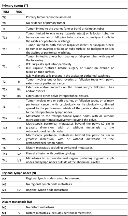
The majority of ovarian tumors are surgically staged to determine the presence of extraovarian disease, classified according to the terms of the staging scheme (I to IV) developed by the Fédération Internationale de Gynécologie et d’Obstétrique (FIGO) and the classification system prognosis helps to define treatment (Table 1.1). Staging is performed by examining histological sections of tissue samples and cytological assessment of fluid samples95.
1.1.4 Treatment of ovarian cancer
Ovarian cancer is highly curable when it is confined to the ovaries, with an expected 80–95% in 5-year survival73. Only about 20% of affected patients are found at
this stage, and the diagnosis is often made incidentally during the study of another medical condition. Current therapies for advanced ovarian carcinomas are limited and not curative and the search for new therapeutic agents in the treatment of the disease and recurrence, have been disappointing22.
The standard treatment for ovarian cancer patients includes cytoreductive surgery, during which adequate staging is performed by pathological examination of ovarian and other tissues to define the histological nature of the tumor and its stage. In patients diagnosed with early disease, surgery alone may be sufficient but in advanced disease, debulking surgery followed by chemotherapy is recommended96.
Chemotherapy after surgery is referred to as ‘first-line’ treatment and involves a combination of a platinum and taxane-based chemotherapy (usually six cycles of carboplatin and paclitaxel) delivered intravenously (IV) or intraperitonealy (IP)97,98.
Patients with advanced ovarian cancer who aren’t initially able to undergo surgery due to unressectable tumors can be treated with chemotherapy, being considered for surgery (neoadjuvant treatment)9 and then followed by platinum-based combination
15
Table 1. 1 - TNM and FIGO classifications for Ovarian, Fallopian Tube and Peritoneal Cancer Staging System
Adapted from the Fédération Internationale de Gynécologie et d’Obstétrique2, 2014.
Primary tumor (T)
TNM FIGO
TX Primary tumor cannot be assessed
T0 No evidence of primary tumor
T1 I Tumor limited to the ovaries (one or both) or fallopian tubes
T1a IA
Tumor limited to one ovary (capsule intact) or fallopian tube; no tumor on ovarian or fallopian tube surface; no malignant cells in the ascites or peritoneal washings.
T1b IB
Tumor limited to both ovaries (capsules intact) or fallopian tubes; no tumor on ovarian or fallopian tube surface; no malignant cells in the ascites or peritoneal washings.
T1c IC
Tumor limited to one or both ovaries or fallopian tubes, with any of the following:
IC1: Surgically spill intraoperatively.
IC2: Capsule ruptured before surgery or tumor on ovarian or fallopian tube surface.
IC3: Malignant cells present in the ascites or peritoneal washings.
T2 II Tumor involves one or both ovaries or fallopian tubes with pelvicextension or peritoneal cancer.
T2a IIA Extension and/or implants on the uterus and/or fallopian tubesand/or ovaries.
T2b IIB Extension to other pelvic intraperitoneal tissues.
T3 III
Tumor involves one or both ovaries, or fallopian tubes, or primary peritoneal cancer, with cytologically or histologically confirmed spread to the peritoneum outside of the pelvis and/or metastasis to the retroperitoneal lymph nodes.
T3a IIIA Metastasis to the retroperitoneal lymph nodes with or withoutmicroscopic peritoneal involvement beyond the pelvis.
T3b IIIB
Macroscopic peritoneal metastases beyond the pelvic ≤2 cm in greatest dimension, with or without metastasis to the retroperitoneal lymph nodes.
T3c IIIC
Macroscopic peritoneal metastases beyond the pelvic >2 cm in greatest dimension, with or without metastasis to the retroperitoneal lymph nodes.
T4 IV Distant metastasis excluding peritoneal metastases.
T4c IVA Pleural effusion with positive cytology.
T4b IVB Metastases to extra-abdominal organs (including inguinal lymphnodes and lymph nodes outside of the abdominal cavity)
Regional lymph nodes (N)
NX Regional lymph nodes cannot be assessed N0 No regional lymph node metastasis
N1 IIIC Regional lymph node metastasis
Distant metastasis (M)
M0 No distant metastasis
16 Similar to other malignancies, the management of ovarian cancer has evolved from single agent to combination of chemotherapy agents. The most frequently used combination as the first-line chemotherapy for ovarian cancer patients is the combination of platinum and taxane drugs.
Two platinum compounds are most commonly used, cisplatin and carboplatin. Cisplatin is one of the most potent antitumor agent known, displaying clinical activity against a wide variety of solid tumors100,101. The toxic side effects associated with the
use of this drug was the motion for the development of the second-generation platinum chemotherapeutic agent, carboplatin102,103.
Besides their chemical similarity, cisplatin and carboplatin operate by same mechanisms: the formation of protein and DNA adducts (covalent bounds) and the generation of reactive oxygen species (ROS). Labile ligands in the coordination sphere, chloride for cisplatin and 1,1-cyclobutanedicarboxylate (CBDCA) for carboplatin, are displaced by water or other biological nucleophiles, and the activated cis -diammineplatinum(II) moiety bind to purine bases in nuclear DNA104. The resulting
platinum-DNA adducts, mainly 1,2-intrastrand cross-links, leads to death of cancer cells through the induction of errors in DNA, transcription inhibition and subsequent downstream effects105. Because cisplatin and carboplatin have the same NH
3
non-leaving group ligands, the resulting DNA adducts are identical106 and therefore the
drugs exhibit the same spectrum of activity107. Its cytotoxic mode of action is mediated
by its interaction with DNA to form DNA adducts, primarily intra-strand crosslink adducts, which activate several signal transduction pathways, including those involving ATR (serine/threonine-protein kinase ATR), p53, p73 and MAPK, ending with the activation of apoptosis100.
Carboplatin, however, is significantly less toxic than cisplatin. The typical patient dose for carboplatin is approximately ten times greater than cisplatin (400 mg/m2 versus 40 mg/m2), and the dose-limiting toxic side effect of carboplatin is
myelosuppression in contrast to nephrotoxicity for cisplatin102.
17
poor penetration into the central nervous system108 and is eliminated via hepatic
metabolism. Despite the initial effectiveness of the response to paclitaxel in most cases patients in due course become resistant and relapse109. At a higher dose,
paclitaxel is known to suppress microtubule minus ends detachment from centrosomes110–112. The taxanes can also induce apoptosis and have anti-angiogenic
properties113. In various studies it is showed as an effective anticancer agent against
lung, breast, ovarian, leukemia and liver cancer114.
Regarding the mechanism of action of taxanes, they bind to the β-subunit of tubulin, resulting in the formation of stable, non-functional microtubule bundles and thus interfering with mitosis, inducing a cell cycle arrest in G2 for paclitaxel and in S-phase for docetaxel115. Recent studies showed that taxanes are also able to induce ROS
production in cancer cells, and hydrogen peroxide (H2O2) was found to be involved in
induced cancer cell death in vitro and in vivo116,117.
Tumor resistance to chemotherapy may be present at the beginning of treatment, can develop during treatment, or become apparent on recurrence of disease118. Patient response to chemotherapy for ovarian cancer is in fact
heterogeneous and there are no tools to aid the prediction of sensitivity or resistance to chemotherapy, treatment stratification and current use in clinical setting119.
Resistance mechanisms that limit the extent of DNA damage associated with chemotherapy include decrease of intracellular drug accumulation, increased drug inactivation and increased DNA damage repair or inhibition of transmitted DNA damage recognition100. Increased glutathione levels is also a mechanism of resistance,
since thiols are highly nucleophilic and besides being ROS scavengers they also react with platinum derived drugs as carboplatin and cisplatin72.
ROS are naturally produced by cells through aerobic metabolism, and high levels of ROS in the cells are associated with many diseases including cancer120. However,
under certain conditions, ROS increase proapoptotic molecules such as p53 and p38 MAP kinase121. ROS-sensitive signaling pathways are persistently elevated in many
18 as second messengers in cellular signaling123. H
2O2 regulates protein activity through
reversible oxidation of its targets including protein tyrosine phosphatases, protein tyrosine kinases, receptor tyrosine kinases and transcription factors122,123. The
formation of ROS upon chemotherapy depends on the concentration of chemotherapeutic agents and the duration of exposure124. The efficiency of several
chemotherapeutic agents depends on the alteration of redox state through the generation of ROS, inducing cell death125.
One main explanation on tumor cell resistance to paclitaxel is the overexpression of P-glycoprotein (P-gp, MDR-1), which works as a drug efflux pump. However, clinical utility of P-gp inhibitors are often ineffective or toxic at the doses required to attenuate P-gp function126,127. Other possible mechanisms of resistance involve
alterations in the drug-binding affinity of the microtubules128, changes in tubulin
structure and cell cycle deregulation129. Thus, paclitaxel-resistant mechanisms are
complicated and still not entirely clear.
Summing up, ovarian cancer usually responds to chemotherapy, (the combination of carboplatin and paclitaxel is the most used) but, in the majority of cases the cancer recurs resulting in the death of eventually half of patients77 as a
consequence of the development of drug resistance99. Therapeutic resistance can arise
from tumor cells that are intrinsically resistant as well as those that have acquired resistance during treatment130. To date, there are no reliable clinical factors that can
properly stratify patients for suitable chemotherapy strategies.
1.2 Molecular features of ovarian cancer
Alterations in cell cycle regulatory genes, mainly tumor suppressor genes and proto-oncogenes induce an unrestrained growth of cells and serve as precursors of immortality. Of the many regulators, tumor suppressor proteins play a pivotal role in the cell cycle regulation process and are highly deregulated in ovarian carcinomas131.
19
1.2.1 Pathways involved in ovarian cancer
1.2.1.1 Notch Signaling Pathway
Notch signaling pathway is an evolutionary conserved pathway that is required for embryonic development, and in adult tissues acts in homeostasis132. Notch
signaling is a justacrine pathway that has been found to be deregulated in human hematological malignancies and solid tumors133,134. It has multiple roles in cell fate
determination through regulation of proliferation, differentiation, survival and apoptosis135,136. Notch receptors (Notch1-4) as well as their ligands Delta-like (Dll1, 3
and 4) and Serrate-like Jagged1 and 2137–140 are single-pass transmembrane proteins
with extracellular domains that consist of multiple epidermal growth factor (EGF)-like repeats. The extent of EGF-like repeat fucosylation by the fucosyltransferases, such as Fringe, determines the affinity strength between the receptors and their ligands. Notch receptors are synthesized in the endoplasmic reticulum as an inactive single peptide precursor141.
Notch signaling is initiated by the binding of Notch Delta/Jagged ligands to Notch receptors (Figure 1.2). This interaction induces a first cleavage (S1) in the Notch receptor by furin-like convertases producing an extracellular N-terminal fragment and a transmembrane C-terminal fragment that also includes the Notch intracellular domain (NICD). A second cleavage (S2) occurs catalyzed by a member of disintegrin and metalloproteases (ADAM) family (ADAM17 or ADAM10), also known as the metalloproteinase tumor necrosis factor-α-converting enzyme (TACE). The remaining Notch fragment is then accessible to the presenilin-dependent γ-secretase protease complex and this complex carries out the third and final cleavage (S3) of the Notch protein135,142. This final cleavage event releases the active NICD into the cytoplasm and
20 Enhancer of Split (HES) family proteins, HES related proteins (HEY)143 as well as cell
cycle regulators such as p21, cyclin D1 and 3144, c-myc145, and HER2146.
The Notch pathway has also been implicated as interacting with the EGFR/HER2 tyrosine kinase receptor family, as well as phosphatidylinositol 3-kinase/AKT/mTOR signaling, two central growth pathways in the physiological and neoplastic setting147.
Although interaction of Notch signaling with other pathways has been a focus of investigation, the exact mechanisms of this crosstalk remain largely unknown. It is evident that the interaction of Notch signaling with other pathways highly depends on the cellular context, and differs from one cellular microenvironment to another140.
Elements of the Notch pathway are altered in more than 20% of HGSC, mostly as amplifications in the ligand Jagged 1 and 2 and Notch 331. Notch3 (mRNA and protein)
was found to be overexpressed especially in serous ovarian carcinomas compared with the normal ovarian surface epithelium148 and correlation with aggressive tumors with
poor prognosis148,149 have been described.
Notch 3 was found to be active in some ovarian cancer cell lines, but according to observations by Hopfer and colleagues150 there were no differences in the expression
of extracellular Notch3 between ovarian cancers and benign ovarian adenomas. Park et al.151 demonstrated that Notch3 expression is associated with post chemotherapy
recurrence, probably as a result of upregulation of the ATP Binding Cassette Subfamily B Member 1 (ABCB1) that acts as a pump of xenobiotics. Chemoresistance to platinum-based therapy was also associated with Notch3 activation152. In ovarian cancer cell
lines Notch1 overexpression has also been shown to promote tumor cell proliferation153,154.
Targeting the Notch pathway either with small molecule inhibitors primarily with
21
Figure 1. 2 - Ovarian cancer signaling pathways scheme
Scheme adapted from Yap, 2013; Longuespée et al., 2012 and Toss et al., 2013 155–157.
1.2.1.2 Mitogen-activated protein kinases (MAPK) pathway MAPK/ERK pathway activation and subsequent interactions are highly regulated events that become deregulated in cancer cells. The pathway begins with the activation of Kirsten rat sarcoma oncoprotein (KRAS), which initiates a multistep phosphorylation events that leads to the activation of RAF/MEK and MAPK pathways, and in conjunction with mammalian target of rapamycin (mTOR), a target of the protein AK strain thymoma (AKT) pathway, which ultimately regulate the transcription of molecules that are involved in mitogenesis158–160 (Figure 1.2).
Cytoplasm Notch Wnt β-catenin EGF RTK Notch 1-4 Nucleus Frizzled Extracellular PI3K FoxM1 Jag /D el ta NICD ADAM/TACE
γ-secretase