Hormetic Effect of Berberine Attenuates the
Anticancer Activity of Chemotherapeutic
Agents
Jiaolin Bao1☯, Borong Huang1☯, Lidi Zou2, Shenghui Chen1, Chao Zhang1, Yulin Zhang1,
Meiwan Chen1, Jian-Bo Wan1, Huanxing Su1, Yitao Wang1, Chengwei He1*
1State Key Laboratory of Quality Research in Chinese Medicine, Institute of Chinese Medical Sciences, University of Macau, Taipa, Macao, China,2Institute of Chinese Materia Medica, China Academy of Chinese Medical Sciences, Beijing, China
☯These authors contributed equally to this work. *[email protected]
Abstract
Hormesis is a phenomenon of biphasic dose response characterized by exhibiting stimula-tory or beneficial effects at low doses and inhibistimula-tory or toxic effects at high doses. Increasing numbers of chemicals of various types have been shown to induce apparent hormetic effect on cancer cells. However, the underlying significance and mechanisms remain to be eluci-dated. Berberine, one of the major active components ofRhizoma coptidis, has been mani-fested with notable anticancer activities. This study aims to investigate the hormetic effect of berberine and its influence on the anticancer activities of chemotherapeutic agents. Our results demonstrated that berberine at low dose range (1.25 ~ 5μM) promoted cell prolifera-tion to 112% ~170% of the untreated control in various cancer cells, while berberine at high dose rage (10 ~ 80μM) inhibited cell proliferation. Further, we observed that co-treatment with low dose berberine could significantly attenuate the anticancer activity of chemothera-peutic agents, including fluorouracil (5-FU), camptothecin (CPT), and paclitaxel (TAX). The hormetic effect and thereby the attenuated anticancer activity of chemotherapeutic drugs by berberine may attributable to the activated protective stress response in cancer cells trig-gered by berberine, as evidenced by up-regulated MAPK/ERK1/2 and PI3K/AKT signaling pathways. These results provided important information to understand the potential side effects of hormesis, and suggested cautious application of natural compounds and relevant herbs in adjuvant treatment of cancer.
Introduction
Hormesis is defined as a process in which exposure to a low dose of a chemical agent or envi-ronmental factor that is damaging at higher doses induces an adaptive beneficial effect on the cell or organism [1,2]. In biology field, hormetic effect of cells or organisms can be considered an adaptive response to a moderate stress induced by physical, chemical, or biological factors
OPEN ACCESS
Citation:Bao J, Huang B, Zou L, Chen S, Zhang C, Zhang Y, et al. (2015) Hormetic Effect of Berberine Attenuates the Anticancer Activity of
Chemotherapeutic Agents. PLoS ONE 10(9): e0139298. doi:10.1371/journal.pone.0139298
Editor:Salvatore V Pizzo, Duke University Medical Center, UNITED STATES
Received:May 5, 2015
Accepted:August 5, 2015
Published:September 30, 2015
Copyright:© 2015 Bao et al. This is an open access article distributed under the terms of theCreative Commons Attribution License, which permits unrestricted use, distribution, and reproduction in any medium, provided the original author and source are credited.
Data Availability Statement:All relevant data are within the paper.
[2]. Hormetic effect has been widely observed for a long time, but received increasing attention in recent years. Accumulating evidence suggests that hormesis, which mediated by low dose toxin or radiation treatment [3,4], mild heat stress [5] or moderate regular exercise [6], show beneficial action to enhance body immunity and resistant to injury or diseases. In this respect, hormetic effect reveals beneficial implications on disease prevention and treatment. However, hormesis may also exhibit a potential adverse impact on cancer treatment with chemothera-peutic agents, which can often induce oxidative stress in cancer cells. Therefore, low dose cyto-toxic agents may induce hormetic effect to stimulate cancer cell proliferation and tumor growth [7]. During the past 30 years, more than 120 chemical agents have shown hormetic effect on cancer cells from over 30 tissue types [7]. Moreover, low dose chemotherapeutic agents, such as bleomycin, chlorambucil, cisplatin, 5-fluorouracil, and doxorubicin, enhanced the proliferation of drug-sensitive or multidrug resistant cancer cells of various types [8–11]. In addition to chemotherapeutic agents, more often it was found that hormesis was induced by natural compounds isolated from plants [12], such as resveratrol, epigallocatechin, quercetin, genistein and flavonoids. In conventional pharmacological studies on anticancer agents, researchers mostly emphasize the anticancer efficacy of high dose agents, while overlook that low dose agents may stimulate cancer cell proliferation owing to the fact that the threshold dose response model is regarded as dominant dose-response model. However, studies indicated that more often the cytotoxic agents exhibited hormetic dose response model than threshold model in toxicological and pharmacological studies [13]. Thus, hormetic stimulatory effects of cytotoxic agents at low doses should be taken into account in the investigation of anticancer agents.
Rhizoma coptidisis a highly valued traditional Chinese medicine which has been widely applied in complementary and alternative medicines in China, Korea, India, Japan, and other Asian countries, especially for the treatment of dysentery, cancer, diabetes mellitus, and eczema, mostly used in formulas [14]. Berberine is one of the major active components of Rhi-zoma coptidis, which is an isoquinoline alkaloid with abundant pharmacological activities [15]. Berberine hydrochloride has been approved by China Food and Drug Administration as anti-dysentery drug for many years. In recent years, berberine has been demonstrated significant anticancer activities in various types of cancer [16,17]. Moreover, berberine also showed syner-gistic anticancer effects in combination treatment of cancer with chemotherapeutic agents [18,
19] or radiotherapy [20]. The encouraging results of studies suggest that berberine might have potential to be developed as an effective adjuvant anticancer agent. However, the hormetic dose response of berberine has not been evaluated yet. This information could be imperative to provide suggestions for potential clinical applications, particularly in the treatment of cancer.
In this study, we investigated whether berberine at relative low doses stimulates the growth of cancer cells, i.e. hormetic effect, and whether this effect attenuates the anticancer activities of chemotherapeutic agents. The underlying molecular mechanisms of hormesis induced by ber-berine were also investigated.
Materials and Methods
Reagents
Berberine hydrochloride (BER), 5-Fluorouracil (5-FU) camptothecin (CPT), and Paclitaxel (TAX) were purchased from Sigma-Aldrich (St. Louis, MO, USA). WST-1 cell proliferation and cytotoxicity assay kit, PD98059, and LY294002 were purchased from Beyotime (Haimen, Jiangsu, China). RPMI 1640 medium, Dulbecco's modified Eagle's medium (DMEM), penicil-lin-streptomycin, trypsin, fetal bovine serum (FBS), and phosphate buffered saline (PBS) were obtained from Gibco (Carlsbad, CA, USA). Antibodies against phosphor-ERK, ERK,
Sciences, where provided support in the form of salary for author LZ, but did not have any additional role in the study design, data collection and analysis, decision to publish, or preparation of the manuscript. The specific role of this author is articulated in the
‘author contributions’section.
phosphor-AKT, AKT andβ-actin were purchased from Cell Signaling Technology, Inc. (Bev-erly, MA, USA).
Cell culture
B16-F10 murine melanoma cell line was purchased from Cell Bank of Type Culture Collection of Chinese Academy of Sciences (Shanghai, China). Human breast cancer cell lines MDA-MB-231, MDA-MB-468, and MCF-7, and human colon cancer cell line LS-174 cells were obtained from American Type Culture Collection (ATCC, Manassas, VA, USA). According to ATCC guideline, cells were cultured in basic medium supplemented with 10% heat-inactivated fetal bovine serum, 100 U/mL penicillin, and 100μg/mL streptomycin at 37°C in a humidified
atmosphere of 5% CO2. The medium was changed every other day.
WST-1 assay
Cell proliferation was determined by WST-1 colorimetric assay [21]. Cancer cells were plated at a density of 2~6×10³cells per well in 96-well plate. After 16~20 h incubation, the cells were treated with a series of concentrations of berberine for 24, 48, or 72 h at 37°C. WST-1 reagent (10μl) was added to each well at 2 h before the endpoint of incubation. The cells were further
incubated for 2 h at 37°C, then the absorbance was measured by a SpectraMax1M5 microplate reader (Molecular devices, Sunnyvale, CA) with a test wavelength at 450 nm and a reference wavelength at 690 nm. Inhibitory rate of cell proliferation was calculated comparing with OD value of control group. All assays were performed at least three replicates and repeated three times.
Western blotting
After treatment, cells were washed twice with PBS and lysed with RIPA lysis buffer containing 1% phenylmethylsulfonyl fluoride (PMSF) and 1% protease inhibitor cocktail (Thermo, Rock-ford, IL, USA). The concentration of protein samples was determined using a BCA protein assay kit (Thermo, Rockford, IL, USA). Equivalent amounts of proteins from each group were separated using SDS-PAGE electrophoresis, followed by transferring onto an immun-blot PVDF membrane (Bio-Rad, Philadelphia, PA, USA). After being blocked for 1 h in 5% non-fat dried milk with PBST buffer, the membrane was incubated with 1:1000 dilutions of primary antibody at 4°C overnight.β-actin was used as the internal control. PVDF membranes were washed three times in PBST buffer, and followed by incubation with 1: 5000 dilutions of the corresponding second antibody. Specific protein bands were visualized using an ECL advanced Western blotting detection kit (GE Healthcare, Buckinghamshire, UK). The density of the bands was quantified by Quantity One Software.
Statistical analysis
Data were expressed as the mean ± standard deviation. One-way ANOVA and Turkey’s Multi-ple Comparison Test are used in the GraphPad Prism 5.0 software (GraphPad Software, Inc., San Diego, CA). The value of statistical significance is set atp<0.05.
Results
BER induced hormetic effect in various cancer cell lines
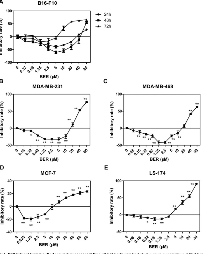
colorimetric assay. We observed the temporal features of hormetic effect induced by BER on B16-F10 cells. BER at relatively low concentrations significantly stimulated cell growth, while high concentration of BER inhibited cell growth of the tested cancer cell lines. This biphasic dose-response phenomenon was consistent with the typical feature of hormesis [22]. The result showed that treatment of BER for 48 h exhibited the greatest growth stimulation on B16-F10 cells with a maximum stimulatory rate of about 70% when BER was at 5μM, comparing to the
groups of treatment for 24 h and 72 h (Fig 1A). As shown inFig 1Bto 1E, the hormetic effects of BER were shown in all tested cancer cells after 72 h treatment. However, the maximum stim-ulatory effects on cell growth and the corresponding range of doses of BER varied in different cancer cell types. BER (1.25μM) maximally stimulated the growth of breast cancer cells
(MDA-MB-231 and MDA-MB-468) by 40%, while BER of the same dose only stimulated the growth of colon cancer LS-174 cells by 12% maximally. These results demonstrated that BER exhibited a typical hormetic dose response in cancer cells.
Hormetic effect of BER attenuated the in vitro anticancer activity of CPT,
TAX and 5-FU in B16-F10 melanoma cells
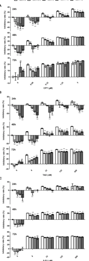
We hypothesized that the hormetic effect of BER could attenuate the anticancer activities of chemotherapeutic agents. To test this hypothesis, low concentrations of BER (2.5–10μM)
com-bined with camptothecin (CPT), paclitaxel (TAX) or 5 fluorouracil (5-FU) were used to treat B16-F10 cells for 24 h, 48 h and 72 h. As shown inFig 2A, CPT inhibited the growth of B16-F10 cells in a dose- and time-dependent manner. Co-treatment of low doses of BER (BER) significantly attenuated the growth inhibition of CPT against B16-F10 cells, for example, the inhibitory rate of 24 h was decreased from 24.3% to 4.9% in the group of CPT (1.25μM) plus
BER (7.5μM) comparing to CPT used alone. Similar results were observed in a longer time
treatment. The growth inhibition of CPT at 0.08μM was even completely reversed by BER
when co-treatment was applied for 24 h and 48 h. Similar results were observed in the com-bined treatment of low doses of BER with TAX or 5-FU (Fig 2B and 2C). The in vitro antican-cer activity of TAX was greatest inhibited by low doses of BER. Particularly, BER reversed the growth inhibition of TAX at all tested concentrations for 24 h treatment. However, the attenua-tion of anticancer activity of chemotherapeutic agents by low doses of BER was greatly
decreased or lost when the co-treatment extended to 48 h or 72 h. These results demonstrated that the hormetic effects of BER significantly attenuated the in vitro anticancer activity of tested chemotherapeutic agents. The degree of attenuation was dependent on the duration of treat-ment, the concentration and type of anticancer agents. The data provided important informa-tion to discern the biomedical significance of hormesis, and suggested a cautious applicainforma-tion of BER both used alone or in combination with other agents in cancer treatment.
Low dose BER up-regulated the MAPK/ERK and PI3K/AKT pathways
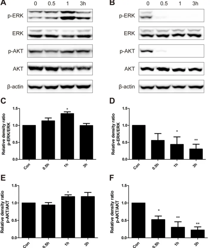
MAPK/ERK1/2 and PI3K/AKT pathways play an important role in cell proliferation and sur-vival [23,24], and adaptive oxidative response [25,26]. We hypothesized that MAPK/ERK and PI3K/AKT signaling pathways were involved in the hormetic effect induced by low dose BER. Hence, we examined the phosphorylated and total protein levels of ERK1/2 and AKT in B16-F10 cells treated with low (5μM) and high doses (40μM) of BER. Representative results
from each group were presented inFig 3. Density analysis showed that ratios of phosphorylated ERK to total ERK were significantly increased with a peak increase of about 40% after treat-ment with 5μM BER for 1 h (Fig 3A). However, the expression of phosphorylated ERK in
B16-F10 cells decreased sharply over time after treatment with high dose of BER (40μM) (Fig
Fig 1. BER induced hormetic effects on various cancer cell lines.B16-F10 cells were treated with various concentrations of BER for 24, 48, or 72 h, respectively. Other cells (MDA-MB-231, MDA-MB-468, MCF-7, and LS-174) were treated with various concentrations of BER for 72 h. Cell viability was determined by WST-1 colorimetric assay. Inhibitory rate was calculated comparing to OD value of untreated control group. Data were expressed as means±SD (n3).*P<0.05,**P<0.01, compared to untreated control.
Fig 2. Low doses of BER attenuated the in vitro anticancer activity of chemotherapeutic agents (CTA) in B16-F10 cells.B16-F10 cells were co-treated with different concentrations of CPT (A), TAX (B), or 5-FU (C) and BER for 24 h, 48 h and 72 h, respectively, before the determination of cell viability by WST-1 assay. Inhibitory rate was calculated comparing to OD value of untreated control group. Data are expressed as mean±SD (n3).*P<0.05,**P<0.01, compared to the group of CTA used alone.
Fig 3. Western blot analysis of protein expression levels of p-ERK, ERK, p-AKT and AKT.B16-F10 cells were treated with low dose of BER (5μM) (A) and high dose of BER (40μM) (B) for 0.5 h, 1 h and 3 h, respectively. Total protein of cell lysates was extracted and subjected to Western blot analysis. The density of each band was quantified by Quantity One Software, and the relative density ratio of each protein was calculated accordingly.β-actin was used as the internal control. Data are expressed as mean±SD (n3).*P<0.05,**P<0.01 compared to untreated control.
treatment of 5μM BER, and had a rapid decline when treated with high dose BER. These
results indicated that low dose BER activated MAPK/ERK and PI3K/AKT signaling pathways, which might be responsible for the hormetic effect and inhibition of anticancer activity of che-motherapeutic agents by BER.
The hormetic effect of BER was reversed by inhibition of MAPK/ERK
and PI3K/AKT signaling pathways
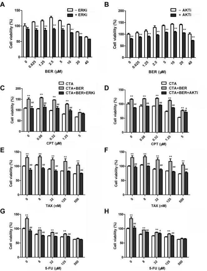
To further validate the role of ERK and AKT signaling pathways in hormetic dose response of BER, we tested whether the pathway inhibitors could affect BER-induced cell growth stimula-tion in B16-F10 cells. As shown inFig 4A and 4B, cell growth of B16-F10 cells was increased after treatment with low dose BER, which was consistent with previous results (Fig 1A). In the presence of 20μM MAPK/ERK kinase (MEK) inhibitor PB98059, the growth stimulation by
low dose BER was completely abolished (Fig 4A). Similarly, PI3K inhibitor LY294002 partially abolished the growth stimulation by low dose BER (Fig 4B). These results demonstrated that MAPK/ERK and PI3K/AKT pathways were involved, at least partially, in the hormetic effect of BER on B16-F10 cells.
MEK and PI3K inhibitors restored the anticancer activity of
chemotherapeutic agents
To investigate whether ERK and AKT activation participates in the attenuation of anticancer activity of chemotherapeutic agents by low dose BER, B16-F10 cells were exposed to chemo-therapeutic agents alone, or in combination with low dose BER (5μM) and MEK or PI3K
inhibitors. As shown inFig 4C–4H, 5μM BER significantly stimulated the growth of B16-F10
cells, and attenuated the in vitro anticancer activity of tested chemotherapeutic agents. In the presence of 20μM MEK inhibitor PD98059, the growth inhibition of CPT, TAX, and 5-FU was
almost completely restored comparing to they were used alone or in combination with BER (Fig 4C, 4E and 4G). Similarly, 5μM PI3K inhibitor LY294002 restored the in vitro anticancer
activity of CPT, TAX, and 5-FU (Fig 4D, 4F and 4H). These results demonstrated that MAPK/ ERK and PI3K/AKT pathways were involved, at least partially, in the attenuation of in vitro anticancer activity of the tested chemotherapeutic agents by low dose BER in B16-F10 cells.
Discussion
Fig 4. ERK and AKT inhibitors reversed the hormetic effect of BER and restored the in vitro anticancer activity of chemotherapeutic agents (CTA). B16-F10 cells were pretreated with 20μM ERK inhibitor (ERKi) PD98059 (A, C, E, G) or 5μM AKT inhibitor (AKTi) LY294002 (B, D, F, H) for 1 h. Cell viability was determined by WST-1 assay after 24 h treatment of 5μM BER alone (A, B) or combined with CTA (C-H). Data were expressed as mean±SD (n3). *P<0.05,**P<0.01 compared to BER (A, B) or CTA (C-H) used alone.#P<0.05,##P<0.01 compared to combination treatment of CTA and low dose BER.
typical hormetic dose response in several cancer cell lines, i.e. relative low doses of berberine stimulated the growth of cancer cells, while high doses of berberine inhibited cell growth. Moreover, low dose berberine remarkably attenuated the in vitro anticancer activity of chemo-therapeutic agents. Our data further indicated that mitogen-activated protein kinases
(MAPK)/ERK and phosphoinositide 3-kinase (PI3K)/AKT signaling pathways were activated by low dose berberine, and involved, at least partially, in the hormetic effect of berberine and its inhibition on the anticancer activity of chemotherapeutic drugs.
The characteristic of hormesis is a biphasic dose-response curve, with stimulatory or benefi-cial effect at low doses and inhibitory or toxic effect at high doses [1,2]. The range of maximum stimulatory response is about 30% to 60% above that of the control, and the width of the stimu-latory dosage is mostly within 100-fold of the threshold value [4]. Our results showed that the maximum stimulatory rate was nearly 70% on B16-F10 cells after 48 h treatment of 5μM
ber-berine, and the range of the stimulatory dosage is about 120-fold below the threshold value (Fig 1A), indicating that the two phasic dose response of berberine is a typical hormetic phe-nomenon. Furthermore, we found that low dose berberine also enhanced the growth of other cancer cells, including breast cancer MDA-MB-231, MDA-MB-468, and MCF-7 cells, and colon cancer LS-174 cells (Fig 1B–1E). The degree of growth stimulation and the dosage range of berberine notably varied among different types of cancer cells. Our results shed light on the universal hormetic phenomenon induced by phytochemical compounds, such as resveratrol, curcumin, epigallocatechin, etc. [12].
In clinical practice, drug combination is a common-use strategy for cancer treatment, which may increase therapy sensitivity, provide multiple drug targets and avoid potential drug resis-tant. Berberine also showed synergistic anticancer activity combined with other chemothera-peutic drugs as reported previously [18,19]. However, in our present study, in contrast to its synergistic anticancer activity at high doses, low dose berberine significantly reduced, or even reversed the anticancer activity of camptothecin (CPT), paclitaxel (TAX), and 5 fluorouracil (5-FU) in B16-F10 cells. As shown inFig 2B, the anticancer activity of TAX was greatest inhib-ited by low doses of berberine. Comparing to groups of 48 h and 72 h, co-treatment of low dose berberine for 24 h exhibited the highest inhibition on the anticancer activity of chemotherapeu-tic agents (Fig 2), which is consistent with the time-dependent manner of hormetic effects induced by berberine (Fig 1). These results confirmed the speculation that hormetic effect of agents could interfere with the anticancer activity of chemotherapeutic drugs [4]. The degree of interference was related to the duration of treatment, the concentration and type of anticancer agents. Therefore, we should be aware of the hormetic dose response of berberine both used alone and in combination with other agents in cancer treatment. An appropriate treatment regime should be carefully arranged and blood concentration of berberine should be monitored in clinical applications to avoid potential unwanted side effects. More studies in this respect are required, and it is interesting to examine the hormetic dose response and the potential adverse effects of more natural compounds and existing anticancer drugs at low doses.
Hormesis was considered a universal phenomenon in many disciplines of biological and medical sciences, such as toxicology, immunology, aging biology, neuroscience, microbiology, radiology, etc. [1]. It has been reported that key signaling pathways of cell survival/ prolifera-tion and oxidative stress response were involved in the processes of hormesis [12], including MAPK/ERK1/2 [28,29], PI3K/AKT [30,31], nuclear factor-erythroid 2p45 (NF-E2)-related factor (Nrf2)/antioxidant response element (ARE) [32,33], etc. Demirovic et al [34] reported that curcumin induced a hormetic dose response in wound healing via oxidative stress
AMP-activated protein kinase (AMPK). In our results (Fig 3), the ratios of phosphorylated ERK1/2 to total ERK1/2 were significantly increased with a peak increase of about 40% after treatment with berberine at 5μM, and decreased rapidly when treated with high dose berberine
at 40μM, indicating that low dose berberine obviously activated both MAPK/ERK1/2 and
PI3K/AKT signaling pathways. MAPK pathways play critical roles in the regulation of cell pro-liferation, differentiation, development, survival, apoptosis, and cellular stresses. Three sub-families of MAPK have been well-characterized in vertebrate: ERK1/2, JNKs, and p38 MAPKs [37]. In MAPK/ERK1/2 signaling pathway, the activation of MAPKs can be provoked by tyro-sine kinase receptors (RTKs), integrins and ion channels in multistep processes [38]. Extracel-lular cell growth factors or cytokines activate Raf via a variety of receptors, followed by the activation of MEK by the phosphorylated Raf, and the phosphorylation of ERK by the activated MEK. Finally, the activated ERK promotes cell proliferation and cell survival by regulating the activity of transcriptional factors and changing target gene expression [23]. The PI3K/AKT pathway also plays important roles in the regulation of cell growth, proliferation, survival, and apoptosis. The serine/threonine kinase Akt is a key mediator of the PI3K signaling pathway. PI3K catalyzes the formation of phosphatidylinositol (3,4,5)-trisphosphate (PIP3), which can stimulates the phosphorylation and activation of AKT. Then, the phosphorylated AKT pro-motes cell survival and proliferation through activating target transcriptional factors[39]. In this study, inhibitors of MEK (PB98059) and PI3K (LY294002) were applied to abolish the acti-vation of ERK and AKT by low dose berberine. Consequently, the hormetic effect induced by berberine was suppressed and the anticancer activities of all three chemotherapeutic drugs were restored. These results demonstrated that MAPK/ERK1/2 and PI3K/AKT signaling path-ways were involved in the hormetic effect and attenuation of anticancer drug activities by low dose berberine. However, the detailed mechanisms for their activation remain to be further investigated.
In summary, our study demonstrated that berberine induced a significant hormetic dose response, in which low dose berberine strongly stimulated the growth of cancer cells, in con-trast to its anticancer activity at high doses. Moreover, low dose berberine greatly attenuated or even reversed the in vitro anticancer activity of chemotherapeutic drugs, including CPT, TAX, and 5-FU. These adverse effects were associated with the activation of signaling pathways for cell proliferation/survival and adaptive oxidative stress response in cancer cells, including MAPK/ERK1/2 and PI3K/AKT. As hormetic effect was considered a universal adaptive response to low dose (sub-lethal) cytotoxic agents, our study suggested cautious application of natural compounds and relevant herbs, which could cause obvious hormetic response, in adju-vant treatment of cancer.
Author Contributions
Conceived and designed the experiments: JB CH JBW YW. Performed the experiments: JB BH LZ. Analyzed the data: JB BH CH MC. Contributed reagents/materials/analysis tools: SC CZ YZ HS. Wrote the paper: JB CH.
References
1. Calabrese EJ, Bachmann KA, Bailer AJ, Bolger PM, Borak J, Cai L, et al. Biological stress response ter-minology: Integrating the concepts of adaptive response and preconditioning stress within a hormetic dose-response framework. Toxicology and applied pharmacology. 2007; 222(1):122–8. doi:10.1016/j. taap.2007.02.015PMID:17459441.
3. Rithidech KN, Scott BR. Evidence for Radiation Hormesis after in Vitro Exposure of Human Lympho-cytes to Low Doses of Ionizing Radiation. Dose-Response. 2008; 6(3):252–71. doi: 10.2203/dose-response.07-024.RithidechISI:000261406100003. PMID:18846261
4. Calabrese EJ. Hormesis and medicine. British journal of clinical pharmacology. 2008; 66(5):594–617.
Epub 2008/07/30. doi:10.1111/j.1365-2125.2008.03243.xPMID:18662293; PubMed Central PMCID: PMC2661975.
5. Li F, Mao HP, Ruchalski KL, Wang YH, Choy W, Schwartz JH, et al. Heat stress prevents mitochondrial injury in ATP-depleted renal epithelial cells. Am J Physiol-Cell Ph. 2002; 283(3):C917–C26. doi:10. 1152/ajpcell.00517.2001WOS:000177573500026.
6. Gomez-Pinilla F. The influences of diet and exercise on mental health through hormesis. Ageing research reviews. 2008; 7(1):49–62. Epub 2007/07/03. doi:10.1016/j.arr.2007.04.003PMID: 17604236; PubMed Central PMCID: PMC3225189.
7. Calabrese EJ. Cancer biology and hormesis: Human tumor cell lines commonly display hormetic (biphasic) dose responses. Critical reviews in toxicology. 2005; 35(6):463–582. doi:10.1080/ 10408440591034502WOS:000233908100001. PMID:16422392
8. Calabrese EJ, Baldwin LA. Chemotherapeutics and hormesis. Critical reviews in toxicology. 2003; 33 (3–4):305–53. Epub 2003/06/18. doi:10.1080/713611041PMID:12809428.
9. Alley MC, Paculacox CM, Hursey ML, Rubinstein LR, Boyd MR. Morphometric and Colorimetric Analy-ses of Human Tumor-Cell Line Growth and Drug Sensitivity in Soft Agar Culture. Cancer Res. 1991; 51 (4):1247–56. ISI:A1991EX82700031. PMID:1705170
10. Vezmar M, Georges E. Reversal of MRP-mediated doxorubicin resistance with quinoline-based drugs. Biochem Pharmacol. 2000; 59(10):1245–52. doi:10.1016/S0006-2952(00)00270-7
ISI:000086605000008. PMID:10736425
11. Vichi P, Tritton TR. Stimulation of growth in human and murine cells by adriamycin. Cancer research. 1989; 49(10):2679–82. PMID:2713852.
12. Calabrese EJ. Hormetic mechanisms. Critical reviews in toxicology. 2013; 43(7):580–606. doi:10. 3109/10408444.2013.808172PMID:23875765.
13. Calabrese EJ, Stanek EJ 3rd, Nascarella MA, Hoffmann GR. Hormesis predicts low-dose responses better than threshold models. International journal of toxicology. 2008; 27(5):369–78. Epub 2008/11/
28. doi:10.1080/10915810802503735PMID:19037807.
14. Tang J, Feng YB, Tsao S, Wang N, Curtain R, Wang YW. Berberine and Coptidis Rhizoma as novel antineoplastic agents: A review of traditional use and biomedical investigations. J Ethnopharmacol. 2009; 126(1):5–17. doi:10.1016/j.jep.2009.08.009WOS:000271790800001. PMID:19686830
15. Lu JJ, Bao JL, Chen XP, Huang M, Wang YT. Alkaloids Isolated from Natural Herbs as the Anticancer Agents. Evid-Based Compl Alt. 2012. Artn 485042 doi:10.1155/2012/485042ISI:000308765000001. 16. Jabbarzadeh Kaboli P, Rahmat A, Ismail P, Ling KH. Targets and mechanisms of berberine, a natural drug with potential to treat cancer with special focus on breast cancer. European journal of pharmacol-ogy. 2014; 740:584–95. doi:10.1016/j.ejphar.2014.06.025PMID:24973693.
17. Ortiz LM, Lombardi P, Tillhon M, Scovassi AI. Berberine, an epiphany against cancer. Molecules. 2014; 19(8):12349–67. doi:10.3390/molecules190812349PMID:25153862.
18. Yu M, Tong X, Qi B, Qu H, Dong S, Yu B, et al. Berberine enhances chemosensitivity to irinotecan in colon cancer via inhibition of NFkappaB. Molecular medicine reports. 2014; 9(1):249–54. Epub 2013/
11/01. doi:10.3892/mmr.2013.1762PMID:24173769.
19. Tong N, Zhang J, Chen Y, Li Z, Luo Y, Zuo H, et al. Berberine sensitizes mutliple human cancer cells to the anticancer effects of doxorubicin in vitro. Oncology letters. 2012; 3(6):1263–7. Epub 2012/07/12.
doi:10.3892/ol.2012.644PMID:22783430; PubMed Central PMCID: PMC3392583.
20. Zhang Q, Zhang C, Yang X, Yang B, Wang J, Kang Y, et al. Berberine inhibits the expression of hypoxia induction factor-1alpha and increases the radiosensitivity of prostate cancer. Diagnostic pathology. 2014; 9:98. doi:10.1186/1746-1596-9-98PMID:24886405; PubMed Central PMCID: PMC4051149. 21. Mizoguchi M, Ishiyama M, Shiga M, Sasamoto K. The development of new type oxidative and reductive
chromogenic reagents in clinical analysis. Bunseki Kagaku. 1996; 45(2):111–24. WOS:
A1996TV40800001.
22. Calabrese EJ, Baldwin LA. Defining hormesis. Human & experimental toxicology. 2002; 21(2):91–7.
Epub 2002/07/10. PMID:12102503
23. Zhang W, Liu HT. MAPK signal pathways in the regulation of cell proliferation in mammalian cells. Cell Res. 2002; 12(1):9–18. doi:10.1038/sj.cr.7290105WOS:000174833300002. PMID:11942415
25. Lee WK, Chakraborty PK, Roussa E, Wolff NA, Thevenod F. ERK1/2-dependent bestrophin-3 expres-sion prevents ER-stress-induced cell death in renal epithelial cells by reducing CHOP. Biochimica et biophysica acta. 2012; 1823(10):1864–76. doi:10.1016/j.bbamcr.2012.06.003PMID:22705154.
26. Robey RB, Hay N. Mitochondrial hexokinases, novel mediators of the antiapoptotic effects of growth factors and Akt. Oncogene. 2006; 25(34):4683–96. doi:10.1038/sj.onc.1209595PMID:16892082.
27. Ortiz LMG, Lombardi P, Tillhon M, Scovassi AI. Berberine, an Epiphany Against Cancer. Molecules. 2014; 19(8):12349–67. doi:10.3390/molecules190812349ISI:000341502600092. PMID:25153862
28. Ranzato E, Boccafoschi F, Mazzucco L, Patrone M, Burlando B. Role of ERK1/2 in Platelet Lysate-Driven Endothelial Cell Repair. Journal of cellular biochemistry. 2010; 110(3):783–93. doi:10.1002/ Jcb.22591WOS:000278837200024. PMID:20512938
29. Ranzato E, Patrone M, Pedrazzi M, Burlando B. HMGb1 promotes scratch wound closure of HaCaT keratinocytes via ERK1/2 activation. Molecular and cellular biochemistry. 2009; 332(1–2):199–205.
doi:10.1007/s11010-009-0192-4WOS:000271503400023. PMID:19588230
30. Berna MJ, Tapia JA, Sancho V, Thill M, Pace A, Hoffmann KM, et al. Gastrointestinal growth factors and hormones have divergent effects on Akt activation. Cell Signal. 2009; 21(4):622–38. doi:10.1016/j. cellsig.2009.01.003WOS:000264226200018. PMID:19166928
31. Qi HY, Han YF, Rong JH. Potential roles of PI3K/Akt and Nrf2-Keap1 pathways in regulating hormesis of Z-ligustilide in PC12 cells against oxygen and glucose deprivation. Neuropharmacology. 2012; 62 (4):1659–70. doi:10.1016/j.neuropharm.2011.11.012WOS:000301221500007. PMID:22146407
32. Juan SH, Cheng TH, Lin HC, Chu YL, Lee WS. Mechanism of concentration-dependent induction of heme oxygenase-1 by resveratrol in human aortic smooth muscle cells. Biochemical pharmacology. 2005; 69(1):41–8. doi:10.1016/j.bcp.2004.09.015WOS:000226268500005. PMID:15588712
33. Szumiel I. Radiation hormesis: Autophagy and other cellular mechanisms. International journal of radia-tion biology. 2012; 88(9):619–28. doi:10.3109/09553002.2012.699698WOS:000308215000001.
PMID:22702489
34. Demirovic D, Rattan SIS. Curcumin induces stress response and hormetically modulates wound heal-ing ability of human skin fibroblasts undergoheal-ing ageheal-ing in vitro. Biogerontology. 2011; 12(5):437–44.
doi:10.1007/s10522-011-9326-7WOS:000295978300006. PMID:21380847
35. Pallas M, Porquet D, Vicente A, Sanfeliu C. Resveratrol: New Avenues for a Natural Compound in Neu-roprotection. Current pharmaceutical design. 2013; 19(38):6726–31. WOS:000326344600005. PMID: 23530512
36. Mattson MP, Cheng AW. Neurohormetic phytochemicals: low-dose toxins that induce adaptive neuro-nal stress responses. Trends Neurosci. 2006; 29(11):632–9. doi:10.1016/j.tins.2006.09.001
WOS:000242294400006. PMID:17000014
37. Johnson GL, Lapadat R. Mitogen-activated protein kinase pathways mediated by ERK, JNK, and p38 protein kinases. Science. 2002; 298(5600):1911–2. doi:10.1126/science.1072682PMID:12471242.
38. Burotto M, Chiou VL, Lee JM, Kohn EC. The MAPK pathway across different malignancies: a new per-spective. Cancer. 2014; 120(22):3446–56. doi:10.1002/cncr.28864PMID:24948110; PubMed Central
PMCID: PMC4221543.