P
HYLOGENETICA
NALYSISANDT
AXONOMICR
EVISIONOFP
HYSODACTYLINAE(C
OLEOPTERA, E
LATERIDAE)
S
IMONEP
OLICENAR
OSA1ABSTRACT
A phylogeny based on male morphological characters and taxonomic revision of the Physodac-tylinae genera are presented. The phylogenetic analysis based on 66 male characters resulted in the polyphyly of Physodactylinae which comprises four independent lineages. Oligostethius
and Idiotropia from Africa were found to be sister groups. Teslasena from Brazil was corrobo-rated as belonging to Cardiophorinae clade. The South American genera Physodactylus and
Dactylophysus were found to be sister groups and phylogenetically related to Heterocrepidius
species. The Oriental Toxognathus resulted as sister group of that clade plus (Dicrepidius ramicornis (Lissomus sp, Physorhynus erythrocephalus)). Taxonomic revisions include di-agnoses and redescriptions of genera and distributional records and illustrations of species. Key to species of Teslasena, Toxognathus, Dactylophysus and Physodactylus are also provided.
Teslasena lucasi is synonymized with T. femoralis. A new species of Dactylophysus is de-scribed, D. hirtus sp. nov., and lectotypes are designated to non-conspecific D. mendax sensu
Fleutiaux and Heterocrepidius mendax Candèze. Physodactylus niger is removed from syn-onymy under P. oberthuri; P. carreti is synonymized with P. niger; P. obesus and P. testaceus
are synonymized with P. sulcatus. Nine new species are described in Physodactylus: P. asper
sp. nov., P. brunneus sp. nov., P. chassaini sp. nov., P. flavifrons sp. nov., P. girardi sp. nov.,
P. gounellei sp. nov., P. latithorax sp. nov., P. patens sp. nov. and P. tuberculatus sp. nov.
Key-Words: Morphology; Click beetles; Neotropical region; Afrotropical region; Ethio-pian region; Oriental region.
INTRODUCTION
The subfamily Physodactylinae was erected by Lacordaire (1857) to include the Brazilian genus
Physodactylus Fischer von Waldheim, 1823 in Ce-brionidae, family currently regarded as subfamily of Elateridae (Lawrence & Newton, 1995; Costa et al.
2010). Posteriorly, several authors included elaterids with apparently burrowing adaptations in
Physodac-tylinae based on the conspicuous features shared by cebrionines and Physodactylus: fossorial legs, falciform mandibles and prosternal anterior lobe (chin piece) not produced.
Fairmaire (1878) erected Toxognathus, from Southeast Asia and related it to Physodactylus. Fleu-tiaux (1892) erected Dactylophysus and Teslasena to include described species in Heterocrepidius Candèze, 1859 and new species from South America, placing
http://dx.doi.org/10.1590/0031-1049.2014.54.18
them with Physodactylus in the tribe Physodactylini (Elaterinae). Schwarz (1906) added to this tribe
Margogastrius Schwarz, 1903 (Tanzania), Idiotropia
Schwarz, 1906 (Algeria), Oligostethius Schwarz, 1906 (South Africa) and Coryssodactylus Schwarz, 1897 (Tanzania), which were excluded by Fleutiaux (1919).
Schenkling (1927) catalogued the group as sub-family, to which he added more three genera: Tharop-sides Fleutiaux, 1918 (Asia), Nullarborica Blackburn, 1911 and Antoligostethus Blackburn, 1911 (Australia).
Fleutiaux (1940a) separated Toxognathus to Tox-ognathinae and in 1947 transferred Tharopsides to Oxynopterinae, as a synonym of Luzonicus Fleutiaux, 1916. Van Zwaluwenburg (1953) described the Aus-tralian genus Patriciella in Physodactylinae. Stibick (1979) presented the group as subtribe of Denticol-linae, including Toxognathus, based on adult charac-ters as prognathous head, truncate prosternal anterior lobe and mesocoxae open to both mesepimeron and mesepisternum. Later, Calder (1996) transferred the Australian genera Nullarborica and Antoligostethus to Elaterinae and Patriciella to Cardiophorinae. Law-rence & Newton (1995) and Costa et al. (2010) listed Physodactylinae (incl. Toxognathinae) as subfamily of Elateridae.
After the modifications made by Fleutiaux (1947), Stibick (1979) and Calder (1996) to the classification of Schenkling (1927), Physodactylinae comprises 28 species in seven genera: Physodactylus, Dactylophysus, Teslasena, Toxognathus, Margogastrius, Idiotropia and Oligostethius. This is currently the composition of the subfamily and is the object of the present work.
Physodactylinae have been assumed to be para-phyletic (Costa et al., 2010), although a comprehen-sive cladistic analysis has not been performed for the group. Douglas (2011) included in his phylogenetic analysis of Elateridae male characters of Physodactylus henningi and Teslasena femoralis and female charac-ters of Margogastrius schneideri. Those physodacty-lines were found to be polyphyletic with T. femora-lis and M. schneideri related to Cardiophorinae and
P. henningi with uncertain relationships, but not as-sociated with the Cardiophorinae.
This work focuses on the cladistic analysis of the Physodactylinae in order to test its monophyly and the taxonomic revision of its genera and species. The physodactylines are poorly represented in the insect collections and several species were described based on one or a few male specimens, with female speci-mens assigned to two species belonged to Physodac-tylus and Dactylophysus. In contrast, Margogastrius is known only from females. Larvae are unknown for
all genera. Therefore, only male characters could be scored. On the other hand, species of the rare phy-sodactyline genera from Africa and Southeast Asia and several species of Physodactylus were included in the matrix. Additionally, many species representing genera traditionally assigned or related to Physodac-tylinae were included as outgroups. The analysis re-sulted in Physodactylinae as polyphyletic and allows some insights into the phylogenetic relationships of its genera.
MATERIAL AND METHODS
Taxonomic revision
Label data of the type specimens with valid names are listed after each species description. A com-plete list of additional material and types of junior syn-onyms examined are listed in the Appendix 1. Each la-bel of all type specimens is transcribed between square brackets whereas notes are in parentheses. The stud-ied material belongs to the following collections, with their respective acronysms and curators in parenthe-ses: Canadian National Collection of Insects, Ontario (CNC, Patrice Bouchard); Coleção Entomológica Pe. J.S. Moure, Universidade Federal do Paraná, Curi-tiba (DZUP, Lúcia Massuti de Almeida); Deutsches Entomologisches Institut, Müncheberg (DEI, Lothar Zerche); Instituto Biológico, São Paulo (IBSP, Sér-gio Ide); Museu de Zoologia da Universidade de São Paulo, São Paulo (MZUSP, Sônia A. Casari); Museu Nacional da Universidade Federal do Rio de Janeiro, Rio de Janeiro (MNRJ, Marcela L. Monné); Museu Paraense Emílio Goeldi, Belém (MPEG, Orlando T. Silveira); Muséum National d’Histoire Naturelle, Paris (MNHN, Antoine Mantilleri); The Natural History Museum, London (BMNH, Max Barclay); Universidade Estadual Paulista “Julio de Mesquita Filho”, Campus de Ilha Solteira, (UNESP-IS, Carlos A.H. Flechtmann).
I prepared drawings with a camera lucida cou-pled to a Stemi SV6 Zeiss stereoscopic microscope or after photos and took photographs using a digital camera Canon A640, a camera Axiocam coupled to Zeiss Imager A1 microscope and a SEM Zeiss LEO 440. Different focal planes were merged using Heli-con Focus 3.10.
Terminology follows Calder (1996) & Costa
& Beutel, 2006) and mouthparts (Casari, 2008). The term “pedunculate anterior sac” is used here in the sense of Douglas (2011). The abdominal marginal plate (Fig. 9L) is understood as the expanded lateral edge of the sternite which fits under epipleura; the lat-eroposterior part of phallobase is the lateral part that extend posteriorly beyond the posterior edge of the median part (Fig. 2U). The free margin of metacoxal plate (Casari, 2008) is the expanded part of metacoxal plate that overlaps part of the metatrochanter and metafemur (Fig. 12O). I referred to antennomeres and abdominal segments with Roman numerals, ab-dominal ventrites with Arabic numbers, abbreviated “index of eye prominence” as IEP (Calder, 1996) and cited characters and character states in the discussion by their numbers separated by hyphen (e.g., 21-0). Polymorphic color patterns were numbered in the description. These numbers are used to refer to color patterns in remark section avoiding repetitions and do not correspond to hypothesis of homology among species.
Measurements were made using the stereoscopic microscope fitted with an ocular micrometer or after drawings. Index of eye prominence (IEP) and length of aedeagus, pronotum and elytra was measured ac-cording to Calder (1996). Other measurements were taken as follows:
— Lengths. Mandible: along a line perpendicular to the base. Prosternum, mesoventrite, meta-ventrite and meta-ventrite 5: along midline; poste-rior edge of prosternum (excluding process) is at level of the anterior edge of procoxal cavity (Fig. 2F). Prosternal process: from anterior edge of procoxal cavity (Fig. 2F) to apex of proster-nal process. Lateral part of ventrite 1: at its most external border; median part of ventrite 1: at the shortest distance between the anterior and posterior margins at middle (Fig. 5K). Phal-lobase: along a longitudinal line from its most anterior part to apex of the lateroposterior part (Fig. 15J); median part of the phallobase: along the midline; lateroposterior part of phallobase: from the level of the posterior margin of the median part to the apex of the lateroposterior part (Fig. 2U). Paramere: in dorsal view from its most anterior margin to the apex. Penis: in dorsal view along the longitudinal line from an-terior tip of the basal strut to apex of penis; basal strut: from its anterior tip to the most posterior edge at middle (Fig. 15I). In Teslasena species, the triangular basal part of penis: in ventral view from the most anterior margin between the
bas-al struts to the point where laterbas-al edges become parallel-sided; posterior part of penis: from this point to the apex (Fig. 2U).
— Widths. Mandible: across its basal part. Prono-tum: at widest point, which is usually at mid-length (in species with rounded pronotal sides) or at base of hind angles (in species with sides convergent anteriad); metaventrite and ven-trite 5: at widest point; prosternum: at smallest transversal line between the notosternal sutures (Fig. 2F). Phallobase: at widest point.
— Diameter of coxal cavity is measured along its widest transversal line. Angle formed by the in-clination of the posterior region of mesoventrite in relation to its anterior region was obtained crossing two lines, each one parallel to anterior and posterior regions in lateral view (Fig. 2K). Degree of inclination of the metacoxa in rela-tion to the transverse body axis in ventral view was measured crossing two lines, a transversal line between the most external points of meta-coxae and an oblique line between the most ex-ternal point of right metacoxa and the apex of metaventrite (Fig. 1H).
Cladistic Analysis
The ingroup included 18 taxa: 11 species of Phy-sodactylus, two species of Dactylophysus, the type spe-cies of Teslasena, two species of Toxognathus and the monotypic genera Oligostethius and Idiotropia. The choice of outgroup taxa was based on the supposed re-lashionships of physodactyline genera with other elat-erid clades indicated by their taxonomic background and the cladistic analysis of Douglas (2011). Out-groups (material examined in Appendix 1) include 20 species: the Eucnemidae Ceratogonis spinicornis
(Fabricius, 1801) and the following elaterid species, with the respective subfamily and tribe in parentheses:
Semiotus distinctus (Herbst, 1806) (Seminotinae), Lis-somus sp. (Lissominae), Scaptolenus lecontei Chevrolat, 1874 (Cebrioninae), Ctenicera silvatica (Van Dyke, 1932) (Dendrometrinae, Ctenicerini), Athous vittatus
(Fabricius, 1792) (Dendrometrinae, Denticollini),
Globothorax latidens Rosa, 2011, Triplonychus pla-giatus (Erichson, 1846), T. crassifemoris Rosa, 2011,
T. cruspinosus Rosa, 2011 and T. tibialatus Rosa, 2011 (Cardiophorinae).
The analysis was based on 66 male morpho-logical characters scored from examination of speci-mens. Female characters and therefore those from the monotypic Margogastrius, were not included because females are unknown for the majority of species. The data matrix (Appendix 2) was constructed in Mesquite version 2.75 (Maddison & Maddison, 2011). Auta-pomorphies were not included, unless they represent states in multistate series. Quantitative and multistate characters that could be ordered by morphological continuity in a linear sequence of intermediate states were treated as additive; otherwise they were treated as non-additive (unordered). The additive characters are identified as ordered in the character list. All char-acters were treated as having equal weight. Specific references are given to some characters that have been employed in previous cladistic analysis (Calder et al.,
1993; Lawrence et al., 2011; Casari, 2008).
The parsimony analysis was performed with T.N.T. software (Goloboff et al., 2003) with heu-ristic search (“traditional search” function) in 5000 random-addition sequences followed by TBR branch swapping, retaining 10 trees per replicate. Branch sup-port was evaluated through Bremer supsup-port (Bremer, 1994) analysis as implemented in TNT, employing the option to save suboptimal trees by 20 steps and collapsing nodes with support less than one step.
RESULTS AND DISCUSSION
List of Characters
1. Color of body setae: [0] yellow; [1] brown; [2] silvery.
2. Frontal carina between antennal insertions: [0] absent (Fig. 5A); [1] present (Fig. 12G) (Calder
et al., 1993 – modified).
3. Frontal carina (contingent on character 2-0): [0] not protruded anteriad (Fig. 22E); [1] pro-truded anteriad, covering the base of labrum (Fig. 26E).
4. Antennal sensory elements beginning on anten-nomere: [0] III; [1] IV. (Calder et al., 1993). 5. Lateral sides of antennomeres IV-XI in dorsal or
ventral view: [0] flat; [1] convex.
6. Shape of the anterior edge of labrum: [0] point-ed (Fig. 2D); [1] straight (Fig. 9D); [2] roundpoint-ed (Fig. 3C).
7. Dorsal surface of labrum: [0] flat; [1] evenly convex; [2] posterior part convex sloping down-wards anteriorly (Fig. 33E).
8. Mandible: [0] not more than 2 times as long as basal width (Fig. 9D); [1] more than 3 times as long as basal width (falciform) (Fig. 2E). (Law-rence et al., 2011).
9. Mandible: [0] unidentate (Fig. 2E); [1] biden-tate with subapical mesal tooth (Fig. 3C); [2] bidentate with subapical dorsal tooth (fig. 106 in Casari, 2008); [3] tridentate.
10. Row of seta on the mesal edge of mandible: [0] absent; [1] present.
11. Extension of the row of seta on the mesal edge of mandible: [0] extended on basal third (fig. 106 in Casari, 2008) to basal half (Fig. 8A); [1] re-stricted to a small basal area (Fig. 2E).
12. Ridge on lateroanterior margin of mandible: [0] absent (Fig. 5A); [1] present (Fig. 9D).
13. Shape of basistipes: [0] triangular as wide as long (fig. 72 in Rosa, 2011); [1] trapezoidal lon-ger than wide (Fig. 9B).
14. Number of long thick setae on basistipes: [0] absent (autapomorphy of Patriciella sp.); [1] one (Fig. 32A); [2] three; [3] more than five (Fig. 12D) (ordered).
15. Setae on galea: [0] fine (Fig. 22B); [1] spiniform (Fig. 30B); [2] spatulate.
16. Shape of the apical maxillary palpomere: [0] slightly expanded and truncate apically (Fig. 12D); [1] securiform, strongly expanded and squarely to obliquely truncate apically; [2] cylindrical to fusiform (narrowed at both ends) (Fig. 9B). (Lawrence et al., 2011).
17. Lateral anterior angles of prementum: [0] not prominent (Fig. 9C); [1] prominent (Fig. 32C). 18. Labial palpigers: [0] separate (Fig. 9C); [1]
con-tiguous (Fig. 32C).
19. Sides of prothorax: [0] convergent anteriad from posterior angles or posterior third (Fig. 12J); [1] subparallel (straight or convex) on major part, convergent only on anterior angles (Fig. 8B), [2] divergent anteriad from posterior third (Fig. 3D).
20. Pronotal lateral carina: [0] present on posterior 1/2-2/3 (Fig. 2H); [1] present on posterior 9/10 (Fig. 25D); [2] complete (Fig. 11C); [3] absent. Ordered.
21. Pronotal punctation: [0] single (Fig. 16B); [1] double (intermixed large and small punctures) (fig. 36 in Rosa, 2011).
with-out notch or concavity (Fig. 3G); [1] with a V-shaped notch (Fig. 2H); [2] with a longer than wide U-shaped notch (Fig. 12L); [3] with a rectangular notch; [4] with a wider than long concave notch.
23. Anterior edge of prosternum: [0] not produced, exposing labium (Fig. 2H); [1] produced for-ward to form “chin piece”, concealing labium (Fig. 12I) (Calder et al., 1993).
24. Procoxal cavities posteriorly to coxae: [0] open (Fig. 3E); [1] closed (Fig. 2F).
25. Posterior region of mesoventrite in relation to its anterior region: [0] inclined ventrad ap-proximately 45° (Fig. 2K); [1] slightly inclined ventrad (at most 30°) (Fig. 3I); [2] inclined 90°. Ordered.
26. Mesocoxal cavity: [0] closed to both mesepim-eron and mesepisternum (Fig. 2J); [1] open to mesepisternum only; [2] open to both mese-pimeron and mesepisternum (Fig. 3H). Or-dered. (Calder et al., 1993).
27. Mesotrochantin: [0] not visible (Fig. 2J); [1] visible (Fig. 5H) (Calder et al., 1993).
28. Mesometaventral suture: [0] not grooved (Fig. 22K); [1] grooved (Fig. 3L).
29. Ratio of width of metaventrite to its length: [0] 1.3-1.6 (Fig. 2I); [1] 1.9 (Fig. 3L).
30. Free margin of metacoxal plate: [0] present (Fig. 7B); [1] absent (Fig. 2I).
31. Degree of inclination of the metacoxa in rela-tion to the transverse body axis in ventral view: [0] 17°-20° (Fig. 22K); [1] 25°-30° (Fig. 3L). 32. Elytral striae: [0] absent; [1] present.
33. Punctation of elytral striae with: [0] one row of punctures (Fig. 33N); [1] two regular rows of homogeneous punctures (equal sizes) (fig. 91 in Rosa, 2011); [2] 3-4 irregular rows of homoge-neous punctures (fig. 94 in Rosa, 2011); [3] two rows of heterogeneous punctures (different siz-es); [4] three rows of heterogeneous punctures (Figs. 32F, 33O).
34. Hind wings: [0] present; [1] absent or reduced. I examined the base of mesonotum without elytra of one specimen of Idiotropia henoni and observed that it had no trace of hind wings. Oli-gostethius capensis has elytra fused at midlength which cannot be opened; therefore I could not examine the base of mesonotum of this species. Nevertheless, there were no hind wings in ven-tral view of elytra without abdomen (about 4/5 of elytral length).
35. Apex of hind wing with sclerotizations present on: [0] anterior, median and posterior fields
(Fig. 2M); [1] anterior and posterior fields (Fig. 7F); [2] anterior field only. Ordered. 36. Hind wing with sclerotization between radial
cell and anterior field sclerotization: [0] absent (Fig. 2M); [1] present (Figs. 9K, 26Q).
37. Transverse vein CuA1: [0] absent (Fig. 2M); [1]
present (Fig. 8E).
38. Notch in the edge of anal area of hind wing: [0] absent (Fig. 7F); [1] present (Fig. 2M) (Calder
et al., 1993).
39. Wedge cell of hind wing: [0] absent (Fig. 7F); [1] present (Fig. 26Q) (Calder et al., 1993). 40. Tarsal claws: [0] simple (Fig. 38B); [1] bifid
(Fig. 32L); [2] trifid (fig. 89 in Rosa, 2011); [3] pectinate (with more than six teeth).
41. Lamella on protarsomere I: [0] absent; [1] pres-ent (Fig. 33F). (Casari, 2008).
42. Lamella on protarsomere III: [0] absent; [1] present (Fig. 33F). (Casari, 2008).
43. Lamella on metatarsomere I: [0] absent (Fig. 33J); [1] present (Fig. 33H) (Casari, 2008). 44. Metafemur laterally: [0] flat (Fig. 3O); [1]
con-vex (Fig. 33J).
45. Metafemur dorsoventrally: [0] slender; [1] broadened.
46. Protibia: [0] slender (Fig. 3M); [1] broadened apically (Fig. 33F).
47. Dorsal edge of protibia [0] straight; [1] sinuous. 48. Length of the lateral part of ventrite 1 in relation
to its median part: [0] 1.1-1.3x longer; [1] 2-3x longer (Fig. 5K); [2] 5-7x longer (Fig. 12R); [3] 9-10x longer; [4] more than 10x, ventrite 1 al-most divided (Figs. 22L, 25H). Ordered. 49. Abdominal marginal plate of the posterior angle
of ventrite 1: [0] not protruded (Fig. 8F); [1] protruded (Fig. 25H).
50. Marginal plate of posterior angle of ven-trites 2-3: [0] not prominent (Fig. 5K); [1] prominent (Fig. 9L).
51. Ventrite 5 at apex: [0] gradually narrowed; [1] abruptly narrowed.
52. Posterior part of the sternite VIII: [0] with two lateral sclerotizations (Fig. 6A); [1] with three sclerotizations (Fig. 13A); [2] evenly sclerotized. Ordered.
53. Position of fusion between tergite and ster-nite IX: [0] anterior (Fig. 10C); [1] posterior (Fig. 6D).
54. Fringe of setae at apex of tergite X: [0] absent (Fig. 2S); [1] present (Figs. 10D, 13D).
56. Anterior edge of phallobase in ventral view: [0] rounded (Fig. 4F); [1] marginate, forming two lateral spines (Figs. 2T, 7D, 14D); [2] bisinu-ous.
57. Position of the lateral spines on anterior edge of phallobase: [0] in the same plane as the remain-ing phallobase (Figs. 7D, 14D); [1] on different plane (dorsal) (Fig. 2T).
58. Lateroposterior parts of phallobase in ventral view: [0] shorter than median part (Fig. 4F); [1] longer than median part (Figs. 2T, 7D).
59. Length of phallobase in relation to length of dorsal surface of parameres: [0] 0.4-0.6x shorter (Fig. 2U); [1] 0.8-1.1x as long as (Fig. 7C); [2] more than 2x longer. Ordered.
60. Parameres articulated to penis: [0] on the ante-rior margin of parameres (Fig. 4E); [1] at mid-length of parameres (Fig. 2U).
61. Paramere at apical region: [0] tapered to apex (Fig. 2T); [1] with a lateral expansion (Fig. 8G). 62. Shape of lateral expansion on paramere at api-cal region (contingent on character 61-1): [0] pointed with apex acute or rounded (Figs. 7D, 6F); [1] convex (Fig. 4F). The apical region pointed has rounded apex only in Idiot-ropia henoni.
63. Contiguous sclerotized band of parameres in the anteromedian region: [0] absent (Fig. 17F); [1] present (Fig. 18L).
64. Apical part of penis: [0] dorsoventrally flattened (Fig. 8G); [1] cylindrical; [2] laterally com-pressed (Fig. 2U). Ordered.
65. Area of articulation between parameres and pe-nis: [0] membranous (Fig. 19K); [1] sclerotized (Fig. 8G).
66. Ventral sclerite of penis: [0] absent (Fig. 2T); [1] present (Fig. 4F).
Phylogenetic analysis
The phylogenetic analysis resulted in four most parsimonious trees with 281 steps, consistency index 0.35 and retention index 0.68. The strict consensus tree demonstrates the polyphyly of Physodactylinae (Fig. 36). The cladogram also indicates the sister-rela-tionship between Oligostethius and Idiotropia, Teslase-na nested with species of Cardiophorinae and Dacty-lophysus closely related to Physodactylus, both forming a clade related to Heterocrepidius species. Toxognathus
is the sister-group of the clade comprising the later three genera, Dicrepidius ramicornis, Lissomus sp. and
Physorhinus erythrocephalus.
The low branch support (Fig. 36) for most in-clusive clades indicates that further investigations on phylogenetic relationships of physodactyline genera within Elateridae are required. On the other hand, the monophyly of Physodactylus and Toxognathus are well supported (Bremer support 17 and 11 respective-ly) as well as the close relationship of Idiotropia and
Oligostethius (Bremer = 11). The data analysed allow some insights into the phylogenetic relationships of those genera and may provide some clues to future research. The resulting synapomorphies indicated by the present analysis are plotted in the Figure 36. The main synapomorphies and their implications to the taxonomic position of the “physodactylines” are dis-cussed below.
Teslasena: The relationship between Teslasena and Cardiophorinae genera was investigated through the inclusion as outgroups of species of Cardiophorus, Horistonotus and Australian and South American spe-cies with fossorial legs belonging to the genera Patrici-ella, Triplonychus and Globothorax. Teslasena and Car-diophorinae species are nested in a clade moderately supported (Bremer = 3) by at least seven exclusive sy-napomorphies: basistipes triangular (13-0), one long thick seta on basistipes (14-1), procoxal cavities closed (24-1), posterior region of mesoventrite inclined ven-trad about 45° (25-0), mesocoxal cavity closed to both mesepimeron and mesepisternum (26-0), loss of the transversal vein CuA1 (37-0) and parameres
ar-ticulated to penis at midlength of parameres (60-1). Douglas (2011) in a cladistic analysis pointed out that many characters found in Cardiophorinae genera, like closed mesocoxa, hind wing with anal area notched and wedge cell absent and paramere articulation apicad of base (at midlength of parameres), are not ex-clusive of the group, but are shared with Negastriinae and Hypnoidinae genera, which were not represented in the present analysis. Yet, the results here indicate that Teslasena belongs to Cardiophorini clade, as also found by Douglas (2011).
metaven-trite short and metacoxae with lateral 2/3 reduced. The group has a worldwide distribution and has been referred either as subfamily or tribe of Dendrometri-nae (Schenkling, 1927; Golbach, 1964; Schimmel & Plattia, 1992; Schimmel, 1996; Bouchard et al.,
2011).
Species of Dimini was not available for this cladistic analysis, nevertheless species belonged to Dendrometrinae genera Ctenicera and Athous were in-cluded. A close relashionship between Idiotropia and
Oligostetius were found, nevertheless the relashionship among those genera and the Dendrometrinae species was not confirmed. This result might be due to the small taxonomic sample, taking into account that Dendrometrinae is a highly diverse and possibly poly-phyletic group that includes about 10 tribes, 150 gen-era and 1500 species (Stibick, 1979; Calder, 1998). Therefore, a more comprehensive phylogenetic analy-sis is required to clarify the taxonomic position of Id-iotropia and Oligostethius.
The sister-group relationship between Idiotro-pia henoni and Oligostethius capensis is supported by the following synapomorphies: mandibles more than three times as long as basal width (8-1), mandible with a row of minuscule setae restricted to a small basal area (11-1), mesometaventral suture grooved (28-1), metaventrite short (1.9x wider than long) (29-1), loss of free margin of metacoxal plate (30-1), loss of hind wings (34-1), anterior edge of phallobase rounded (56-0) and lateral parts of phallobase shorter than median part (58-0).
Toxognathus: Toxognathus was assumed to belong to Physodactylinae (Fairmaire, 1878; Schwarz, 1906; Schenkling, 1927) based on its anterior prosternal edge not prominent and tibiae widened apicad. Fleu-tiaux (1940a) considered that the pectinate claws and simple tarsomeres were consistent characters to sepa-rate Toxognathus species and placed them in Toxog-nathinae. Pectinate claws and simple tarsomeres are also found in Melanotini (Elaterinae) genera, there-fore a species of the genus Melanotus Eschscholtz, 1829 was included in the cladistic analysis. The re-sults demonstrated that Toxognathus is more closely related to Dicrepidiini (Elaterinae) species included in this analysis than to Melanotus similis. Therefore the pectinate claws and the ridge on lateroanterior surface of mandible, observed in both M. similis and Toxogna-thus species, may have convergently evolved.
The sister-group relationship between the clade of Toxognathus species and ((Dicrepidius ramicornis + (Lissomus sp. + Physorhinus erythrocephalus)) + ( Het-erocrepidius + (Dactylophysus + Physodactylus))) is
sup-ported by complete frontal carina (2-1), hind wing with a sclerotization between radial cell and anterior field sclerotization (36-1), fringe of short setae at apex of tergite X (54-1) and phallobase as long as the dorsal surface of parameres (59-1).
Only specimens of Toxognathus beauchenei, T. coomani and T. costulatus were available to dissec-tion of wings and could be studied in details. The lat-ter two were included in the cladistic analysis because they represent two species group separated in the first pair of statements of the key under taxonomic revi-sion below. According to the data present here, those species share the following synapomorphies: dorsal surface of labrum concave and sloping downwards an-teriorly (7-2), mandible with a ridge on lateroanterior surface (12-1), apical maxillary palpomere cylindrical to fusiform (16-2), posterior edge of hypomeron with a V-shaped notch (22-1), loss of wedge cell (39-0) and tarsal claws pectinate (40-3). The clustering of Toxog-nathus with Dicrepidius, Heterocrepidius, Dactylophy-sus and Physodactylus species suggests that this genus may belong to Elaterinae. Nevertheless, the Elaterinae species included in the present analysis were not re-covered as a monophylum. Toxognathus species also share several similarities with species of Melanotini genera (discussed below under taxonomic revision) which should be considered in future studies with focus on this tribe. A comprehensive cladistic analy-sis of Elaterinae would provide higher support to the phylogenetic relationships and taxonomic position of
Toxognathus. For the moment, it should be considered
incertae sedis within Elaterinae.
Dactylophysus: Dactylophysus was defined and in-cluded in Physodactylinae by Fleutiaux (1892) based on two species formerly in Heterocrepidius Candèze, 1859, which possess tibiae widened apicad. He dif-ferentiated Dactylophysus species from Physodactylus
edge of prosternum produced to form “chin piece” (23-1), loss of the hind wing sclerotization between radial cell and apical anterior field (36-0) and posterior part of sternite VIII with three sclerotizations (52-1).
Physodactylus: Physodactylus henningi was included by Douglas (2011) in his phylogenetic analysis of 50 Elat-eridae taxa. The analyses with different parameters and analytical methods resulted in P. henningi clustered ei-ther with several genera in a large polytomy, with Semi-otus furcatus Fabricius, 1775 or as basal branch of the clade including species of Lissominae, Thylacosterni-nae and CebrioniThylacosterni-nae. The present phylogenetic analy-sis found Physodactylus clade as sister-group of Dactylo-physus. They were clustered with Heterocrepidius species in a clade supported by protibia broadened apically (46-1), lateral part of ventrite 1 5-7x longer than me-dian part (48-2) and posterior angle of ventrite 1 with marginal plate protruded (49-1). The monophyly of
Physodactylus is corroborated in this analysis by the fol-lowing synapomorphies: lateral sides of antennomeres convex (5-1), dorsal surface of labrum with anterior part convex sloping downwards anteriorly (7-2), loss of free margin of metacoxal plate (30-1), metacoxa in-clined 17° (31-0), metafemur laterally convex (44-1), dorsal edge of protibia sinuous (47-1) and ventrite 1 almost divided in the median line (48-4).
Taxonomic Revision
Margogastrius Schwarz, 1903
Gastrimargus Schwarz, 1902: 309, preocc. by Gastri-margus Saussure, 1884 (Orthoptera).
Margogastrius Schwarz, 1903: 80; Schwarz, 1906: 310, 312; Schenkling, 1927: 509; Fleutiaux, 1919: 106.
Type species (by monotypy): Gastrimargus schneideri
Schwarz, 1902.
Diagnosis (female): Mandible falcate unidentate, pro-notal sides rounded without lateral carina, procoxae open externally, prosternal process with dorsal surface curved dorsad to apex; free margin of metacoxal plate short and reduced laterally, tibiae widened apicad with dorso-apical angles straight and not produced, metatrochanter strongly convex, metafemur convex weakly widened dorso-ventraly; tarsomeres and claws simple.
Distribution: TANZANIA.
Margogastrius schneideri (Schwarz, 1902) (Figs. 1, 34A)
Margogastrius schneideri Schwarz, 1903: 80; Schwarz, 1906: 312; Schenkling, 1927: 509.
Gastrimargus schneideri Schwarz, 1902: 310.
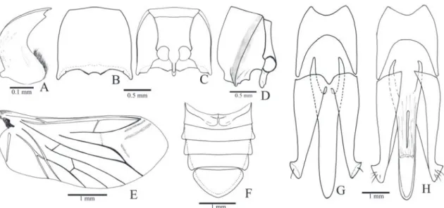
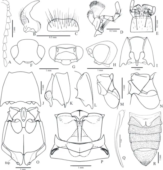
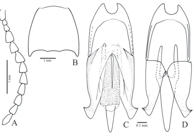
Redescription (female, Fig. 34A): Integument light brown with antennae, elytral base, ventral pterotho-rax and ventrites lighter; covered with short, fine and decumbent yellow setae, denser on ventral surface. Length 13.0 mm; elytral base as wide as prothorax; elytra 2.5 times longer than pronotum.
Head (Figs. 1B-C) with frons concave, fron-tal carina complete medially; frontoclypeal region steeply declivous to base of labrum, rectangular, about 3.0x wider than long; punctation fine and umbilicate, sparse at middle, dense on lateral and anterior margins. Antenna (Fig. 1A) with 11 an-tennomeres, serrate from antennomere III, nearly reaching the posterior half of the pronotum; an-tennomere III 2.0x longer than the II; IV as long as III. IEP 0.10. Mouthparts directed ventrally; labrum (Fig. 1B) semicircular, convex. Mandible (Fig. 1B) falcate, unidentate with laterodorsal sur-face with long setae; mesal margin at base with a row of short setae. Maxilla with galea trapezoidal, widened apicad, densely pilose; lacinia narrow, tongue-like, densely pilose; medistipes fused to the basistipes, corresponding area of the medistipes with one long stiff seta and several shorter and finer setae. Labium with mentum covered with fine and short setae. Maxillary and labial apical palpomeres elliptical.
Pterothorax (Figs. 1H, I): Mesoventrite with posterior region abruptly inclined ventrad about 43° in relation to anterior region, with anterior articulating surfaces
weakly concave; borders of mesoventral cavity curved. Mesocoxal cavity open to mesepisternum (Fig. 1I) (closed in the right side of the lectotype);
chantin not visible externally; mesocoxae separated by 0.7x their diameters; mesepisternum with a carina on inner anterior angle (Fig. 1I); mesometaventral su-ture distinct. Scutellum (Fig. 1J) cordiform, notched medially on anterior margin, abruptly elevated above the level of mesoscutum. Metaventrite (Fig. 1H) 1.7x wider than long, 1.6x longer than mesoventrite, finely and densely punctate; metepisternum about 5.0x longer than wide. Elytral sides parallel-sided to apical third then gradually tapering to apices; striae with a row of punctures, interstices convex, glabrous and non-punctate; epipleura abruptly narrowed near metacoxae. Hind wings (Fig. 1M) 1/4 shorter than elytra, unfolded; cross-vein r3 very short, radial cell
3.5x longer than wide, CuA1 absent; MP4 linked to
CuA2 (it is linked to MP3 in the other wing of the
same specimen); wedge cell absent; apex with anterior and posterior field sclerotizations; anal notch present.
Metacoxa (Fig. 1H) inclined 17° in relation to transverse axis of body; free margin of metacoxal plate short, absent on outer third. Metatrochanter and metafemur laterally convex, metafemur 2.2x longer than wide (Fig. 1L). Tibia (Figs. 1K, L) compressed laterally, 2.1x wider at apex than at base, with two spurs, a row of 10-13 spiniform setae along each outer and inner apical border and several spiniform setae on dorsal and ventral margins; tarsomeres simple, with fine and spiniform setae, decreasing in length from I-IV, V as long as III and IV together; claws simple.
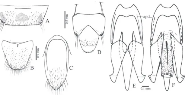
Abdominal ventrites with punctures umbili-cate, 0.5-1 diameters apart; posterior angles of ven-trites 2-4 with acute and prominent marginal plates; ventrite 5 pentagonal 1.5x wider than long. Tergite VIII (Fig. 1O) sparsely setose on anterior margin, anterior sclerotized margin rounded with a V-shaped notch medially. Sternite VIII (Fig. 1N) with base tri-angular with anterolateral angles acute directed later-ad, apex shortly emarginate, partly membranous with a pair of longitudinal sclerotizations; spiculum 4.75 times longer than base. Sternite X (proctiger) as long as paraprocts; paraprocts 2.24x as long as coxites.
Ovipositor (Figs. 1P, Q): baculi 4.6 times lon-ger than coxites, strongly sclerotized; coxites evenly strongly sclerotized, without setae, with apices acute and divergent, without styli. Reproductive tract (Figs. 1P, R): bursa copulatrix with a pair of narrow sclerotizations; pedunculate anterior sac spherical and weakly sclerotized; duct of spermathecal gland open-ing at base of the pedunculate anterior sac.
Lectotype: [Coll. Schwarz], [Africa or., Micindani, ex coll. F. Schneider], [Margogastrius schneideri Schw., Schneideri schneideri Schw], [Dtsch. Entomol.
Insti-tut Berlin], [coll. DEI, Müncheberg], [Margogastrius schneideri Schwarz, C. Girard vid. 1974], [S/F ?? Cardiophorinae, C. Girard det. 1974], [Lectotype], [Lectotype Margogastrius schneideri Schwarz, design. Douglas 2006], female (DEI).
Paralectotype: [Coll. Schwarz], [Africa or., Micinda-ni, ex coll. F. Schneider], [Margogastrius schneideri Schw.], [Dtsch. Entomol. Institut Berlin], [coll. DEI, Müncheberg], [Margogastrius schneideri Schwarz, C. Girard vid. 1974], [S/F ?? Cardiophorinae, C. Girard det. 1974], [Paralectotype], [Paralectotype Margo-gastrius schneideri Schwarz, design. Douglas 2006] 1 female (DEI).
Distribution: TANZANIA.
Remarks: Margogastrius present several characters at-tributed as synapomorfies to Cardiophorinae, such as cordiform scutellum, hind-wing without CuA1 and
wedge cell, wing anal area notched, female genitalia with a “glandular reservoir” (pedunculate anterior sac) entering the bursa copulatrix (Calder, 1996) and par-allel-sided prosternum (Douglas, 2011). The phylo-genetic analysis carried out by Douglas (2011) found a close phylogenetic relationship between Margogas-trius and Cardiophorinae species.
view seem to be the unique character that separate
Margogastrius from most Cardiophorus species and
Coptostethus globulicollis. Margogastrius also differs from those species in the mesocoxal cavity open to mesepisternum, neverthelles this is probably a labile character, since one specimen present one closed and one open mesocoxal cavity.
Margogastrius is similar to the fossorial genus
Patriciella Van Zwaluwenburg, 1953 from Australia (Calder, 1996) in its pronotum with lateral prono-tal carina absent, prosternal process tapered in lat-eral view, procoxal cavities open, scutellum cordate, metatrochanter strongly convex and tibiae widened apically. It differs from this Australian cardiophorine in (the later in parentheses): median occipital carina absent (present), free margin in metacoxal plate pres-ent (abspres-ent), the pronotum more convex, tibiae with dorso-apical angle straight (acute and produced) and metafemur slender. Margogastrius is also distinguish-able by its short antenna, elytral interstices convex and body ventrally and dorsally strongly convex.
Margogastrius is also similar to the fossorial car-diophorine Blaiseus in the tibiae expanded apicad, the unidentate mandibles and the prosternal process curved dorsad (Douglas, 2009). It differs from this genus in (the later in parentheses): lateral pronotal carina absent (present), scutellum notched anterome-dially (concave), elytral interstices convex (costate).
Blaiseus species occurred in Oriental Region, Central America and South Africa and the only known female belonged to B. zamoranoensis Douglas, 2009 from Honduras. This female shares with Margogastrius the small eyes and the short hind wing.
Teslasena Fleutiaux, 1892
Anelastes Kirby, 1857 (pars); Lucas, 1857: 71.
Physodactylus Fischer von Waldheim, 1823 (pars); Bonvouloir, 1875: 711.
Teslasena Fleutiaux, 1892: 405, 410; Schwarz, 1906: 310, 313; Schenkling, 1927: 509; Golbach, 1994: 27; Chassain, 2005: 65.
Type species (by monotypy): Anelastes femoralis Lucas, 1857.
Diagnosis (male): Mandible falcate, unidentate, an-tennae reaching or surpassing the posterior angles of pronotum, pronotal sides rounded with lateral carina directed dorsad anteriorly, not reaching the anterior margin of pronotum, prosternal process strongly curved with apex sagittiform in dorsal view, tapered in
lateral view; scutellum pentagonal; metafemur strong-ly convex with dorsal margin rounded, tibiae widened apicad, protibia with dorsal apical angle acute and produced, tarsomeres simple, claws bifid, elytral striae with a median and two lateral rows of punctures, the lateral ones smaller and each one bearing a seta.
Distribution: BRAZIL.
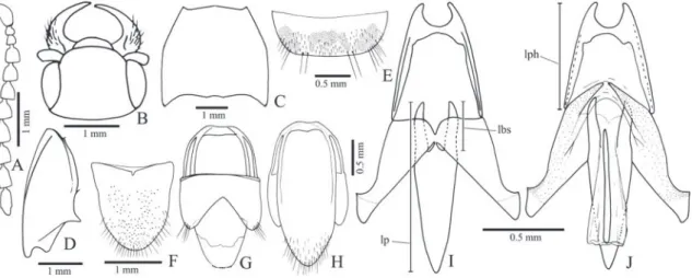
Redescription (male): Integument (Figs. 34B, C) shiny, evenly light brown to black or colored in variable pat-terns; dorsal surface glabrous, except for the scutel-lum, base of pronotum, elytral base and striae, which are covered with yellow setae in most, ventral surface densely covered by decumbent to semi-erect yellow setae. Total length: 7.5-12.0 mm; elytral base as wide as prothorax, elytra 2.6-3.1x longer than pronotum.
Head (Fig. 2B) directed anteroventrally, ante-rior margin of frons straight or rounded; frontal ca-rina complete, not produced anteriad; frontoclypeal region (Fig. 2C) steeply declivous to base of labrum 3.0-3.7x wider than long; punctation double, smaller punctures heterogeneously distributed, larger punc-tures sparse anteriorly, 0.2-0.5 diameters apart pos-teriorly. Antenna (Fig. 2A) with 11 antennomeres, antennomere II globular, III-XI serrate, III 2.5-2.7x longer than II. IEP 0.21-0.45. Labrum (Fig. 2D) con-vex, 2.5 times wider than long, densely and coarsely punctate, covered with long setae. Mandible (Fig. 2E) falcate, unidentate, laterodorsal face punctate with long and short setae, lateral edge evenly or abruptly curved to apex, mesobasal margin translucent and covered with microsetae. Maxilla (Fig. 32A, 32B) with medistipes partly fused to basistipes; medistipes triangular wider than long with several short and fine setae and one long and stiff seta; galea oval, outer an-terior part covered with long setae, inner part with denser and shorter setae; lacinia elongate, acutely nar-rowed apicad, densely pilose. Labium (Fig. 32C): pre-mentum with anterolateral angles strongly produced laterad, anterior margin fringed by short and fine se-tae; palpigers contiguous, palpomere II with 2-3 long and stiff setae. Maxillary and labial apical palpomere securiform.
punctures sparse; anterior angles indistinct; posterior angles non-carinate, narrow, acute and divergent. Hypomeron (Fig. 2H) with heterogeneous double punctation 1-3 diameters apart anteriorly, gradually sparser posteriad, smooth on anterior 1/5-1/4, poste-rior margin with an inverted V-shaped notch near the posterior angle. Notosternal suture (Fig. 2F) straight, hypomera not margined. Prosternum (Fig. 2F) 1.0-1.1x longer than wide, with homogeneous double punctation, 0.5-1.0 diameter apart, smaller punctures predominant; anterior margin straight, covering men-tum. Prosternal process (Figs. 2F, H) 2.2-2.4x longer than diameter of procoxae; ventral surface laterally compressed and ascending at about 45° to apex. Pro-coxal cavity closed.
Pterothorax (Figs. 2I-K): Mesoventrite with posterior region inclined about 60° above of the anterior region (Fig. 2K), with anterior articulating surfaces short and weakly concave; lateral lobes narrow, directed lateroposteriad; borders of mesoventral cavity straight divergent posteriad on anterior 2/3, convergent on apical third. Mesocoxal cavity closed, free trochantin absent; mesepisternum with a weak carina antero-medially (Fig. 2J). Mesometaventral suture distinct. Metaventrite 1.40-1.48x wider than long, 1.57-1.62x longer than mesoventrite, with double punctation; metepisternum 6 times longer than wide. Scutellum pentagonal 1.0-1.3 longer than wide with anterior margin straight, abruptly elevated above the level of mesoscutum. Metanotum (Fig. 2L) with prescutum separated medially from the scutum by a median membranous; posterior part of the scutellum elon-gate, with a longitudinal apodeme. Elytra with api-ces conjointly rounded, striae with a median row of punctures without seta and a pair of lateral row of mi-nuscule puncture each one bearing a seta (Fig. 32F); interstices flat on humeral region, convex posteriorly, smooth; epipleura gradually narrowed posteriad to metacoxa (Fig. 32G). Hind wing (Fig. 2M) with ra-dial cell about 2x longer than wide, CuA1 and wedge
cell absent; apex with anterior, median and posterior field sclerotizations; anal notch present.
Metacoxa inclined 28°-30° in relation to the transverse axis of body; inner third 1.7-2.1x longer than the dorsal outer 2/3; ventral part abruptly nar-rowed laterally with inner third 3.9-5.7x longer than outer 2/3; free margin of metacoxal plate absent (Fig. 2I). Tibiae with 8-14 spiniform setae along each outer and inner apical border (Fig. 32K), dorsal mar-gin covered with several irregular rows of spiniform setae, outer and inner surface covered with fine setae. Protibia 2.6-2.9x wider at apex than at base, with
dor-sal margin rugose, slightly curved or sinuous with api-cal angle acute and produced inwards (Fig. 32H). Me-sotibia 2.2-2.6x wider at apex than at base, with dorsal apical angle acute (Fig. 32I). Metatibia 2.7-3.1x wider at apex than at base with apical dorsal angle acute and produced laterad (Fig. 32J). Pro- and mesofemur sub-rectangular and convex; metafemur strongly convex with dorsal margin curved 1.7-1.9x longer than wide; metatrochanter less convex than metafemur. Tarso-meres (Fig. 32K) simple decreasing in length from I-IV, V shorter than III and IV together, densely cov-ered with short setae; ventral surface of tarsomere I and apical borders of tarsomeres I-IV with long spini-form setae. Claws bifid (Fig. 32L).
Abdominal ventrites (Figs. 2N, O) densely cov-ered with short and decumbent setae on lateral 3/4, setae short to moderately long and semidecumbent medially, with punctation double homogenously distributed 0.5-1.0 diameter apart; marginal plate absent laterally, present on the posterior corner of ventrites 1-4 forming an acute and produced angle di-rected posteriad; ventrite 1 with lateral part 3.6-7.0x longer than the median part, 2-3 subequal in length, 4 shorter than 3 medially; ventrite 5 flat, pentago-nal 1.5-2.1x wider than long. Pregenitalic segments and aedeagus covered with yellow setae. Tergite VIII (Fig. 2P) evenly sclerotized V-shaped with short se-tae on lateroposterior margin, anterior sclerotized margin curved with or without a median V-shaped notch; sternite VIII (2Q) evenly sclerotized or with a pair of circular membranous areas anteromedially, triangular to pentagonal with apex rounded, margin-ate or notched, covered with modermargin-ately to long se-tae posteriorly, anterior sclerotized margin straight or sinuous; sternite IX (Fig. 2R) fused to tergite IX at midlength of sternite IX, with anteromedian region membranous, latero-anterior margins sclerotized and strut-like; lateroposterior margins parallel or gradu-ally narrowed posteriad, apex rounded with short and long setae; tergite IX (Fig. 2S) with anterior margin curved, apical lobes rounded, glabrous; tergite X (Fig. 2S) with apex membranous, suboval, smooth and glabrous, evenly sclerotized in most.
translucent membrane. Penis posterior to struts with basal part triangular, posterior part narrow, parallel-sided 0.77-1.47x as long as the triangular basal part; with or without a very small ventral sclerite (about 1/8 the penis length); basal struts about 0.5x the total length of penis.
Remarks: The autapomorphies identified from Tes-lasena femoralis in the cladisitc analysis are shared by
Teslasena foucarti, making them apparent synapomor-phies of both species. Teslasena species are most simi-lar to Globothorax latidens by the pronotum convex with sides strongly rounded. Nevertheless the com-bination of several characters (elytral striae in three rows, apex of hind wing with three sclerotizations, tar-sal claws bifid, protibiae broadened apicad with dor-sal apical angle strongly prominent and the marginal plates of abdominal ventrites 2-3 strongly prominent) distinguishes Teslasena species from Globothorax latidens. Besides, Teslasena differs from G. latidens in (G. latidens characters in parentheses): procoxae and mesocoxae closed (open), mesoventrite more inclined posteriorly (almost horizontal), prementum with latero-anterior angles prominent (non-prominent), frons carinate (non-carinate), mandible smaller, la-brum without anterior fringe of setae (fringed with setae), shape of metatibiae and hind wing with three sclerotizations (two) (illustrations of G. latidens in Rosa, 2011).
I have examined only photos of Globothorax chev-rolati Fleutiaux, 1891, the type-species of the genus (figs. 105-107 in Rosa, 2011). This species is known only by female, which make comparisons with Teslase-na species difficult, since these species are known only by males. Nevertheless, some differences not observed elsewhere to have been related to sexual dimorphism in elaterids can be pointed out between Teslasena and
G. chevrolati (the latter in parentheses): labrum pen-tagonal (semi-elliptical), pronotum without notch between hind angle and median line (notched), pros-ternal process tapered in profile (truncate), free margin of metacoxal plate absent (present), claws bifid (trifid) and elytral striae with three rows of heterogeneous punctures (two rows of homogeneous punctures).
Teslasena species are similar to Patriciella from Australia (Calder, 1996 and specimen examined) shar-ing falcate mandibles, pronotum convex with sides rounded, prosternal process curved posteriad of coxae and tapered in lateral view, the shape of protibia, with dorsal margin sinuous and dorso-apical angle acute and produced, the metatrochanter and metafemur strongly convex and widened dorsoventrally, the ab-sence of free margin of metacoxal plate and abdominal marginal plate well developed. Teslasena differs from this fossorial cardiophorine in the presence of a lateral carina on pronotum, absence of the median occipital carina, procoxal cavity closed, meso and metatibia with dorso-apical angle less produced and tarsal claws bifid.
Key to species for male Teslasena
1. Metatibial inner spur straight (Fig. 32J), pronotum with disc, anterior and lateral borders glabrous or with a few small setae (Figs. 32D, E), posterior border covered with short setae ...T. femoralis (Fig. 34B) — Metatibial inner spur with tip curved, pronotum nearly entirely pilose with setae moderately long,
shorter on posterior border ...T. foucarti (Fig. 34C)
Teslasena femoralis (Lucas, 1857) (Figs. 2, 32, 34B)
Anelastes femoralis Lucas, 1857: 71.
Physodactylus femoralis; Bonvouloir, 1875: 711.
Teslasena femoralis; Fleutiaux, 1892: 411; Schwarz, 1906: 313; Schenkling, 1927: 509; Chassain, 2005: 66.
Teslasena lucasi Fleutiaux, 1899: 206; Schenkling, 1927: 509; syn. nov.
Redescription (male, Fig. 34B): Integument with variable color patterns: 1) entirely light brown or with antennomeres III-XI black; 2) brown to black
pronotum light brown with the median longitudinal third darker; 11) black with antenna, tibiae and tar-sus light brown. Total length 7.5-12.0 mm; elytra 2.6
times longer than pronotum. Head (Figs. 2B, C) con-vex or with a median circular depression, frontal carina straight or rounded; IEP 0.21-0.45 antenna reaching
the base of pronotal hind angle or surpassing it by one antennomere. Pronotum 1.11-1.3x wider than long, with double punctation on disc, larger punctures 1-3 diameters apart, denser on lateral borders; discal, an-terior and lateral borders glabrous, posan-terior border pilose with short setae; some specimens with a few pronotal discal or lateral punctures with a minuscule seta (Fig. 32E). Scutellum covered with short setae. Elytra tapered apicad from humerus or anterior third.
Aedeagus (Figs. 2T-V): Phallobase 0.27-0.32x the total length of aedeagus, 0.84-1.13x longer than wide, ra-tio between lateroposterior and median parts 3.8-6.7; penis: basal triangular part with sides straight, apical part 0.77-1.47 as long as the triangular basal part; with or without ventral sclerite.
Homeotype of T. femoralis: [Jatahy, prov. Goyas Brèsil, Sept. a Nov. 97], [Teslasena femoralis Luc, comparé 1899, Collection FLEUTIAUX], [Muséum Paris, Coll. E. Fleutiaux]; [Teslasena femoralis (Lucas) J. Chassain det. 05], male (MNHN).
Material examined: BRAZIL. Rondônia: 3 exs. (MZUSP); Mato Gosso: 65 exs. (MNHN), 50 exs. (MNRJ), 32 exs. (MZUSP); Mato Grosso do Sul:
66 exs. (MZUSP); Goiás: 1 ex. (IBSP), 2 exs. (DZUP), 7 exs. (MNHN), 6 exs. (MZUSP).
Distribution: BRAZIL. Rondônia: Vilhena; Mato Grosso: Utiariti, Chapada dos Parecis, Chapada dos Guimarães, Campo Verde; Goiás: Mineiros, Jataí;
Mato Grosso do Sul: Cassilândia, Costa Rica, Três La-goas, Maracaju, Porto Murtinho.
Remarks: Fleutiaux (1899) distinguished Teslasena lu-casi from T. femoralis by its “body short and black, sides of pronotum narrowed near the base but not sin-uous and punctation weaker”. The holotype of T. lu-casi and similar specimens from Jataí and Chapada dos Guimarães examined bear ratio between the body length and width (without head) 2.73-3.04. These are the smaller measurements of a ratio that varies from 2.73-3.29, with relatively short and long specimens found in populations from same region, as those from Três Lagoas with body ratio 2.95-3.29. Body en-tirely black as in T. lucasi holotype is found in some specimens from Chapada dos Guimarães, which also includes specimens with the color pattern 8. Some specimens with long body from Três Lagoas, Campo Verde and Mineiros also exhibited body entirely black. The specimens from Vilhena are black as T. lucasi
specimens, but they have body relatively longer and
larger punctation. The holotype and two specimens of T. lucasi identified by Fleutiaux have the smallest punctation and the smallest eyes (IEP 0.21-0.26), however these features were also found polymorphic in specimens of T. femoralis of a same region. There-fore, specimens of T. lucasi do not present any unique combination of characters that differentiates them from specimens of T. femoralis examined. For this rea-son I propose synonymy T. lucasi under T. femoralis.
Besides the polymorphisms in the body color pattern, pronotal punctation, eye size and relative body length, specimens of T. femoralis exhibit high variability in the antennal length, shape of the ster-nite VIII and in the ratio between the apical and basal parts of the penis. Specimens from Vera, Nova Ma-moré and a specimen of T. lucasi have the shortest penis apical part (0.77-0.96x the basal length), while specimens from Três Lagoas have apical part ranging from 0.77-1.47x the basal length. Ventral sclerite of penis is usually absent in T. femoralis, except in the specimens from Chapada dos Guimarães and Cha-pada dos Parecis, which present a small sclerite.
Teslasena foucarti Chassain, 2005 (Fig. 34C)
Teslasena foucarti Chassain, 2005: 66.
Redescription (male, Fig. 34C): Integument brown to black, legs lighter. Total length 9.0-12.0 mm; elytra 2.3-2.4 times longer than pronotum. Head concave or with a median circular depression, frontal carina straight between antennal insertions; IEP 0.25-0.35 antenna reaching or surpassing the hind angle of pro-notum by one antennomere. Propro-notum 1.05-1.10x wider than long, with double punctation on disc, larger punctures 0.5-1.0 diameters apart, denser on lateral borders; densely pilose with long decumbent setae. Scutellum covered with short setae. Elytra ta-pered apicad from humerus.
Aedeagus: Phallobase 0.31x the total length of ae-deagus, 1.10x longer than wide, length ratio between lateroposterior and median parts 4.3; penis: basal tri-angular part with sides straight, apical part 0.71x as long as the triangular basal part; with a ventral sclerite.
Paratypes: same data as holotype, 4 exs. (MNHN), 1 ex. (MZUSP).
Distribution: BRAZIL. Mato Grosso: Chapada dos Parecis.
Remarks: T. foucarti (Fig. 34C) was detailed described by Chassain (2005), who distinguished this species from T. femoralis in the shape of the anterior margin of frons, which is straight in T. foucarti, its antenno-meres relatively longer, the pronotum narrower ante-riorly with punctation larger on lateral and anterior borders and the metatibial inner spur with tip curved. As discussed above, the analysis of a large series of
T. femoralis revealed that those features, with the ex-ception of the curved metatibial spur, are highly vari-able among the specimens and do not constitute con-sistent diagnostic characters. On the other hand, the curved tip of the metatibial inner spur was observed only in T. foucarti specimens, which also present body pilosity longer. The most evident difference in the pilosity of these species was observed in the pro-notum, which is moderately long and usually dense on T. foucarti and absent or minuscule and scarce in
T. femoralis. Their aedeagus do not present significant differences.
Idiotropia Schwarz, 1897
Athous Eschscholtz, 1829 (pars); Abeille de Perrin, 1894: 91.
Idiotropa Schwarz, 1897: 63 (misspelling recognized by Schwarz, 1906: 314).
Idiotropia Schwarz, 1906: 310, 314; Schenkling, 1927: 510.
Type species (by monotypy): Athous henoni Abeille de Perrin, 1894.
Diagnosis (male): Frontal carina absent, mandibles long, bidentate directed anteriorly; pronotum quadrate, weakly convex, sides widened anteriorly, anterior angles right not produced, posterior angle without carina; scutellum cordiform wider than long, convex and perpendicular to mesoscutum at base; elytra subparallel to posterior third, not fused to each other, wings absent; legs narrow, tibia and tarsomeres 1-2 with spiniform setae, tibial spurs robust, longer than setae, tarsomeres and claws simple.
Distribution: ALGERIA.
Idiotropia henoni (Abeille de Perrin, 1894) (Figs. 3, 4, 34D)
Athous henoni Abeille de Perrin,1894: 91.
Athus henoni; Schwarz 1897: 63 (misspelling).
Idiotropa henoni; Schwarz, 1897: 63 (misspelling).
Idiotropia henoni; Schwarz 1906: 314; Schenkling, 1927: 510.
Redescription (male, Fig. 34D): Body weakly convex; integument light to dark brown, legs and epipleura lighter; covered with short, fine and yellow setae, erect with curved apex. Total length: 4.0-4.5 mm; elytral base 0.9-1.0x as wide as prothorax; elytra 2.3-2.4x longer than pronotum.
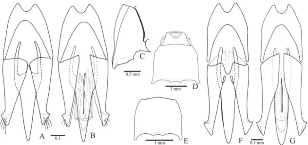
Head (Fig. 3B) with frons flattened or slightly concave at middle, frontal carina absent; frontoclypeal region 45° declivous to base of labrum; punctures um-bilicate, 0.5-1.0 diameter apart. Antenna (Fig. 3A) with 11 antennomeres, serrate from antennomere IV, surpassing pronotal posterior angles by two an-tennomeres; antennomere III 1.1x longer than the II; IV 1.7x longer than III; antennomere IV 1.4x lon-ger than wide. IEP 0.31-0.33. Mouthparts directed anteriorly. Labrum (Fig. 3C) semielliptical, convex, covered with long setae. Mandible (Fig. 3C) long, falcate, bidentate, apical tooth long and acute, sub-apical tooth short and acute; laterodorsal face at base densely punctate, with long setae; lateral edge evenly curved apicad; mesal margin at base with a row of short setae. Maxilla with galea membranous, oboval, densely pilose; lacinia elliptical, densely pilose; me-distipes trapezoidal, convex, longer than wide, with several long and short setae; Labium with mentum trapezoidal with a lateroposterior pair of long setae, parpifers contiguous. Maxillary and labial apical pal-pomeres elliptical.
Pterothorax: Mesoventrite with posterior region in-clined ventrad about 25° in relation to anterior region (Fig. 3I), anterior articulating surfaces concave mar-ginate posteriorly by a curve carina (Fig. 3L); borders of mesoventral cavity divergent from base to posterior margin of posterior lobes then curved and convergent. Mesocoxal open to mesepisternum and mesepim-eron, mesotrochantin visible externally (Figs. 3H, I); mesepisternum (Fig. 3H) without carina, with inner and outer angles rounded. Mesoventrite separated from metaventrite at middle by a superficial groove (Figs. 3I, L). Metaventrite (Fig. 3L) 1.91x wider than long, 1.0-1.1x as long as mesoventrite, with puncta-tion 0.5-1.0 diameter apart; metepisternum about 11 times longer than wide. Elytra weakly convex with
sides parallel to posterior third, apices conjointly rounded; striae with a row of punctures surrounded by smaller punctures; interstices convex, epipleurae abruptly narrowed near metacoxae. Hind wings ab-sent. Scutellum (Fig. 3J) cordiform 1.6-1.7x wider than long, convex, basal part perpendicularly elevated above the level of mesoscutum (Fig. 3K). Metanotum (Fig. 3J) with apodemes and allacrista reduced, scutel-lum and postnotum indistinct.
Metacoxa (Fig. 3L) about 30° inclined in rela-tion to transverse axis of body; outer half of the ven-tral part reduced to a line; inner half of the venven-tral part about 3 times longer than the dorsal lateral part; free margin of metacoxal plate absent to very short. Legs (Figs. 3M-O) narrow, sparsely pilose. Metafemur 2.8x
longer than wide, tibiae 2.0-2.6 wider at apex than at base, with two subequal spurs stout and longer than setae, with a row of spiniform setae along the dorsal margin and 5-7 spiniform setae along apical outer and inner borders; spiniform setae decreasing in width and length from pro- to metatibia; tarsomeres simple, with dense, long pilosity on ventral face, tarsomeres V longer than III and IV together; claws simple.
Abdominal ventrites with punctures umbilicate, 1.0-2.0 diameters apart; posterior angles of ventrites right, without marginal plates; ventrite 1 as wide as 2, gradually decreasing in width from 3 to 5; lateral part of ventrite 1 about 4.0x longer than its median part; 5 semioval 1.5-1.7x wider than long, convex at apex. Spiracles of tergite I small (Fig. 3J). Tergite VIII (Fig. 4D) suboval, evenly sclerotized, anterior margin curved, with minuscule setae on posterior margin; sternite VIII (Fig. 4C) 1.84 wider than long, sclero-tized along anterolateral region, anterior margin pro-duced anteriad and emarginate at middle; lateropos-terior angles rounded and produced with a few short setae. Sternite IX (Fig. 4A) partly sclerotized, fused to tergite IX at anterior region of sternite IX, with anterior margin straight, sides subparallel, tapering to apex on posterior third; apex sparsely pilose. Tergite IX (Fig. 4B) evenly sclerotized, apex of posterior lobes with a few long setae; tergite X (Fig. 4B) semioval with apex membranous, glabrous.
Aedeagus (Figs. 4A, B): Phallobase suboval, convex with anterior margin folded dorsad, 0.33x length of aedeagus; length ratio between lateroposterior and median parts 0.2. Parameres dorsoventrally flattened, apex securiform elongate with lateral angle acute
slightly produced, weakly sclerotized, without setae. Penis with sides gradually tapering from base of basal struts to apex, basal struts 0.16x length of penis; dor-sal articulation of penis with a short narrow process between the basal struts fused to the parameres; ven-tral sclerite narrow with apex upturned.
Holotype: [Athous Henoni Abeille], [Collection Hé-non], [Type], [Constantine, coll. HéHé-non], [Muséum Paris Coll. E. Fleutiaux] male, (MNHN).
Material examined: Without locality: 5 exs. (MNHN). ALGERIA, Constantine: 1 ex. (MNHN).
Distribution: ALGERIA.
Remarks: Idiotropia henoni presents the mesometaster-nal suture grooved which makes the meso- and meta-ventrite seem to be separated at middle, which was also observed by Candèze (1857) in South African species of Beliophorus Eschscholtz, 1829 (Dimini). Idiotropia henoni is similar to the Neotropical monotypic Aptero-elater Golbach, 1964 (Dimini) in the metacoxal plate reduced laterally, in the convex scutellum wider than long, pronotal shape, general punctation and absence of hind wings (figs. 3, 5 in Golbach, 1964).
Oligostethius Schwarz, 1906
Idiotropia Schwarz, 1897: 63 (pars); Schwarz, 1903: 375.
Oligostethius Schwarz, 1906: 310, 314; Schenkling, 1927: 510.
Type species (by monotypy): Idiotropia capensis
Schwarz, 1903.
Diagnosis (male): Frontal carina absent between an-tennal insertions, mandibles long, bidentate; protho-rax elongate, convex, sides rounded, wider than ely-tra, pronotum coarsely punctate, with anterior angles strongly produced over the eyes, posterior angles carinate; posterior margin of pronotum with a pair of lateral incisions; scutellum elliptical wider than long and strongly convex; elytra about twice longer than pronotum fused to each other, wings absent; legs nar-row, tarsomeres and claws simple.
Distribution: SOUTH AFRICA.
Oligostethius capensis (Schwarz, 1903) (Figs. 5, 6, 34E)
Idiotropia capensis Schwarz 1903: 375.
Oligostethius capensis; Schwarz, 1906: 315; Schen-kling, 1927: 510.
Redescription (male, Fig. 34E): Body convex. Integu-ment brown or dark brown with antennae and legs light brown, covered with very short, fine, decumbent silvery setae. Total length: 9.6-10.0 mm; elytral base 0.83x as wide as prothorax, elytra 1.91x times longer than pronotum.
Head (Fig. 5C) with frons (Fig. 5A) concave at median line, frontal carina absent medially; fron-toclypeal region gradually declivous to base of la-brum; punctures umbilicate, 0.5-1.0 diameter apart; antennal insertion (Figs. 5A, B) placed within saucer-like impression. Antenna with antennomere II cylin-drical 1.3x longer than wide; antennomeres III-VI convex and serrate; III 1.7x longer than II and 1.6x wider than long; IV-VI subequal 0.92x as long as III and 1.1x wider than long. IEP 0.24; mouthparts di-rected ventrally. Labrum (Fig. 5A) semielliptical, con-vex. Mandible (Fig. 5A) narrow and long, bidentate, apical tooth long and acute, subapical tooth smaller; laterodorsal surface at base coarsely punctate with long setae, lateral edge evenly curved apicad; mesal margin at base with a row of short setae. Maxilla with galea oboval, densely pilose, lacinia elliptical, densely pilose; medistipes trapezoidal longer than wide with several long stiff setae and a few finer and shorter setae. Labial palpigers separate; mentum trapezoidal with a lateroposterior pair of long setae and sparsely short setae.
Pronotum (Fig. 5D) dorsally convex, 1.17-1.19x longer than wide, sides rounded, lateral carina com-plete (Fig. 5F), with punctures umbilicate, coalescent forming irregular striae; anterior angle produced an-teriorly, wide and rounded covering half eye (Fig. 5F); posterior angle acute, short, with tip upturned (Fig. 3F), carinate; posterior margin adjacent to pos-terior angle with a short incision contiguous to a
na. Hypomeron (Fig. 5G) with punctures umbilicate, 0.5-1.0 diameter apart; posterior margin straight and contiguous to tip of posterior angle, without notch. Notosternal suture (Fig. 5E) margined by a shiny band along hypomeron margin at posterior half, grooved anteriorly. Prosternum (Fig. 5E) parallel-sided on pos-terior half, with divergent sides on anpos-terior half, 2.31x longer than wide, with punctures umbilicate, 0.5-1.0 diameters apart; anterior prosternal lobe rounded and produced, covering mentum. Prosternal process (Figs. 5E, F) with ventral surface laterally compressed and ascending at about 45° to apex, 2.8x longer than procoxal diameter. Procoxae open.
Pterothorax: Mesoventrite gradually inclined ventrad posteriorly about 30°, anterior articulating surfac-es (Figs. 5H, I) concave marginate posteriorly by a prominent carina; borders of mesoventral cavity di-vergent from base to anterior margin of mesocoxae then curved and convergent. Mesoventral cavity deep, floor with a smooth median band bordered by minus-cule punctation (Fig. 5H). Mesocoxal cavity (Fig. 5I) open to mesepisternum and mesepimeron, mesepim-eron long, closing the major part of cavity; mesotro-cantin visible; mesepisternum with a carina on the outer angle and a prominent carina on inner anterior angle contiguous to the anterior carina of mesoven-trite. Mesoventrite separated from the metaventrite at middle by a weak groove. Metaventrite (Fig. 5H) 1.91x wider than long, 0.9x as long as mesoventrite, with punctation 0.5-1.0 diameter apart; metepister-num about 7x longer than wide. Elytra fused to each other along the median suture, sides divergent from
humerus to posterior third then rounded to apex; stri-ae with a row of punctures, interstices flat with dense, rasp-like punctation, epipleura abruptly narrowed near metacoxae. Scutellum (Fig. 5J) elliptical wider than long, strongly convex, abruptly elevated above the level of mesoscutum. Hind wings absent.
Metacoxa (Figs. 5H, I) inclined 25° in relation to transverse axis of body, median region of the dorsal surface about 15x longer than outer region; free mar-gin of metacoxal plate very small. Legs densely pilose, setae moderately long and curved apically. Metatro-chanter and metafemur compressed laterally, metafe-mur 3.5x longer than wide. Tibiae compressed later-ally, 2x wider at apex than at base, with two subequal spurs shorter than setae, with a row of 10-13 stiff setae along apical border; tarsomeres simple, densely setose on ventral face, decreasing in length from I-IV, V as long as III and IV together; claws simple.
Abdominal ventrites (Figs. 5K, L) with punc-tures umbilicate, 0.5-1.0 diameter apart, marginal plates absent; lateral part of ventrite 1 2.0x longer than its median part, ventrite 1 narrower than 3, 3-4 subequal, 5 semioval 1.12x wider than long. Sternite VIII (Fig. 6A) subrectangular 2.73x wider than long with posterior margin emarginate, predominantly membranous with a pair of dark sclerotizations along the anterior margin and a pair of lateroposterior light scletotizations; tergite VIII (Fig. 6B) semioval, pilose, evenly sclerotized except for a membranous triangular region on anteromedian margin; sternite IX (Fig. 6C) partly sclerotized, fused to tergite IX at half length of sternite IX, with anterior margin bilobed and pro-duced dorsad; posterior margin rounded, apex with