439
Two new cis-Andean species of the South American catfish genus
Megalonema
allied to trans-Andean
Megalonema xanthum
, with
description of a new subgenus (Siluriformes: Pimelodidae)
John G. Lundberg and Wasila M. Dahdul
A revised diagnosis of the pimelodid catfish genus Megalonemais given based on synapomorphic features of the Weberian complex and gas bladder. Megalonema xanthum from the Magdalena River is redescribed. Two new cis-Andean species of Megalonemaare described, M. amaxanthum n. sp. from the Amazon River basin, and M. orixanthum n. sp. from the Orinoco River basin. These three species are differentially diagnosed by shape and size of the supraoccipital posterior process, adipose-fin shape, vertebral counts, eye size, premaxillary bone shape and dentition, length of the anal-fin base, width between the posterior nostrils and presence/absence of dentations on the pectoral spine. Eretmomegalonema new subgenus is established for M. xanthum, M. amaxanthum and M. orixanthum and supported by the uniquely synapomorphic paddle-like structure of its pelvic fin and hypertrophied basipterygium. Unambiguous synapomorphies indicate a sister-group relationship between M. amaxanthum and M. orixanthum, with M. xanthum basal to this pair. This topology is congruent with the Neogene origins of separate Magdalena, Amazon and Orinoco basins suggesting vicariant control of diversification of Eretmomegalonema.
Uma diagnose do gênero Megalonema é fornecida baseada em caracteres sinapomórficos do aparelho de Weber e da bexiga natatória. Megalonema xanthum do rio Magdalena, é redescrita. Duas novas espécies cis-Andinas de Megalonema são descritas:M.amaxanthum sp. n. da bacia Amazônica, e M. orixanthumda bacia do rio Orinoco. Estas três espécies são diagnosticadas pela forma e tamanho do processo supraoccipital posterior, forma da nadadeira adiposa, contagem do número de vértebras, tamanho do olho, forma do premaxilar e dentição, comprimento da base da nadadeira anal, distância entre as narinas posteriores, e presença/ausência de dentições no espinho da nadadeira peitoral. Eretmomegalonema, novo subgênero, é estabelecido para M.xanthum,M.amaxanthumeM.orixanthum e suportado pelas únicas estruturas sinapomórficas da nadadeira pélvica em forma de remo, e do basipterígio hipertrofiado. Claras sinapomorfias indicam uma relação de grupo irmão entre M.amaxanthum e M.orixanthum, com M.xanthum basal a esse grupo. Esta topologia é congruente com a origem Neogênica das distintas bacias do Magdalena, Amazonas e Orinoco, sugerindo um evento vicariante de diversificação de Eretmomegalonema.
Key words: Eretmomegalonema,Megalonema amaxanthum,Megalonema orixanthum.
Department of Ichthyology, The Academy of Natural Sciences, 1900 Benjamin Franklin Parkway, Philadelphia, PA 19103-1195, USA. lundberg@ansp.org (JGL), dahdul@ansp.org (WMD)
Introduction
The pimelodid catfish genus Megalonema has an ample distribution in South America throughout the Paraná, Amazon, Essequibo, Orinoco, Maracaibo and Magdalena basins. The species of Megalonema inhabit medium to large rivers mostly in flowing marginal reaches and over fine substrates of mud or sand. These are streamlined catfishes of modest size with
from the río Magdalena, Colombia (Eigenmann, 1912b). Subsequent species descriptions and taxonomic transfers have raised the number of presumed valid species in Megalonema to six (Lundberg & Littmann, 2003). The existence, however, of at least one undescribed cis-Andean species has been known for over 25 years and our recent study of Amazon and Orinoco populations indicates additional undescribed species. The purpose of this paper is to describe two new species that are related to M. xanthum, and to recognize these three as a monophyletic subgroup of Megalonema characterized by oddly hypertrophied pelvic fins. In a subsequent paper we are revising the remaining species of the genus.
Material and Methods
Measurements and counts follow Lundberg et al. (1991), Lundberg & Parisi (2002) and Lundberg & Akama (2005) with the addition of the distance from snout tip to insertion of outer pelvic-fin ray, and least distance from orbital margin to posterior nostril. Measurements are straight-line distances taken with a digital caliper to the nearest 0.1 mm from a sample of 136 specimens ranging in standard length (SL) from 38.2 to 120.0 mm. Measurement ratios are expressed in thousandths (mils) in text and Table. We used least squares regression to analyze variation in log10 transformed variables, and t-tests of the residuals from regression lines to assess taxonomic differences in proportional measurements. Vertebral counts include the five elements united in the Weberian complex; the first caudal vertebra is that immediately posterior of the visceral cavity and recognized by its fully developed hemal spine; the compound centrum of the caudal skeleton is counted as one. Radiographed (R), cleared and stained (C&S), and articulated dry skeletal (Sk) specimens were used for counts of vertebrae and median-fin rays. Institutional acronyms are as listed on the Catalog of Fishes website at http:// www.calacademy.org/research/ichthyology/catalog/ abtabr.html.
Results
The genus Megalonema possesses nearly all of the diagnostic synapomorphies for the following nested groups (Lundberget al., 1991: 842, fig. 1; see also Hardman & Lundberg, 2006): Pimelodidae sensu Lundberg & Littmann (2003), non-phractocephaline pimelodids (i.e., all Pimelodidae except Steindachneridion, Phractocephalus, Leiarius, Perrunichthys), and the Calophysus-Pimelodus clade. Megalonema is exceptional among Pimelodidae in lacking a deep sutural joint between the fifth and sixth centra. In the
1986; Lundberg et al., 1991).
Beyond the aforementioned, the relationships of Megalonema are unsettled. Driver (1919) interpreted similarities of the reduced gas bladders, encapsulating Weberian complexes and external morphology among Megalonema,Luciopimelodus and Pinirampus (as Perugia and including M. xanthum) as indicative of relationship, placing these taxa together as the Luciopimelodinae. Alternatively, Stewart (1986) described a number of unambiguous synapomorphies supporting his Calophysus group: Calophysus, Pinirampus, Luciopimelodus, Pimelodina and Aguarunichthys. Stewart found that Megalonema exhibits unique features of the gas bladder and Weberian vertebral structure that set it apart from the Calophysus group, although he pointed out other possible synapomorphies in the reduced structure of the pectoral spine, elevated pectoral-fin ray count and loss of the posterior cleithral process. Lundberg et al. (1991) tentatively leaned toward placing Megalonema basal to the Calophysus group. We are pursuing the question of the systematic position of Megalonema with both morphological and molecular data.
MegalonemaEigenmann, 1912
Megalonema Eigenmann, 1912a: 150. Gender neuter. Type species.Megalonema platycephalum Eigenmann, 1912a, by original designation.
Diagnosis.Eigenmann’s (1912a) diagnosis of Megalonema is a mixture of derived and plesiomorphic character states, each with a checkered distribution among pimelodids. Accordingly, we offer an alternative diagnosis emphasizing unique or unambiguous synapomorphies. Thus, Megalonema is characterized by the following. A unique structure of the gas bladder and its encapsulating processes of the Weberian complex (Fig. 1) including: reduced, dumbbell-shaped gas bladder with distended lateral lobes connected by a tubular commissure ventrally crossing Weberian compound centrum; paired, ventrally incomplete bony capsules surrounding lateral lobes of gas bladder formed by deeply down-curved 4th
transverse processes of Weberian complex; a prominent ventral flange of compound centrum immediately anterior to tubular commissure of gas bladder and sutural joint with 5th
typical intervertebral joint without interdigitating sutures, processes or superficial ossification (Fig. 1).
Some other pimelodids possess modifications of the fourth transverse processes for encapsulation of gas bladder: Hypophthalmus (Howes, 1983) and the genera of the Calophysus group (Stewart, 1986; Driver, 1919). Many osteological details of the Weberian complex, however, differ among these groups and Megalonema. Further, the gas bladder morphologies differ strikingly among them: bilobed
inMegalonema, small median sac with anterior projections in theCalophysus group, and a pair of small separate sacs in Hypophthalmus.
We also note that Megalonema may have a possibly diagnostic dark caudal spot embedded within the base of upper lobe of the caudal fin, internal to uppermost 4-5 branched fin rays. Some specimens show a matching spot on the lower lobe. We have observed the upper caudal spot in specimens of all species except M. argentina (no specimens examined) andM. xanthum for which the only available specimens are the faded types collected nearly a century ago. It is possible thatM. xanthum has the caudal spot in life but it is bleached out as it is in faded specimens of other species. Upper lobe caudal spots in a similar position are also found in Pimelod-idae in Hemisorubim,Platysilurus and Platystomatichthys, but in these the pigmentation is chromatophores in the skin, external to the fin rays.
Eretmomegalonema, new subgenus
Type Species. Megalonema xanthum Eigenmann, 1912, by original designation. We establish this taxon for a subgroup of the genus Megalonema containing M. xanthum, M. amaxanthum new species and M. orixanthum new species. Diagnosis.Eretmomegalonemais uniquely characterized by the paddle-like structure of its pelvic fin and hypertrophied basipterygium (Figs. 2a-b). Both sexes and juveniles exhibit these features. The outer three pelvic-fin rays (1st-3rd) are
elongated and a little thickened to form a prominent paddle-like lobe. These rays exceed twice the length of the inner-most (6th) pelvic ray, and about 1.4 times the length of the 4th.
The 1st pelvic ray is proximally unbranched and unsegmented
for about half its length and its segments are foreshortened (square). The 2nd pelvic ray is similarly unbranched and
unsegmented for about half its length, then distally it is asymmetrically branched and segmented as follows. Distal to its first division, the 2nd ray’s outer primary ramus does not
further divide and its segments are square; the inner primary ramus divides into secondary rami of which the outer ramus remains undivided and with short segments, whereas the inner secondary ramus divides into thin, tertiary rami with more elongated (rectangular) segments. The 3rd ray is unbranched
and unsegmented for about 40% of its length; its primary rami further divide twice and have slightly elongated segments. In contrast to the outer rays, the inner three rays are slender, branched before their midlength, and their segments are elongate rectangles.
The basipterygium is elongated with an overall bony length nearly twice the combined width of both basipterygia. Also, the midline symphysis is complete along the entire length of the anteromedial process, basal plate and posterior process. The posterior process is long and has a triangular shaped tip. On the ventral side, the anteromedial process has a proximal lateral-margin crest. Dorsally, the anterolateral Fig. 1. a, Weberian complex, post-Weberian vertebrae,
basioccipital region and upper bones of shoulder girdle of Megalonema cf. platycephalum, ANSP 178515. b, Weberian complex and gas bladder of M. orixanthum, ANSP 187450. aoc = bony aortic canal; bocc = basioccipital; ccfl = ventral flange of compound centrum; tcom = position of gas bladder tubular commissure posterior to ventral flange; clr = cleithral ring; cpcv = position of bony canal for passage of posterior cardinal vein; gbld = lateral distention of gas bladder; gbtc = transverse commissure of gas bladder; ij5-6 = typical intervertebral joint between c5 and c6; tp4 = ventrally incomplete bony capsules surrounding lateral lobes of gas bladder formed by deeply down-curved 4th transverse
process has on its lateral-margin a prominent high crest that curves medially near its tip. The crests are associated with the arrector, adductor and abductor muscles of the elongated outer pelvic-fin rays.
In marked contrast to Eretmomegalonema, other species of Megalonema (Figs. 2 c-d) and other pimelodids lack pelvic-fin lobes, although in some the pelvic pelvic-fins may be generally elongated. Also, no other Megalonema(Figs. 2 c-d) or other pimelodids have the high dorsal crest on the anterolateral process (compare Fig. 2b). Also, other species of Megalonema have a wide gap across the midline between the anteromedial processes (Figs. 2 c-d), but a completely bony basipterygial symphysis including the anteromedial processes (compare Figs. 2a-b) is found in some other pimelodids (Parisi et al., 2006; Rocha et al., 2007): Propimelodus, Exallodontus, Pimelodus altissimus,Duopalatinus peruanus,Cheirocerus and the Calophysus group.
Etymology. Subgenus Eretmomegalonema, from Greek eretmon, oar, in reference to the paddle-like pelvic fins, and megalonema, name of the catfish genus to which the new taxon belongs. Gender Neuter.
Key to the species of Subgenus Eretmomegalonema 1. Supraoccipital posterior process narrow, its basal
width less than half its length, its sides gently tapering (Fig. 3b); adipose fin rising steeply and terminating
posterior to vertical through tips of adpressed anal-fin rays; eye small, horizontal diameter contained 5-6 times in head length; pectoral spine with numerous, erect dentations along posterior margin; premaxillary tooth band broad with about 8-10 rows of teeth; 18-20 gill rakers on first arch. Magdalena River Basin ……… Megalonema xanthum 1’. Supraoccipital posterior process broad based, its basal width equal to or greater than its length, and its sides abruptly tapering (Figs. 3c,d); adipose fin rising at a shallow angle from back and terminating at or anterior to vertical through tips of adpressed anal-fin rays; eye large, horizontal diameter contained 3-5 times in head length; premaxilla extremely narrow with one row of teeth or none; 25 or more gill rakers on first arch. Cis-Andean distribu-tion………2 2. Supraoccipital posterior process bell-shaped in outline, its tip blunt and separated by a distinct gap from supraneural (Fig. 3c); pectoral spine lacking dentations, premaxilla with a single row of teeth. Amazon River Basin ……… Megalonema amaxanthum 2’. Supraoccipital posterior process triangular in outline,
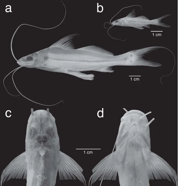
Megalonema xanthum Eigenmann, 1912 Fig. 4
Megalonema xanthum Eigenmann, 1912b: 16 [original description; type locality: Colombia, Cudinamarca Department, Girardot, Magdalena River]. -Stewart, 1986; 670 [anatomy]. -Maldonado-Ocampo et al., 2005: 170, fig. 160, map 162 [description, distribution, specimen list]. Perugia xanthus. -Driver, 1919:453, pl. 2, figs. A, C, D, pl. 3,
fig. 5 [anatomy of Weberian complex and gas bladder, in key, relationships]. -Eigenmann, 1922: 35, pl. 3, fig. 3 [ho-lotype photograph].
Diagnosis. A species of the subgenus Eretmomegalonema distinguished from others by the following combination of features. Supraoccipital posterior process (Figs. 4b, 3b) narrow, its basal width less than half its length, its sides gently tapering to a sharp tip separated by a distinct gap from supraneural; adipose-fin (Fig. 4a) large, its margin anteriorly rising steeply and straight or in a broad convex curve to a gently curved apex; adipose fin terminating posterior to ver-tical through tips of adpressed anal-fin rays; total vertebrae modally 47, range 45-48 (Table 1); eye relatively small, hori-zontal diameter 154-195 mils of HL; anal-fin base relatively short, 94-117 mils of SL (Fig. 5a); width between posterior nostrils relatively narrow, 138-179 mils of HL (Fig. 5b); pecto-ral spine with numerous, erect dentations along posterior margin; premaxillary tooth band broad with about 8-10 rows of teeth; and 18-20 gill rakers on first arch.
Description. Meristic data for 5 to 23 specimens are in Table 1, and morphometric data for 20 to 23 paratypes are in Table 2. Megalonema xanthum (Fig. 4) is a medium sized pimelodid with a maximum length known to us of 159.3 mm SL in the holotype (note that the length of 202 mm reported by Eigenmann (1912b) is consistent with Total Length). Dorsal
profile of head and nape gently convex from snout tip to vertical at insertion of maxillary barbel, then nearly straight to origin of dorsal fin, then body profile scarcely convex along dorsal-fin base, straight to posterior insertion of adipose fin, and a little concave across caudal peduncle. Ventral profile slanting convexly ventrally from snout tip to mental barbels, nearly straight to pectoral-fin origin, straight or convex across abdomen to anal-fin origin, then slanting dorsally to caudal peduncle and slightly concave along caudal peduncle.
Cross-sectional shape roughly trapezoidal from snout to supraoccipital, then deeply and broadly triangular to dorsal-fin origin, and increasingly compressed to caudal dorsal-fin. Maximum body depth at dorsal-fin origin contained 4.5-6.3 times in SL. Maximum body width across cleithra in front of pectoral spine insertion, contained 5.8-6.8 times in SL.
Head of moderate length, contained 4.1-4.8 times in SL and relatively deep, its depth at base of supraoccipital posterior process slightly less than body width. Head covered Fig. 3. Details of supraoccipital processes of a,Megalonema cf. platycephalumANSP 178450,b,M. xanthumCAS 63674, c, M. amaxanthum, holotype, CBF 11896 (ex FMNH 106769), and d,M. orixanthum ANSP 148180, paratype. Paired lines on each image show the gap between the tip of supraoccipital process and supraneural anteromedial point in or just below skin.
soft pectoral rays 11 12 13 14
M.xanthum 1 6 5
M.amaxanthum 5 9 2
M.orixanthum 1 6 3 gill rakers on first branchial arch
18 19 20 / 25 26 27 28 29
M.xanthum 1 3 1
M.amaxanthum 1 1 2
M.orixanthum 1 3
total vertebrae
42 43 44 45 46 47 48
M.xanthum 1 1 18 3
M.amaxanthum 21 21 14 5
M.orixanthum 12 21 2
by thin skin, skull roofing bones relatively smooth, not orna-mented with tubercles or ridges. Snout moderately long, con-tained 2-2.3 in head length, and with broadly rounded margin in dorsal and ventral views. Snout and upper jaw projecting; with mouth closed less than half of premaxillary dentition exposed. Anterior nostril behind snout margin in a shallow, circular depression, its aperture dorsally directed and encircled
posterior nostrils equal to or a little greater than distance between posterior nostril and eye.
Eye relatively small, about 1.5-2 times longer than deep, placed dorsolaterally and centered on midpoint of bony head length; interorbital space scarcely convex. Interorbital wide, containing horizontal eye diameter 1.2-1.6 times. Anterior cra-nial fontanelle narrow, parallel-sided, originating behind level of posterior nostrils and terminating between eyes at epiphy-seal bar. Posterior cranial fontanelle closed anteriorly, per-sisting as oval aperture in center of supraoccipital. Supraoc-cipital posterior process narrow-based (its basal width con-tained about 3 times in its length), dorsally flat, not angular or keeled, but prominent with sides scarcely tapering to pointed tip, failing by about half of its own length to contact supraneural (anterior nuchal plate obsolete).
Mouth subterminal, opening anteriorly and widely; gape broad, its width across inner surface of ricti twice greater than interorbital width. Lips thin-skinned and smooth. Rictal fold well defined but not fleshy or swollen, subtended above and below by deep folds respectively reaching base of maxillary barbel and about one third distance to mandibular symphysis. Premaxillary tooth band of uniform length along transverse width, with bluntly rounded posterolateral corner. Premaxillary teeth conical, fine, in about 8-10 irregular rows, more numerous in larger specimens. Dentary teeth similarly slender, arranged in about 6-8 rows at symphysis and fewer posterolaterally. No prevomerine, metapterygoid or accessory
tooth plates present on the palate.
Origin of maxillary barbel in depression close to base of anterior nostril and above rictus, continuing to below eye; maxillary barbel reaching just beyond posterior end of adipose fin to base of caudal fin. Outer mental barbel reaching near or onto anal fin, not beyond adipose fin. Tip of inner mental barbel reaching near pelvic fin insertion. Branchiostegal membranes anteriorly united to isthmus and overlapping before diverging. Gill rakers long, slender and bladelike, 18-20 on first gill arch (in 5 specimens): 4-5 rakers on upper limb, 14-15 on lower limb.
Dorsal-fin lepidotrichia I,7; spinelet absent or vestigial; dorsal-spine base with much reduced articulating processes. Dorsal spine and first branched fin ray distinctly longer than remaining, progressively shorter rays. Dorsal spine slender, its distal half or more segmented and flexible, its shaft smooth and non-serrate. Dorsal-fin origin at vertical through middle of adpressed pectoral fin; its posterior insertion above pelvic-fin origin. Adpressed dorsal pelvic-fin reaches onto adipose pelvic-fin. Adipose-fin origin located at vertical before or at tip of last dorsal-fin ray. Adipose fin expansive, anteriorly rising straight or convexly to its gently curved apex at vertical about midway between pelvic-fin base and anal-fin origin; terminating posterior to tips of adpressed anal-fin rays; 1.5-2 times longer than head, relatively high, its height 4.7-6 times in its base.
Caudal fin deeply forked with sharply pointed lobes, the lower lobe shorter, the uppermost two principal rays a little
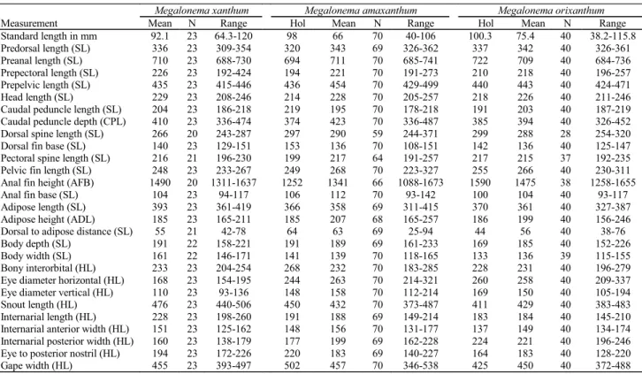
Megalonema xanthum Megalonema amaxanthum Megalonema orixanthum
Measurement Mean N Range Hol Mean N Range Hol Mean N Range Standard length in mm 92.1 23 64.3-120 98 66 70 40-106 100.3 75.4 40 38.2-115.8
Predorsal length (SL) 336 23 309-354 320 343 69 326-362 337 342 40 326-361 Preanal length (SL) 710 23 688-730 694 711 70 685-741 722 709 40 684-736 Prepectoral length (SL) 226 23 192-424 194 221 70 191-273 210 218 40 196-257 Prepelvic length (SL) 435 23 415-446 436 454 70 429-499 440 443 40 424-471 Head length (SL) 229 23 208-246 214 228 70 205-257 218 226 40 211-246 Caudal peduncle length (SL) 204 23 186-218 219 195 70 178-218 191 203 40 187-219 Caudal peduncle depth (CPL) 410 23 336-474 374 423 70 336-487 385 394 40 326-452 Dorsal spine length (SL) 266 20 243-287 297 290 59 244-371 299 288 28 254-320 Dorsal fin base (SL) 140 23 129-151 153 136 70 108-151 142 136 40 125-147 Pectoral spine length (SL) 216 21 196-230 199 217 64 191-257 217 215 37 192-235 Pelvic fin length (SL) 248 23 233-267 249 268 70 223-327 255 266 40 230-311 Anal fin height (AFB) 1490 20 1311-1637 1252 1341 66 1088-1673 1590 1475 38 1258-1655 Anal fin base (SL) 104 23 94-117 106 112 70 93-142 100 104 40 93-117 Adipose length (SL) 393 23 361-419 366 358 69 311-415 370 361 40 327-387 Adipose height (ADL) 185 23 165-211 185 207 68 165-257 186 199 40 156-246 Dorsal to adipose distance (SL) 55 21 42-78 64 63 69 25-94 44 56 40 38-76 Body depth (SL) 191 22 158-221 191 189 69 161-233 169 185 40 152-226 Body width (SL) 161 22 146-171 141 139 70 118-165 133 136 39 115-155 Bony interorbital (HL) 233 23 204-254 268 232 70 183-285 228 231 40 196-279 Eye diameter horizontal (HL) 168 23 154-195 244 263 70 214-321 260 258 40 209-337 Eye diameter vertical (HL) 110 23 93-136 148 158 70 112-214 169 150 40 105-194 Snout length (HL) 476 23 440-506 450 432 70 373-487 411 429 40 383-483 Internarial length (HL) 228 23 198-260 191 188 69 149-214 183 184 40 145-210 Internarial anterior width (HL) 151 23 125-162 148 156 70 131-177 137 149 40 134-174 Internarial posterior width (HL) 160 23 138-179 177 199 69 162-228 224 221 40 196-246 Eye to posterior nostril (HL) 194 23 172-226 220 183 69 140-227 164 183 40 128-220 Gape width (HL) 455 23 393-497 502 457 70 346-538 425 450 40 372-488
margin of anal fin slightly concave. Anal-fin rays 13-16, mod-ally 14, in 19 specimens.
Pectoral fin I, 12-14, modally I, 13, in 11 specimens. Pecto-ral spine slender, its distal half or more segmented and flex-ible, its shaft anteriorly smooth, posteriorly with small erect dentations, one per segment where segments are discernable. Pectoral-spine base with much reduced articulating (fin-locking) processes. Pectoral spine and outer few branched pectoral rays prolonged but none filamentous. Pectoral-fin margin concave. Pectoral axillary gland pore absent. Posterior cleithral process obsolescent.
Pelvic fin as described for subgenus. Also, pelvic-fin insertion below posterior insertion of dorsal fin. Tip of pelvic fin not reaching anal fin.
Urogenital papilla small, located behind anus near base of inner pelvic-fin rays; no indication of sexual dimorphism.
Lateral line straight with side branches alternating dorsally and ventrally, complete and reaching at least onto caudal-fin base.
Total vertebrae in 23 specimens (Table 1), modally 47, range 45-48 including Weberian complex; in 20 specimens 18-19 precaudal vertebrae, modally 18, and 27-30 caudal vertebrae, modally 29.
Coloration in alcohol. All available specimens are types collected in 1912 that have faded or darkened in preservative. In addition to the dark retina of the eye, small, dark chromatophores are scattered over the dorsum of head and body. Many specimens show a subcutaneous silvery layer along the lower flanks. No specimens show the upper caudal lobe spot that is present in all other species of the genus, but this may be due to its loss in preservative.
In the original description, Eigenmann (1912b: 17), who collected several specimens, described the color of the species as “Plumbeous, yellow in life.” An artist’s illustration of M. xanthum in Maldonado-Ocampo et al. (2005: 291, fig. 160) shows the ground color as gray and otherwise without pigment pattern.
Distribution.Endemic to the Magdalena River basin, Colombia. Maldonado-Ocampoet al. (2005: 341, map 162) plot two localities for the species in or near the río Magdalena mainstem above the 90 m elevation contour. All of the types are from localities above 90 m.
Material examined. 48 specimens, all from Colombia,
Cundinamarca Department. FMNH 56032, holotype, 159.3 mm SL, Girardot (see digital images and radiographs online at http:// acsi.acnatsci.org/base/index.html); BMNH 1920.12.20.112-113, paratypes 2, 74-77 mm SL; CAS 63674 [ex IU 12681-82], paratypes 11, 70-118 mm SL (4, 70-118 mm SL), Girardot; FMNH 10285, paratype 1, 107 mm SL, Apulo; FMNH 10289, paratype 1, 69 mm Fig. 5. Scatter plots illustrating: a, anal-fin base length relative
SL, Apulo; FMNH 77890, 2, 101-126.5 mm SL, Apulo; FMNH 77906, 1, 108 mm SL, Apulo; USNM 76930, paratypes 24, 64-120 mm SL (8, 64-111 mm SL), Apulo; USNM 167852, paratypes 5, 88-114 mm SL, Apulo.
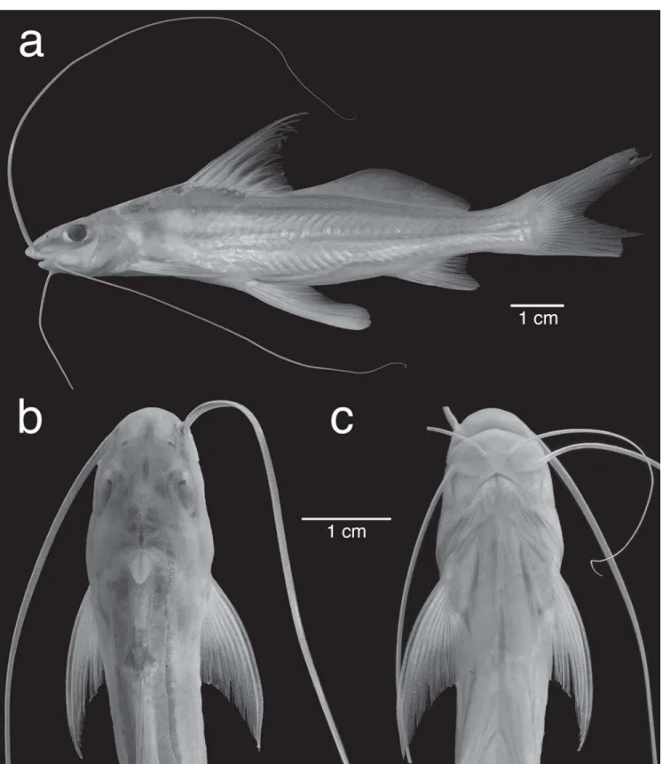
Megalonema amaxanthum, new species Fig. 6
Holotype.CBF 11896 (ex FMNH 106769), 98.0 mm SL, Bolivia,
Pando State, río Tahuamanu from Boca Nareuda to below Cachuelita, 11°18' S, 68°44' W, 13 Sep 1996, H. Ortega et al.
Paratypes.All records within the Amazon drainage basin. Bolivia, Pando State: ANSP 187452, (ex FMNH 106769), 2, 39-42 mm SL, FMNH 106769, 48, 47-98 mm SL (8, 47-97 mm SL), USNM 393558, (ex FMNH 106769), 2, 81-95 mm SL, all collected with the holotype; AUM 23777, 3, 685 mm SL, río Acre at Cobija, 7-9 Feb, 17-982, C. K. Swing et al.Brazil, Acre State: MCP 36198, 1, 85 mm SL, rio Acre, city of Xapuri, at sand extraction encampment, 10°39’51" S, 68°30’45" W, 22 Jul 2004, R. Reis et al. Amazonas State: ANSP 179213, 1, 81 mm SL, rio Solimões (Amazonas Dr.) below rio Purus, upriver of Manacapuru, collected with 3 m bottom trawl in channel, 3°27’02" S, 60°45’07" W, 1 Aug 1996, A. Zanata
et al. ANSP 187453, 2, 42-77 mm SL, rio Solimões below rio Içá, collected with 3 m bottom trawl in 1 m channel, 3°00’26.4" S, 67°52’27.1" W, 23 Nov 1993, J. Lundberg et al., field number JGL 93-122; ANSP 187454, 1, 65 mm SL, rio Solimões below rio Jutaí, collected with 3 m bottom trawl in 2.5-5.5 m channel, 2°31’32.7" S, 66°36’34.8" W, 12 Nov 1993, J. Lundberg et al., field number JGL 93-45; ANSP 187455, 6, 32-60 mm SL (2, 40-59 mm SL), rio Jutaí above rio Solimões, collected with 3 m bottom trawl in 5-10 m channel, 2°52’55.7" S, 66°57’37.2" W, 16 Nov 1993, J. Lundberg et al., field number JGL 93-69; ANSP 187456, 10 alcohol and 2 C&S, 35-64 mm SL (4, 49-64 mm SL), rio Jutaí above rio Solimões, collected with 3 m bottom trawl in 3.7-12.7 m channel, 2°57’03.8" S, 67°00’26.8" W, 16 Nov1993, J. Friel et al., field number JPF 93-76; ANSP 187457, 1, 50 mm SL, rio Jutaí above rio Solimões, collected with 3 m bottom trawl in channel, 2°49’33.3" S, 66°55’06.8" W, 13 Nov 1993, J. Sullivan et al., field number JPS 93-48; CU 84562, 3, 47.1-64.4 mm SL, rio Jutaí above rio Solimões, collected with 3 m bottom trawl in 8.2-11.1 m channel, 2°53’42.7" S, 67°00’34.5" W, 16 Nov 1993, J. Friel et al., field number JPF 93-82; INPA 29490, 1, 48 mm SL, rio Solimões below rio Içá, collected with 3 m bottom trawl in channel, 2°57’58.3" S, 67°49’44.4" W, 23 Nov 1993, J. Sullivan et al., field JPS 93-77; INPA 29491, 3, 48-62 mm SL, rio Negro, collected with 3 m bottom trawl in 3.6-5.6 m channel, 2°21’44" S, 49°28’54" W, 10 Dec 1993, M. Garcia et al., field number MG 93-37; MUSM 32629, 1, 63 mm SL, rio Japurá above rio Solimões, collected with 3 m bottom trawl in 2.9-6.7 m channel, 3°02’58.3" S, 64°46’39.2" W, 29 Oct 1993, O. Oyakawa
et al., field number OTO 93-1; MZUSP 99744, 1, 65 mm SL, rio Solimões, collected with 3 m bottom trawl in channel, Nov 1993, J. Frielet al., field number JPF 93-143; MZUSP 99745, 1, 51 mm SL, rio Negro below Curidique, collected with 3 m bottom trawl in 5-8.5 m channel, 1°58’40.6" S, 61°14’15.6" W, 5 Dec 1993, J. Friel et al., field number JPF 93-154; MZUSP 99746, 1, 70 mm SL, rio Negro, collected with 3 m bottom trawl in 7.7-9.8 m channel, 1°41’37.0" S, 61°26’50.1" W, 6 Dec 1993, J. Friel et al., field number JPF 93-158; MZUSP 99747, 4, 42-67 mm SL (3, 51-67 mm SL), same data as CU 84562. Pará State: ANSP 187458, 1, 48 mm SL, rio Tocantins above rio Pará, collected with 3 m bottom trawl in 6-15 m channel, 2°21’44" S, 49°28’54" W, 19 Nov 1994, A.
Zanataet al., field number AMZ 94-140; ANSP 187459, 4, 40-45 mm SL (2, 40-45 mm SL), rio Tocantins above rio Pará, collected with 3 m bottom trawl in 5.5-6.9 m channel, 2°23’40.2" S, 49°28’06.3" W, 19 Nov 1994, J. Lundberg et al., field number JGL 94-116; ANSP 189096, 4, 29.5-38.8 mm SL, rio Tocantins above rio Pará, collected with 3 m bottom trawl in 3-5 m channel, 2°24’17.5" S, 49°27’55.5" W, 19 Nov 1994, J. Lundberg et al., field number JGL 94-114; IAvH-P 11020, 14, 30-54 mm SL (2, 50-54 mm SL), same data as ANSP 189096; MBUCV V-35377, 4, 54-60 mm SL, rio Tocantins above rio Pará, collected with 3 m bottom trawl in 15.5-25.5 m channel, 2°26’52" S, 49°26’38" W, 19 Nov 1994, A. Zanataet al., field number AMZ 94-145. Rondônia State: MCP 36200, 1, 69 mm SL, rio Mamoré, frente ao bairro Cristo Rei no município de Guajará-Mirim, collected with 3 m bottom trawl in channel, 10°47’03" S, 65°20’58" W, 25 Jul 2004, R. Reis et al.
Roraima State: AMNH 246010, 5, 60-68 mm SL, rio Branco above rio Negro, collected with 3 m bottom trawl in 3.7-7.9 m channel, 1°15’29.0" S, 61°50’37.1" W, 8 Dec 1993, J. Baskin et al., field number JNB 93-17; FMNH 117835, 1, 51 mm SL, rio Branco above rio Negro, collected with 3 m bottom trawl in 4.3-6.7 m channel, 1°18’31.4" S, 61°51’52.3" W, 8 Dec 1993, J. Lundberg et al., field number JGL 93-170; USNM 393559, 1, 67 mm SL, rio Solimões below rio Içá, collected with 3 m bottom trawl in 5.5-9.2 m channel, 1°17’55.4" S, 61°51’21.0" W, 23 Nov 1993, J. Lundberg
et al., field number JGL 93-174. Guyana, Rupununi: ANSP 180494, 2, 72-83 mm SL, Takutu River (R. Branco-Negro Dr.), 3.77 km SSW of Lethem.1 Nov 2003, M. Sabaj et al. Peru, Loreto Department: ANSP 182732, 10, 44-69 mm SL (7, 54-69 mm SL), río Nanay (Amazonas Dr.), large left bank beach upstream from mouth, N of Iquitos, 15 Aug 2005, M. Sabaj et al.; INHS 36643, 1, 49 mm SL, río Napo (río Amazonas Dr.) mouth of río Mazan, near town of Mazan, 20 Jul 1995, L. Page et al.; INHS 44116, 1, 74 mm SL, río Napo and Quebrada (río Amazonas Dr.), Mazan, 33.3 km NE Iquitos at bearing 34°, 3°29’32.5" S, 73°05’11.9" W, 2 Aug 1997, M. Sabaj et al.; MUSM 32833, 3, same data as ANSP 182732. Madre de Dios State: ANSP 180628, 2, 79.8-81 mm SL, MUSM 32834, 1, 77 mm SL, río Tahuamanu (Orton - Madre de Dios Dr.), vicinity of San Lorenzo, 1 Aug 2004, M. Sabaj et al.; ANSP 180633, 1, 106 mm SL, AUM 47794, 1, 93.4 mm SL, INPA 30248, 1, 90 mm SL, MUSM 32835, 1, 97.5 mm SL, all río Acre (Purus Dr.), at town of Inapari on border with Brazil, 2 Aug 2004, M. Sabaj et al.
Diagnosis.A species of the subgenus Eretmomegalonema distinguished from others by the following combination of features. Supraoccipital posterior process (Fig. 3c) bell-shaped in outline, broad, its basal width equal to or greater than its length, and its sides convexly tapering to a blunt tip separated by a distinct gap from supraneural; adipose-fin (Figs. 6a,b) margin anteriorly rising at a shallow angle from back and usu-ally in a weakly concave curve to its apex, and adipose-fin
terminating at or anterior to vertical through tips of adpressed anal-fin rays; total vertebrae modally 42-43, range 42-45 (Table 1); eye large, horizontal diameter 214-321 mils of HL; anal-fin base relatively long in larger individuals, 93-142 mils of SL (Fig. 5); width between posterior nostrils relatively narrow, 162-228 mils of HL (Fig. 5); pectoral spine lacking dentations; premaxilla extremely narrow with a single row of teeth; and 25-29 gill rakers on first arch.
Description. Meristic data for 4 to 61 specimens are in Table 1, and morphometric data for 59 to 70 specimens are in Table 2. Megalonema amaxanthum (Fig. 6) is a medium-sized pimelodid with a maximum length known to us of 106 mm SL. Dorsal profile of head and nape convex from snout tip to vertical at posterior nostril, then nearly straight to origin of dorsal fin, scarcely convex along dorsal-fin base, then less convex to posterior insertion of adipose fin, and gently concave across caudal peduncle. Ventral profile slanting convexly ventrally from snout tip to end of gill region, slightly convex to straight to pelvic-fin origin, abruptly stepped dorsad posterior to pelvic girdle, then straight or convex onto anal-fin base, slanting dorsally to caudal peduncle and slightly concave along caudal peduncle.
Cross-sectional shape roughly trapezoidal from snout to supraoccipital, then deeply and broadly triangular to dorsal-fin origin, and increasingly compressed to caudal dorsal-fin. Maximum body depth at dorsal-fin origin contained 4.3-6.2 times in SL. Maximum body width across cleithra in front of pectoral spine insertion, contained 6.1-8.5 times in SL.
Head of moderate length, contained 3.9-4.9 times in SL and relatively deep, its depth at base of supraoccipital posterior process equal to body width. Head covered by thin skin, revealing smooth, unornamented skull roofing bones. Snout moderate, contained 2.1-2.7 in head length, and with broadly rounded margin in dorsal and ventral views. Snout and upper jaw projecting; with mouth closed less than half of premaxillary dentition exposed. Anterior nostril behind snout margin in a shallow, circular depression, its aperture dorsally directed and encircled with a low fleshy rim; width between anterior nostrils contained 1.0-1.4 times in distance between anterior and posterior nostrils. Posterior nostril aperture ovoid to subtriangular, preceded by thin, semicircular membrane as large as its aperture; width between posterior nostrils contained 0.8-1.2 times in distance between anterior and pos-terior nostrils. Distance between anpos-terior and pospos-terior nos-trils equal to or a little greater than distance between poste-rior nostril and eye.
Eye large and somewhat bulging, about 1.5-2 times longer than deep, placed more laterally than dorsally, centered on middle of bony head length; interorbital space convex. Interorbital narrow, containing horizontal eye diameter 0.7-1.2 times. Anterior cranial fontanelle narrow, parallel-sided, originating behind level of posterior nostrils and terminating between eyes at epiphyseal bar. Posterior cranial fontanelle closed anteriorly, persisting as oval aperture in center of supraoccipital. Supraoccipital posterior process broad-based (its basal width greater than in its length), dorsally slightly convex, not angular or keeled, bell-shaped in outline, with a blunt or rounded tip, failing by about half or more of its own length to contact supraneural (anterior nuchal plate obsolete). Mouth subterminal, opening anteriorly and widely; gape broad, its width across inner surface of ricti twice greater than interorbital width. Lips thin-skinned and smooth. Rictal fold well defined but not fleshy or swollen, subtended above
and below by deep folds respectively reaching base of maxil-lary barbel and about one third distance to mandibular sym-physis. Premaxillary dentition reduced to a single narrow row of fine, conical teeth. Dentary teeth similarly slender, arranged in about 3 rows at symphysis and one row posterolaterally.
Origin of maxillary barbel in depression close to base of anterior nostril and above rictus, continuing to below eye; maxillary barbel reaching onto but not beyond caudal fin. Outer mental barbel reaching at least posterior half or just beyond adipose fin. Tip of inner mental barbel reaching pelvic fin insertion to half of the length of depressed pelvic fin. Branchiostegal membranes anteriorly united to isthmus and overlapping before diverging. Branchiostegal rays 8. Gill rakers long, slender bladelike, 25-29 on first gill arch (in four speci-mens): 6-7 rakers on upper limb, 1 at cartilaginous angle, 18-22 on lower limb.
Dorsal-fin lepidotrichia I,7; spinelet absent or vestigial; dorsal-spine base with much reduced articulating processes. Dorsal-fin spine and first branched fin ray distinctly longer than remaining, progressively shorter rays. Dorsal spine slender, its distal half or more segmented and flexible, its shaft smooth and non-serrate. Dorsal-fin origin at vertical through middle of adpressed pectoral fin; its posterior insertion above pelvic-fin origin. Only the longest anterior fin rays and dorsal-fin spine of adpressed dorsal dorsal-fin reach adipose dorsal-fin. Adipose-fin origin located at vertical posterior to tips of last four dorsal-fin rays. Adipose dorsal-fin large, anteriorly rising with a straight or barely convex margin at a low angle to its base, its apex at vertical near anal-fin origin; terminating at or near tips of adpressed anal-fin rays; 1.4-1.8 times longer than head, rela-tively high, its height 3.9-6.1 times in its base.
Caudal fin deeply forked; pointed upper lobe with filamentous unbranched principal ray, the lower lobe narrowly rounded and a little shorter, and none of its rays prolonged; inner margins of both lobes straight to concave. Principal caudal-fin rays 1+7+8+1. Anal-fin origin below middle of adi-pose-fin base, posterior insertion before end of adipose fin, distal margin of anal fin slightly concave. Anal-fin rays 12-16, modally 13, in 40 specimens.
Pectoral fin I, 11-13, modally I, 12, in 15 specimens. Pecto-ral spine slender, its distal half or more segmented and flex-ible, its shaft entirely smooth without dentations. Pectoral-spine base with much reduced articulating (fin-locking) pro-cesses. Pectoral spine and outer few branched pectoral rays prolonged but none filamentous. Pectoral-fin margin concave. Pectoral axillary gland pore absent. Posterior cleithral pro-cess obsolescent. Pelvic fin as described for subgenus. Also, pelvic-fin insertion below posterior insertion of dorsal fin. Tip of pelvic fin close to or reaching anal fin.
Urogenital papilla small located behind anus near base of inner pelvic-fin rays; no indication of sexual dimorphism.
Lateral line straight with side branches alternating dor-sally and ventrally, complete, and terminating near the ends of middle caudal-fin rays.
Coloration in alcohol. Head and body yellowish to tan in background color. Top and upper sides of head and body lightly to moderately covered with small brownish chromatophores. Top of head with dense concentrations of pigment at and posterior to maxillary barbel insertion, on the midline above mesethmoid between light olfactory organs, and along anterior cranial fontanelle except between eyes. Oval posterior cranial fontanelle dark or covered with hyaline skin. Deep lying, quadrangular dark spot across midline between level of eyes and posterior cranial fontanelle. Supraoccipital posterior process pallid, otherwise nape darker. Dark area of variable extent near or on supraneural, middle and posterior nuchal plates, and at insertion of dorsal fin. A variable series of dark spots at bases of dorsal-fin rays. Tympanic area often darker than surrounding sides behind pectoral girdle. Sclerotic coat of eyes dark or silvery. Upper caudal spot generally present. Rayed fins with hyaline rays and transparent membranes; some specimens with chromatophores along dorsal-, caudal-, anal-, pectoral and pelvic-fin rays and on membranes of dorsal and caudal fins. Adipose fin hyaline, with small, widely dispersed chromatophores.
Distribution. Amazon River basin, Brazil, Guyana (Takutu River), Peru, Bolivia, and probably Colombia and Ecuador. Etymology. The name amaxanthumrefers to the distribution of the species in the Amazon basin and its relationship to the speciesM. xanthum.
Megalonema orixanthum, new species Fig. 7
Holotype.ANSP 187449 (ex ANSP 148143), 100.3 mm SL,
Co-lombia, Meta State, río Metica, ca. 3 km SE of Hacienda Mozambique, 3°57' N, 73°02' W, 24 Mar 1975, J. Böhlke, et al.
Paratypes.All records within the Orinoco drainage basin. Colom-bia, Meta State: ANSP 148143, 23, 90-114 mm SL (8, 90-114 mm SL), FMNH 117836, 2, 95-97 mm SL, IAvH-P 11021, 2, 93-96 mm SL, MBUCV V-35380, 2, 90-97 mm SL, MZUSP 99748, 2, 92-96 mm SL, USNM 393560, 2, 85-95 mm SL, all collected with the holotype; ANSP 131337, 1, 92 mm SL, río Metica, ca. 1.5 km E of Rajote (Plancha 267), 3°56' N, 73°03' W, 19 Mar 1973, W. Saul and W. Smith-Vaniz; ANSP 148142, 2, 77-116 mm SL, río Metica at El Aviso, 3°59' N, 72°59' W, 30 Mar 1975, W. Saul and L. Fuiman; ANSP 148180, 1 C&S, 98 mm SL, río Metica, ca. 3 km SE Hacienda Mozambique, 3°57' N, 73°02' W, 30 Mar 1975, J. Böhlke et al.; ANSP 148181, 1, 95 mm SL, río Metica, ca. 3 km SE Hacienda Mozambique, 3°57' N, 73°02' W, 30 Mar 1975, J. Böhlke et al.
Venezuela, Amazonas State: ANSP 160744, 1, 77 mm SL, río
Orinoco: shores of Isla de Raton, 5°05' N, 67°48' W, 14 Nov 1985, B. Chernoff et al.; ANSP 162484, 3, 29-77 mm SL, río Casiquiare
río Orinoco (Atlantic Dr.), island W of Puerto Venado, 4.5 km S of Samariapo, 56.5 km SW of Puerto Ayacucho, 5°12’25" N, 67°48’32" W, 28 Feb 2005, M. Sabaj et al.; AUM 43048, 13, 47-50 mm SL (2, 47-50 mm SL), same data as ANSP 185144; AUM 43845, 2, 79-94 mm SL, río Orinoco, beach, 16.1 km E of La Esmeralda, 25 Mar 2005. Apure State: ANSP 160186, 2, 84-87 mm SL, río Meta, ca 40 min upstream from confluence of río Orinoco, 6°18' N, 67°37' W, 27 Nov 1985, B. Chernoff et al.; ANSP 165236, 5, 61-78 mm SL, río Apure: between río Portuguesa mouth and S.Fernando de Apure airport, 7°54’00" N, 67°32’00" W, 4 Nov 1989, S. Schaefer et al.; ANSP 187450, 4 alcohol, 1 C&S, 59-73 mm SL, río Apure between San Fernando and río Portuguesa, collected with 3 m bottom trawl, 19 Jul 1984, Lundberg et al. Bolivar State: ANSP 160407, 2, 66-71 mm SL, río Orinoco - río Caura confluence beaches, canals, lagoons & islands, vicinity Puerto Las Majadas, 7°38’36" N, 64°50’W, 23 Nov 1985, B. Chernoff et al.; ANSP 160857, 1, 57 mm SL, río Cuchivero at Cuchivero ferry crossing, 7°29' N, 65°53' W, 17 Nov 1985, B. Chernoff et al.; ANSP 187451, 1, 71 mm SL, río Orinoco at Ciudad Bolivar, collected with 3 m bottom trawl in 10 m channel, 8°09' N, 63°32' W, 8 Nov 1979, E. Marsh et al., field number ECM 1-79.
Diagnosis.A species of the subgenus Eretmomegalonema distinguished from others by the following combination of features. Supraoccipital posterior process (Fig. 3d) broadly triangular in outline, its basal width equal to or greater than its length, and its sides tapering straight to a nearly pointed or bifid tip nearly reaching supraneural; adipose fin (Fig. 7a) smaller, its margin anteriorly rising at a shallow angle from back and usually in a weakly concave curve to its apex, and adipose fin terminating at or anterior to vertical through tips of adpressed anal-fin rays; total vertebrae modally 44, range 43-45 (Table 1); eye large, horizontal diameter 209-337 mils of HL; anal-fin base relatively short, 93-117 mils of SL (Fig. 5); width between posterior nostrils relatively broad, 196-246 mils of HL (Fig. 5); pectoral spine with numerous erect dentations along posterior margin; premaxilla extremely narrow with a single row of teeth or teeth absent; and 28-29 gill rakers on first arch.
then convex onto anal-fin base, slanting dorsally to caudal peduncle and concave along caudal peduncle.
Cross-sectional shape roughly trapezoidal from snout to supraoccipital, then deeply and broadly triangular to
dorsal-fin origin, and increasingly compressed to caudal dorsal-fin. Maxi-mum body depth at dorsal-fin origin contained 4.4-6.6 times in SL. Maximum body width across cleithra in front of pecto-ral spine insertion, contained 6.5-8.7 times in SL.
Snout moderate, contained 2.1-2.6 in head length, and with broadly rounded margin in dorsal and ventral views. Snout and upper jaw projecting; with mouth closed less than half of premaxillary dentition exposed. Anterior nostril behind snout margin in a shallow, circular depression, its aperture dorsally directed and encircled with a low fleshy rim; width between anterior nostrils contained 0.9-1.5 times in distance between anterior and posterior nostrils. Posterior nostril aperture ovoid to subtriangular, preceded by thin, semicircular membrane as large as its aperture; width between posterior nostrils con-tained 0.7-1.0 times in distance between anterior and posterior nostrils. Distance between anterior and posterior nostrils equal to distance between posterior nostril and eye.
Eye large and somewhat bulging, about 1.5-2 times longer than deep, placed more laterally than dorsally, centered on middle of bony head length; interorbital space convex. Interorbital narrow, containing horizontal eye diameter 0.6-1.2 times. Anterior cranial fontanelle narrow, parallel-sided, originating behind level of posterior nostrils and terminating between eyes at epiphyseal bar. Posterior cranial fontanelle closed anteriorly, persisting as oval aperture in center of supraoccipital. Supraoccipital posterior process broad-based (its basal width equal to or greater than its length), dorsally slightly convex, not angular or keeled, parabolic in outline, failing by less than half its own length to contact supraneural (anterior nuchal plate obsolete).
Mouth subterminal, opening anteriorly and widely; gape broad, its width across inner surface of ricti twice greater than interorbital width. Lips thin-skinned and smooth. Rictal fold well defined but not fleshy or swollen, subtended above and below by deep folds respectively reaching base of maxillary barbel and about one third distance to mandibular symphysis. Premaxillary toothless or with a single row of a few fine, conical teeth. Dentary teeth slender, arranged in about 3 rows at symphysis and one row posterolaterally.
Origin of maxillary barbel in depression close to base of anterior nostril and above rictus, continuing to below eye; maxillary barbel reaching onto but not beyond caudal fin. Outer mental barbel reaching posterior end of or just beyond adipose fin. Tip of inner mental barbel reaching pelvic-fin insertion to half of the length of depressed pelvic fin. Branchiostegal membranes anteriorly united to isthmus and overlapping before diverging. Branchiostegal rays 8. Gill rakers long, slender bladelike, 28-29 on first gill arch (in four speci-mens): 6-8 rakers on upper limb, 1 at cartilaginous angle, 20-21 on lower limb.
Dorsal-fin lepidotrichia I,7; spinelet absent or vestigial; dorsal-spine base with much reduced articulating processes. Dorsal-fin spine and first branched fin ray distinctly longer than remaining, progressively shorter rays. Dorsal spine slender, its distal half or more segmented and flexible, its shaft
fin origin located at vertical posterior to tips of last four dorsal-fin rays. Adipose dorsal-fin large, anteriorly rising with a straight or barely convex margin at a low angle to its base, its apex at vertical near anal-fin origin; terminating at or near tips of adpressed anal-fin rays; 1.4-1.7 times longer than head, rela-tively high, its height 4.1-6.4 times in its base.
Caudal fin deeply forked; pointed upper lobe with filamentous unbranched principal ray, the lower lobe narrowly rounded and a little shorter, and none of its rays prolonged; inner margins of both lobes straight to concave. Principal caudal-fin rays 1+7+8+1. Anal-fin origin below middle of adi-pose-fin base, posterior insertion before end of adipose fin, distal margin of anal fin slightly concave. Anal-fin rays 12-15, modally 13, in 27 specimens.
Pectoral fin I,11-13, modally I,12, in 9 specimens. Pectoral spine slender, its distal half or more segmented and flexible, its shaft smooth anteriorly and with numerous erect dentations along posterior margin. Pectoral-spine base with much reduced articulating (fin-locking) processes. Pectoral spine and outer few branched pectoral rays prolonged but none filamentous. Pectoral-fin margin concave. Pectoral axillary gland pore absent. Posterior cleithral process obsolescent. Pelvic fin as described for subgenus. Also, pelvic-fin insertion below posterior insertion of dorsal fin. Tip of pelvic fin close to or reaching anal fin.
Urogenital papilla small located behind anus near base of inner pelvic-fin rays; no indication of sexual dimorphism.
Lateral line straight with side branches alternating dorsally and ventrally, complete, and terminating near the ends of middle caudal-fin rays.
Total vertebrae in 35 specimens (Table 1), modally 44, range 43-45 including Weberian complex; in 36 specimens 16-18 precaudal vertebrae, modally 17, and 26-29 caudal vertebrae, modally 27.
pectoral girdle. Sclerotic coat of eyes dark or silvery. Upper caudal spot generally present. Rayed fins with hyaline rays and transparent membranes; some specimens with chromatophores along dorsal-, caudal-, anal-, pectoral and pelvic-fin rays and on membranes of dorsal and caudal fins. Adipose fin hyaline, with small, widely dispersed chromatophores.
Distribution. Orinoco River basin, Colombia and Venezuela. Etymology. The name orixanthumrefers to the distribution of the species in the Orinoco basin and its relationship to the speciesM. xanthum.
Discussion
Two unambiguous synapomorphies described in the spe-cies diagnoses indicate a sister-group relationship between M. amaxanthum and M. orixanthum within Eretmomegalo-nema: broad and short supraoccipital posterior process (Figs. 3c,d) and greatly reduced premaxilla and upper jaw dentition. As inferred from its similarities to species of Megalonema not belonging to Eretmomegalonema, M. xanthum has the plesiomorphic conditions of these elements, i.e. long, narrow supraoccipital posterior process (Fig. 3b, compare Fig. 3a) and broad, well-toothed premaxilla. Thus, trans-Andean M. xanthum is considered to be phylogenetically basal within Eretmomegalonema to the cis-Andean species pair in the Amazon and Orinoco. The sequence of two cladistic events in the tree topology of Eretmomegalonema matches the se-quence of origin of drainage divides separating the Magdalena, Amazon and Orinoco basins (Lundberg et al., 1998; Lundberg, 2005). The Magdalena watershed was formed and isolated from the Amazon and Orinoco by about 10 Ma in the Middle to Late Miocene when the Eastern Andes was uplifted. The low divides that now separate the Amazon and Orinoco originated later in the Miocene at about 8 Ma. The picture that emerges is one of vicariant control of cladogen-esis and divergence of the three species in Eretmomegalo-nema, and the persistence of their allopatric distributions.
Comparative material.Megalonema platycephalum Eigenmann,
1912,Guyana, Demerara-Berbice: ANSP 179249, 1 Sk, 145 mm SL, Essequibo River (east bank) at Kurukupari, 04°39’41"N, 058°40’31"W. Rupununi: ANSP 179697, 2, 92-133 mm SL, Rupununi River (Essequibo Dr.) at Kwatamang, 4 km SE of Annai, 03°55’03"N, 059°06’01"W. Megalonema cf. platycephalum.Peru, Loreto Department: ANSP 178450, 2 alcohol, 1 C&S, 105-110 mm SL, río Nanay (tributary río Amazonas) at Pampa Chica, village 4.54 km W of Iquitos (large beach along N bank), 3°45’09"S, 73°17’00"W; ANSP 178515, 1 Sk, río Napo, near town of Mazan, 3°29’10"S, 73°06’24"W. Madre de Dios State: ANSP 187323, 1 Sk, 240 mm SL, río Tahuamanu (Orton–Madre de Dios Dr.), vicin-ity of San Lorenzo. Colombia, Meta State: ANSP 131341, 45-81 mm SL, río Metica, upstream from entrance to Lake Mozambique, halfway to entrance to Laguna ‘Arrotas.’ Venezuela, Apure State: ANSP 189040 (ex DU F998), 6 alcohol, 1 C&S, 53-76 mm SL, río
Apure between San Fernando and río Portuguesa, collected with 3 m bottom trawl, 19 Jul 1984, Lundberg et al. Megalonema platanum
(Günther, 1880), Brazil, Rio Grande do Sul: MZUSP 78465, 1 Sk, 210 mm SL, rio Uruguai, em frente ao porto de São Borja. Paraguay, Asunción: UMMZ 207635, 2, 180-205 mm SL, Pettirossi fish market (=Mercado Cuatro), Asunción (from nearby río Paraguay).
Acknowledgements
For loans of specimens we thank David Catania (CAS, San Francisco), Osvaldo Oyakawa (MZUSP, São Paulo), Francisco Provenzano (MBUCV, Caracas), Mary Anne Rogers (FMNH, Chicago), Roberto Reis (MCP, Porto Alegre), Mike Retzer (INHS, Champaign), David Werneke and Jonathan Armbruster (AUM, Auburn), and Jeff Williams and Richard Vari (National Museum of Natural History). We are most grateful to Kyle Luckenbill (ANSP) for his expert preparation of the illustrations, and to Mark Sabaj Pérez (ANSP) for his assistance with spotting and management of specimens, and many helpful comments on this study. Two anonymous reviewers provided valuable corrections and comments on the manuscript, and we are indebted to the one who voluntarily translated our abstract into Portuguese. Support for travel and publication was provided by the All Catfish Species Inventory (ACSI, US National Science Foundation DEB-0315963) and a research award to JGL (US National Science Foundation, DEB-0089612).
Literature Cited
Driver, C. S. 1919. On the Luciopimelodinae, a new subfamily of the South American Siluridae. Proceedings of the American Philosophical Society, 58:448-456.
Eigenmann, C. H. 1912a. The freshwater fishes of British Guiana, including a study of the ecological grouping of species, and the relation of the fauna of the plateau to that of the lowlands. Memoirs of the Carnegie Museum, 5:i-xxii + 1-578, pls. 1-103. Eigenmann, C. H. 1912b. Some results from an ichthyological reconnaissance of Colombia, South America. Part I. Indiana University Studies, 16[sic. 8]:1-27.
Eigenmann, C. H. 1922. The fishes of western South America, Part I.—The fresh-water fishes of northwestern South America, including Colombia, Panama, and the Pacific slopes of Ecuador and Peru, together with an appendix upon the fishes of the Rio Meta in Colombia. Memoirs of the Carnegie Museum, 9:1-346, pls. 1-38.
Hardman, M., and J. G. Lundberg. 2006. Molecular phylogeny and a chronology of diversification for “phractocephaline” catfishes (Siluriformes: Pimelodidae) based on mitochondrial DNA and nuclear recombination activating gene 2 sequences. Molecular Phylogenetics and Evolution, 40:410-418.
Howes, G. J. 1983. Problems in catfish anatomy and phylogeny exemplified by the Neotropical Hypophthalmidae (Teleostei: Siluroidei). Bulletin of the British Museum of Natural History (Zoology), 45(1):1-39.
446. In: Reis, R. E., S. O. Kullander & C. J. Ferraris, Jr. (Eds.). Check list of the Freshwater Fishes of South and Central America. Porto Alegre, Edipucrs, 729p.
Lundberg, J. G., F. Mago-Leccia & P. Nass. 1991. Exallodontus aguanai, a new genus and species of Pimelodidae (Pisces: Siluriformes) from deep river channels of South America, and delimitation of the subfamily Pimelodinae. Proceedings of the Biological Society of Washington, 104:840-869.
Lundberg, J. G., L. G. Marshall, J. Guerrero, B. Horton, M. C. Malabarba & F. Wesselingh. 1998. The Stage for Neotropical Fish Diversification: A History of Tropical South American Rivers. Pp. 13-48. In: Malabarba, L. R., R. E. Reis, R. P.Vari, Z. M. S. Lucena & C. A. S. Lucena (Eds.). Phylogeny and Classification of Neotropical Fishes. Porto Alegre, Edipucrs, 603p.
Lundberg, J. G. & B. M. Parisi. 2002. Propimelodus, new genus, and a description of Pimelodus eigenmanni van der Stigchel, 1946, a long recognized yet poorly-known South American catfish (Pimelodidae: Siluriformes). Proceedings of the Academy of Natural Sciences, Philadelphia, 152:75-88.
Maldonado-Ocampo, J. A., A. Ortega-Lara, J. S. Usma Oveido, G. Galvis Vergara, F. A. Villa-Navarro, L. Vásquez Gamboa, S. Prada-Pedreros & C. A. Ardila-Rodríguez. 2005. Peces de los Andes de Colombia: Guía de Campo. Bogotá, Instituto de Investigación de Recursos Biológicos Alexander von Humboldt, 346p.
Parisi, B. M., J. G. Lundberg & C. DoNascimiento. 2006.
Propimelodus caesius a new species of long-finned pimelodid catfish (Teleostei: Siluriformes) from the Amazon Basin, South America. Proceedings of the Academy of Natural Sciences of Philadelphia, 155:67-78.
Rocha, M. S., R. R. de Oliveira & L. H. Rapp Py-Daniel. 2007. A new species of Propimelodus Lundberg & Parisi, 2002 (Siluriformes: Pimelodidae) from rio Araguaia, Mato Grosso, Brazil. Neotropical Ichthyology, 5(3):279-284.
Stewart, D. J. 1986. Revision of Pimelodina and description of a new genus and species from the Peruvian Amazon (Pisces: Pimelodidae). Copeia, 1986:653-672.