Discovery and Characterization of Auxiliary Proteins Encoded by
Type 3 Simian T-Cell Lymphotropic Viruses
Jocelyn Turpin,a,b,c,d,eChloé Journo,a,b,c,d,eNga Ling Ko,fFlore Sinet,a,b,c,d,e,gAlexandre Carpentier,hAmandine Galioot,a,b,c,d,e,g Dustin Edwards,iAnne-Mieke Vandamme,jLouis Gazzolo,d,kMadeleine Duc Dodon,d,kAntoine Gessain,fFatah Kashanchi,l Ivan Balansard,mRomain Lacoste,nRenaud Mahieuxa,b,c,d,e
Equipe Oncogenèse Rétrovirale, Lyon, Francea; Equipe Labellisée Ligue Nationale Contre le Cancer, Lyon, Franceb; International Center for Research in Infectiology, INSERM U1111-CNRS UMR5308, Lyon, Francec; Ecole Normale Supérieure de Lyon, Lyon, Franced; Université Lyon 1, Lyon, Francee; Epidémiologie et Physiopathologie des Virus Oncogènes, CNRS UMR 3569, Pasteur Institute, Paris, Francef; Master Biosciences ENS Lyon, Lyon, Franceg; Molecular and Cellular Epigenetics (GIGA) and Molecular Biology (Gembloux Agro-Bio Tech), University of Liège, Liège, Belgiumh; Virus Tumor Biology Section, Center for Cancer Research, National Cancer Institute, NIH, Bethesda, Maryland, USAi; University of Leuven, Department of Microbiology and Immunology, Rega Institute for Medical Research, Clinical and Epidemiological Virology, Leuven, Belgium, and Centro de Malária e outras Doenças Tropicais and Unidade de Microbiologia, Instituto de Higiene e Medicina Tropical, Universidade Nova de Lisboa, Lisbon, Portugalj
; Laboratoire de Biologie Moléculaire de la Cellule, Unité Mixte de Recherche 5239, Centre National de la Recherche Scientifique, UMS3444 Biosciences Lyon-Gerland, Lyon, Francek
; National Center for Biodefense and Infectious Diseases, George Mason University, Manassas, Virginia, USAl
; UMS 3537 CNRS-AMU, Marseille, Francem; Station de Primatologie-UPS846-CNRS, Rousset sur Arc, Francen
ABSTRACT
Human T-cell lymphotropic virus type 1 (HTLV-1) and HTLV-2 encode auxiliary proteins that play important roles in viral
replica-tion, viral latency, and immune escape. The presence of auxiliary protein-encoding open reading frames (ORFs) in HTLV-3, the latest
HTLV to be discovered, is unknown. Simian T-cell lymphotropic virus type 3 (STLV-3) is almost identical to HTLV-3. Given the lack of
HTLV-3-infected cell lines, we took advantage of STLV-3-infected cells and of an STLV-3 molecular clone to search for the presence of
auxiliary transcripts. Using reverse transcriptase PCR (RT-PCR), we first uncovered the presence of three unknown viral mRNAs
en-coding putative proteins of 5, 8, and 9 kDa and confirmed the presence of the previously reported
RorfII
transcript. The existence of
these viral mRNAs was confirmed by using splice site-specific RT-PCR with
ex vivo
samples. We showed that p5 is distributed
through-out the cell and does not colocalize with a specific organelle. The p9 localization is similar to that of HTLV-1 p12 and induced a strong
decrease in the calreticulin signal, similarly to HTLV-1 p12. Although p8, RorfII, and Rex-3 share an N-terminal sequence that is
pre-dicted to contain a nucleolar localization signal (NoLS), only p8 is found in the nucleolus. The p8 location in the nucleolus is linked to a
bipartite NoLS. p8 and, to a lesser extent, p9 repressed viral expression but did not alter Rex-3-dependent mRNA export. Using a
trans-formation assay, we finally showed that none of the STLV-3 auxiliary proteins had the ability to induce colony trans-formation, while both
Tax-3 and antisense protein of HTLV-3 (APH-3) promoted cellular transformation. Altogether, these results complete the
character-ization of the newly described primate T-lymphotropic virus type 3 (PTLV-3).
IMPORTANCE
Together with their simian counterparts, HTLVs form the primate T-lymphotropic viruses. HTLVs arose from interspecies
transmission between nonhuman primates and humans. HTLV-1 and HTLV-2 encode auxiliary proteins that play important
roles in viral replication, viral latency, and immune escape. The presence of ORFs encoding auxiliary proteins in HTLV-3 or
STLV-3 genomes was unknown. Using
in silico
analyses,
ex vivo
samples, or
in vitro
experiments, we have uncovered the
pres-ence of 3 previously unknown viral mRNAs encoding putative proteins and confirmed the prespres-ence of a previously reported viral
transcript. We characterized the intracellular localization of the four proteins. We showed that two of these proteins repress
vi-ral expression but that none of them have the ability to induce colony formation. However, both Tax and the antisense protein
APH-3 promote cell transformation. Our results allowed us to characterize 4 new retroviral proteins for the first time.
T
ogether with their simian counterparts (simian T-cell
lympho-tropic virus type 1 [STLV-1], STLV-2, STLV-3, and STLV-4),
human T-cell lymphotropic virus type 1 (HTLV-1), HTLV-2,
HTLV-3, and HTLV-4 form the primate T-lymphotropic virus
(PTLV) family. Phylogenetic analyses have demonstrated that
HTLVs arose from interspecies transmission that occurred in the
past and may still occur between Old World nonhuman primates
(NHPs) and humans as well as among NHPs (
1–8
; for a review, see
reference
9
). While HTLV-1 and HTLV-2 are found throughout
the world (
10
,
11
), PTLV-3 and -4 seem restricted to Africa so far
(
12–18
).
HTLV-3 was recently discovered (
6
,
7
,
19
,
20
), a decade after
STLV-3 was first isolated (
21
,
22
) and a few years after other
STLV-3 strains were reported (
23–27
). Additional
PTLV-3-in-Received24 July 2014 Accepted20 October 2014 Accepted manuscript posted online29 October 2014
CitationTurpin J, Journo C, Ko NL, Sinet F, Carpentier A, Galioot A, Edwards D, Vandamme A-M, Gazzolo L, Duc Dodon M, Gessain A, Kashanchi F, Balansard I, Lacoste R, Mahieux R. 2015. Discovery and characterization of auxiliary proteins encoded by type 3 simian T-cell lymphotropic viruses. J Virol 89:931–951. doi:10.1128/JVI.02150-14.
Editor:S. R. Ross
Address correspondence to Renaud Mahieux, [email protected]. Copyright © 2015, American Society for Microbiology. All Rights Reserved. doi:10.1128/JVI.02150-14
on May 14, 2018 by B-ON FCCN TRIAL
http://jvi.asm.org/
fected individuals were later reported (
28–36
; for a review, see
reference
12
). While HTLV-1, thanks to its Tax (Tax-1) and
HTLV-1 basic leucine zipper (HBZ) proteins, causes leukemia
after a long period of clinical latency (
37
), other HTLVs have not
been associated with oncogenic processes. However, the number
of PTLV-3 and -4-infected individuals identified so far is very low
(
7
,
19
,
30
,
31
,
33
), thus precluding epidemiological analyses.
Nev-ertheless, we previously demonstrated that the HTLV-3 Tax
(Tax-3) amino acid sequence contains at least one domain, a
PDZ-binding motif, that is absent from HTLV-2 Tax (Tax-2) and
is critical for cellular transformation (
38
). More recently, using a
high-throughput transcriptomic approach, we demonstrated that
the Tax-3 protein was phenotypically related to Tax-1, thus
sug-gesting that HTLV-3 might indeed be pathogenic (
39
). Others
have also shown that HTLV-3 and -4 encode antisense transcripts
(APH-3 and APH-4, respectively) that repress viral expression
(
40
), as is the case for the HTLV-1 and HTLV-2 HBZ and APH-2
proteins, respectively (
41–43
). The ability of APH-3 and -4 to
Turpin et al.
932 jvi.asm.org Journal of Virology January 2015 Volume 89 Number 2
on May 14, 2018 by B-ON FCCN TRIAL
http://jvi.asm.org/
drive cellular proliferation and/or transformation has not yet been
investigated.
In addition to its Tax and HBZ proteins, HTLV-1 also encodes
the p12, p13, and p30 auxiliary proteins (for a recent review, see
reference
44
). These proteins arise after complex splicing of their
respective mRNAs and have important roles in viral latency, viral
transmission, and viral escape from immune responses. HTLV-2
also encodes the auxiliary proteins p10, p11, and p28, which share
some functional properties with HTLV-1 p12 and p30 despite low
sequence similarity.
HTLV-1 p30 is translated from a doubly spliced mRNA
tran-scribed from open reading frame II (ORF II) (for a review, see
references
45
and
46
), while HTLV-2 encodes p28, a protein that is
similar to p30 (
47
,
48
). When ectopically expressed, p30 is
de-tected within nucleoli and nuclei. p30 harbors two nucleolar
retention signals (NoRSs) and four nuclear localization signals
(NLSs) (
49
). The p30 protein also contains a Rex-binding domain,
a p300-binding domain, and a DNA-binding domain. At the
post-transcriptional level, p30 counteracts the positive effects of Tax
and Rex. p30 specifically binds to and retains the doubly spliced
tax/rex
mRNA in the nucleus, resulting in decreased expression
levels of these regulatory proteins. Thus, p30 is a negative
regula-tor of viral replication (
50
) and promotes viral latency (
51
,
52
).
p30 also contributes to Toll-like receptor 4 (TLR4)
downregula-New PTLV Auxiliary Proteins
on May 14, 2018 by B-ON FCCN TRIAL
http://jvi.asm.org/
tion at the cell surface and decreases proinflammatory cytokine
production by human macrophages (
53
). Consequently, p30 also
promotes decreased immune responses against HTLV-1
infec-tion.
HTLV-1 p13 corresponds to the C-terminal 87 amino acids
(aa) of p30 (for a recent review, see reference
54
). It accumulates in
the inner membrane of mitochondria but can also be found in the
cell nucleus, where it binds Tax, thereby preventing its interaction
with CBP/p300 (
55
). As a result, p13 inhibits Tax-mediated viral
transcription and, hence, also contributes to viral latency. p13 also
causes mitochondrial swelling (
56
,
57
). By promoting apoptosis,
p13 might represent a way to escape immune surveillance and
favor clonal expansion of latently infected cells.
HTLV-1 p12 is translated from a singly spliced mRNA
tran-scribed from ORF I (
58
). p12 is a transmembrane protein and
accumulates in the endoplasmic reticulum (ER) and
cis
-Golgi
ap-paratus (
59
). p12 contains two putative transmembrane domains,
four putative proline-rich (PXXP) Src homology 3
(SH3)-binding domains, two putative leucine zipper (LZ) motifs, a
putative adaptin motif, a calcineurin-binding motif, and a
noncanonical ER retention/retrieval motif. p12 activates
STAT5 signaling and increases T-cell proliferation (
60
). p12 also
controls calcium release from the ER (
61
) and T-cell activation
(
62
) and promotes immune escape. Depending on the viral
sub-type, p12 can be cleaved near the amino terminus to remove the
ER retention signal to form the p8 protein (
63
). This cleavage
allows p8 to traffic to the cell surface (
63
). Interestingly, HTLV-1
p8 modulates lymphocyte function-associated antigen 1 (LFA-1)
clustering at the cell surface, thus promoting the formation of
cell-to-cell contacts and favoring virus transmission (
64
,
65
).
Here, using
ex vivo
and
in cellulo
approaches, we characterized
three auxiliary proteins (p5, p8, and p9) encoded by STLV-3 and
confirmed the existence of a fourth one (RorfII). We showed that
p8 and p5 sequences are well conserved among all PTLV-3
iso-lates. RorfII is present in subtypes B and A but is absent from
subtype D. p9 was encoded only by subtype B strains. Using
im-munofluorescence assays, we observed that p9 and p8 have the
same localization as HTLV-1 p12 and p30, respectively. Luciferase
assays demonstrated that p8 expression is linked to the
down-regulation of long terminal repeat (LTR) activation, which
does not involve competition with Rex-3-dependent export.
Colony formation assays showed that none of the STLV-3
aux-iliary proteins have the ability to transform cells, while the
expression of both Tax-3 and, to a certain extent, APH-3 can
lead to focus formation. Altogether, these results demonstrate
that all PTLVs have evolved to encode auxiliary proteins,
which, in spite of having different sequences, share some
bio-logical properties.
MATERIALS AND METHODS
Cell culture.293T, HeLa, and Cos cells were maintained in Dulbecco’s modified Eagle medium–Glutamax-I (Gibco, Invitrogen) plus 10% fetal bovine serum (FBS) (Gibco, Invitrogen) and 100g/ml penicillin-strep-tomycin (Gibco, Life Technologies). PH969-infected cells (22) were maintained in Roswell Park Memorial Institute (RPMI) medium plus 10% FBS (Gibco, Invitrogen) and 100g/ml penicillin-streptomycin (Gibco, Life Technologies).
Papio hamadryas papioPBMCs.Heparinized blood specimens were drawn from twoPapio hamadryas papiobaboons (animal identification numbers PPA-F3 and PPA-F11) housed at the primate center of the Cen-tre National de la Recherche Scientifique in Rousset sur Arc and cared for in compliance with French regulations and guidelines of the Federation of European Laboratory Animal Science Associations (FELASA). The STLV-3 status of these baboons was described previously (24). Peripheral blood mononuclear cells (PBMCs) were purified by Ficoll (Histopaque; Sigma) gradient centrifugation and frozen in liquid nitrogen. Cells were then thawed and grown in the presence of phytohemagglutinin (PHA) (1
g/ml) (Sigma) and interleukin-2 (IL-2) (150 U/ml) for 1 to 3 days.
RT-PCR and sequence analyses.mRNAs were extracted from STLV-3PH969-infected cells or from 293T cells that had been transfected for 48 h with the STLV-3PPA-F3molecular clone by using TRIzol reagent (66). A reverse transcriptase (RT) reaction was achieved with SuperScript II RT (Invitrogen), using primers located upstream of thetax/rexacceptor site in order to prevent amplification of thetax/rexmRNA (Fig. 1A). PCRs were then performed with different combinations of primers, allowing the specific detection of singly and doubly spliced viral mRNAs present in the PH969 and PPA-F3 samples. Forward PCR primers were located in the 5= LTR or in the second exon oftax/rex(Fig. 1B). Reverse PCR primers were the same as those used for the RT reaction. After migration on agarose gels, bands were purified, cloned by using the TA cloning kit, and se-quenced (GATC Biotech). The names of the newly identified different STLV-3 auxiliary proteins were based on their predicted molecular weights, as determined by using the compute pI/Mwtool of Expasy (67). The primers were named according to their positions on STLV-3 ge-nomes. The primers used were as follows: PPA-F3 forward primers Fwd-280 (5=-CACTTTGCCAATCCCTCCCTC-3=), Fwd-316 (5=-CTGGTCA TCCCTGCTTACCCG-3=), Fwd-388 (5=-GCCCTTCGCCGTCTTCCAC TC-3=), Fwd-742 (5=-CATGGGAAAGACTTATAGCTC-3=), Fwd-4942 (5=-TCCCAGTAGGTGGAAGCCATCTCT-3=), and Fwd-5026 (5=-CCG ACCCAAAAATCAGAGACCATC-3=); PPA-F3 reverse primers Rev-6823
Turpin et al.
934 jvi.asm.org Journal of Virology January 2015 Volume 89 Number 2
on May 14, 2018 by B-ON FCCN TRIAL
http://jvi.asm.org/
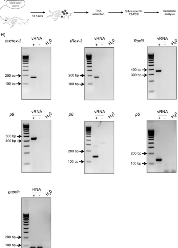
FIG 1Characterization of STLV-3 pX mRNAs by splice-specific RT-PCR. (A) Strategy for amplifying unknown pX transcripts from 293T cells transfected with the STLV-3PPA-F3molecular clone or from STLV-3PH969-infected cells. Viral mRNAs, encoded by the pX region, were specifically reverse transcribed. cDNAs
were amplified by PCR using forward primers (black arrows) located either in the 5=LTR or before thetax/rexsplice donor site, while reverse primers (gray arrows) were located before thetax/rex-3splice acceptor site. (B) RNAs extracted from STLV-3PH969-infected cells (viral RNA [vRNA]) were reverse transcribed
by using Rev-7036 primers. cDNAs were amplified by using either Fwd-4984/Rev-7036 or Fwd-273/Rev-7036 primers. The white arrow indicates RorfII cDNA. (C and D) RNA extracted from 293T cells transfected with the STLV-3PPA-F3molecular clone (viral RNA) were reverse transcribed by using Rev-7173 primers.
(C) cDNAs were amplified by using Fwd-316/Rev-6918 primers. All the bands were purified, cloned, and sequenced. The white arrow indicates the band corresponding to p9 cDNA. (D) cDNAs were amplified by using Fwd-316/Rev-7173 primers. All the bands were purified, cloned, and sequenced. The white arrow indicates the band corresponding to bothp8andp5. (E) Exon composition of the newly identified STLV-3PPA-F3RNAs. Other spliced viral RNAs are also shown.
Numbers and arrows indicate splice sites. Boxes indicate ORFs, and asterisks represent positions of the start codon within each ORF. Putative proteins identified in this report were named according to thein silicoprediction of their molecular weight. (F) Consensus motifs of mammalian splice donor (sd) and splice acceptor (sa) sites. The dinucleotides essential for these sites are underlined. Sequences of the identified functional splice donor and acceptor sites in viral mRNAs are aligned to the consensus. (G) RNA was extracted 48 h after transfection of 293T cells with the STLV-3PPA-F3molecular clone. (H) One-step RT-PCR was
performed on 500 ng RNA extracted from STLV-3-transfected (⫹vRNA) or mock-transfected (⫺vRNA) 293T cells, using primers located on the splice junctions oftax/rex-3,tRex-3,RorfII,p9,p8, andp5viral mRNAs.gapdhwas used as a control.
on May 14, 2018 by B-ON FCCN TRIAL
http://jvi.asm.org/
FIG 2p5 and p8 RNAs are expressedin vivoin STLV-3-infectedPapio hamadryas papiobaboons. (A) RNA was directly extracted from uncultured PBMCs obtained from an STLV-3-infectedPapioanimal (PPA-F3) or from a noninfected animal (PPA-F11) (B) or after 24 to 72 h ofex vivoculture in RPMI medium supplemented with IL-2 and PHA (C). (B and C) RT-PCR was performed as described in the legend ofFig. 1.gapdhwas used as a control. Ctrl, RNA extracted from cells transfected with the STLV-3PPA-F3molecular clone.
Turpin et al.
936 jvi.asm.org Journal of Virology January 2015 Volume 89 Number 2
on May 14, 2018 by B-ON FCCN TRIAL
http://jvi.asm.org/
(5=-GTTGGAGGAAAGGAAGAGGCG-3=), Rev-6885 (5=-CTGCTGAG TTATTGGCGAGAACGA-3=), Rev-6918 (5=-CGGTTGGATGACCTTG CCCAG-3=), Rev-7173AS (5= -GAGACTCCAATCCCAGGAACTGTG-3=), Rev-7399AS (5=-GGTCCCAGGTAATCTGATGTTCG-3=), and Rev-7989AS (5=-GCTGCCATCAGTGAAAGTCCA-3=); PH969 forward primers Fwd-273 (5=-GCTCCTTGCATCTCGCCAA-3=) and Fwd-4984 (5=-TCCCGTGGCGTCTCCTAAAA-3=); and PH969 reverse primers Rev-6642 (5=-GACATTGAGGGGGACTCTTCA-3=), Rev-6725 (5=-GGG AAAACCAGCAGCTACGC-3=), Rev-6747 (5=-GGCTACATAGATTTCT TGGAGCGG-3=), Rev-6763 (5= -CTGTGAATTCCTAGGGGGCTAC-3=), Rev-6936 (5=-CGATTGGATGGCCTTGCCCAG-3=), Rev-7036 (5= -ATCAGGTAGGGATCAAGTGGA-3=), and Rev-7200 (5=-CTAGCCCCG GAGACTTCAATCC-3=).
Splice-specific RT-PCR.Total RNAs were extracted from either 293T cells transfected with the STLV-3PPA-F3molecular clone or PBMCs ob-tained from naturally infectedP. hamadryas papiobaboons (24). RNAs were purified on silica columns (RNeasy minikit; Qiagen). Five hundred nanograms of total RNAs was then used as a matrix for RT-PCR (one-step RT-PCR kit; Qiagen). PCRs were performed according to the manufac-turer’s instructions. The following forward primers were named by the targeted splice junctions: Fwd-RNAp5 (5=-AACTCCATGGGTGTAAGA GG-3=), Fwd-RNAp8 (5=-AACTCCATGGCAAGGTTTCC-3=), Fwd-RNAp9 (5=-TAGCTCCCCGACAAAACCCC-3=), Fwd-RNApRorfII (5= -AACTCCATGGCTCTTCGCGG-3=), Fwd-RNAtax/rex3 (5=-AACTCCA TGGCCCATTTCCC-3=), and Fwd-RNAtRex3 (5=-CTTCCACTCGCCC ATTTCCC-3=). As reverse primers, Rev-7173 (5=-GAGACTCCAATCCC AGGAACTGTG-3=) was used for auxiliary cDNAs, and Rev-7399 (5=-G GTCCCAGGTAATCTGATGTTCG-3=) was used for tax/rex-3 and
tRex-3 cDNAs. As a control,gapdh cDNAs were amplified by using primers Fwd-GAPDH (5=-AGCCACATCGCTCAGACAC-3=) and Rev-GAPDH (5=-GCCCAATACGACCAAATCC-3=). After migration on an agarose gel, PCR products were purified by using the Wizard SV Gel and PCR Clean-Up system (Promega) and sequenced (GATC Biotech).
Sequence conservation.Data were collected from the NCBI database. All available complete PTLV-3 sequences (seeTable 2) were used for sequence comparisons. Multiple nucleotide and amino acid sequence
alignments were performed by using Clustal W running under Bioedit, version 7.0.5.3 (68).In silico-translated ORF sequences were aligned by using the NCBI BLAST Protein tool. The percent identity (PID) was de-termined by dividing the number of identities by the length of the refer-ence strain (i.e., PPA-F3, except for RorfII, where PH969 was used as a reference sequence).
Plasmids.The pSG5M, STLV-3 LTR-Luc (38), STLV-3PPA-F3 molec-ular clone (66), Tax-3 (38), and p12-hemagglutinin (HA) (69) plasmids were previously described. The His- and HA-tagged auxiliary protein-encoding sequences were cloned into the pSG5M vector by using EcoRI/ BamHI restrictions sites. When needed, cDNAs were also cloned into the pEGFP-C3 and -N1 vectors in frame with green fluorescent protein (GFP) cDNA. Rex-3 cDNA was amplified from 293T cells transfected with the STLV-3PPA-F3molecular clone by RT-PCR; Rex cDNA was then cloned into the pSG5M vector by using EcoRI/BamHI restriction sites, with and without a histidine tag.
Single or combined arginine-to-alanine (R¡A) point mutations were made in wild-type p8-His, wild-type p8-GFP, or p8-GFP deletion mu-tants by using the QuikChange mutagenesis kit (Stratagene).
The Rex response element 1 (RxRE-1) sequence was removed from a cytomegalovirus (CMV)–Luc–RxRE-1 reporter plasmid (70) after BamHI digestion and replaced by a sequence that contains a previously predicted RxRE-3 sequence (6,21,32) that was amplified from STLV-3PPA-F3DNA by PCR using the following primers: 5=-AAAAAAAAGGAT CCATAAAGAACCCTGGGCCC-3=and 5=-GGGGGGGGGATCCTGTT TGCTTTCTTCCCTAGGGC-3=.
Immunofluorescence.HeLa and Cos cells were seeded and grown on a coated coverslip. At 36 h posttransfection, cells were fixed in a 4% for-malin solution (Sigma) and permeabilized with 0.5% Triton X-100 (Sigma) for 5 min. Following washes with phosphate-buffered saline (PBS), cells were incubated at room temperature for 1 h with anti-His6 (catalog no. sc-804 [Santa Cruz] [1:100] or ab5000 [Abcam] [1:250]), anti-HA (MMS-101-R [Covance] [1:150] or H6908 [Sigma] [1:150]), an-ticalreticulin (PA3-900 [Affinity BioReagents] [1:100]), and antinucleolin (ab13541 [Abcam] [1:500]) primary antibodies in PBS–5% milk and then incubated for 1 h with the following appropriate conjugated secondary TABLE 1Percent amino acid identities between different STLV-3 and HTLV-3 strainsa
Protein
% amino acid identity
HTLV-3 STLV-3 Pyl43, subtype B 2026ND, subtype B Lobak18, subtype B Cam2013AB, subtype D TGE-2117, subtype A PH969, subtype A CTO604, subtype B CTO-NG409, subtype B PPA-F3, subtype B Cmo86991B, subtype D Structural
Gag 96.21 95.73 96.21 88.63 95.97 95.50 96.21 96.45 100.00 88.63
Pro 90.96 89.83 90.96 76.27 89.27 89.83 90.40 95.48 100.00 76.27
Pol 92.61 93.62 92.61 81.75 91.60 92.05 92.61 95.41 100.00 81.41
Env 95.11 96.95 95.52 85.74 94.09 93.69 95.11 96.33 100.00 85.54
Regulatory
Rex-3 91.21 94.51 91.76 71.43 89.01 83.52 91.76 93.96 100.00 71.43
Tax-3 96.00 97.71 96.00 88.86 96.00 96.00 96.00 96.57 100.00 88.86
tRex-3 88.33 92.50 82.50 60.00 79.17 78.33 89.17 90.83 100.00 60.00
APH-3 32.77 90.64 92.77 77.02 90.64 90.21 92.77 91.06 100.00 76.17
Auxiliary
p5 90.38 94.23 92.31 75.00 94.23 92.31 92.31 88.46 100.00 75.00
p8 pX deletion 89.86 73.91 63.77 76.81 76.81 72.46 81.16 100.00 62.32
p9 pX deletion 83.54 50.63 NC NC NC 50.63 82.28 100.00 NC
RorfII pX deletion 56.47 NC NC 92.94 100.00 NC 60.00 60.00 NC
aHTLV-3 and STLV-3 strains whose full-length sequences are available in GenBank were used. Pyl43, 2026ND, Lobak18, CTO604, CTO-NG409, and PPA-F3 belong to PTLV-3 subtype B; Cam2013AB and Cmo96991B belong to subtype D; and TGE-2117 and PH969 belong to subtype A. The percent amino acid identity was calculated for each full-length protein and compared to those of the proteins of the PPA-F3 or PH969 reference strain. Results for predicted auxiliary ORFs with an early stop codon are labeled NC (not conserved).
New PTLV Auxiliary Proteins
on May 14, 2018 by B-ON FCCN TRIAL
http://jvi.asm.org/
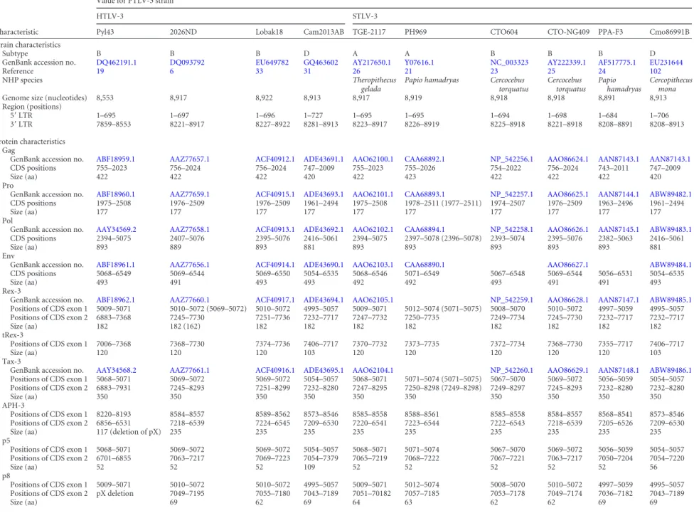
TABLE 2Bioinformatics analysis of PTLV-3 coding sequences and predicted coding sequences, including auxiliary proteinsa
Characteristic
Value for PTLV-3 strain
HTLV-3 STLV-3
Pyl43 2026ND Lobak18 Cam2013AB TGE-2117 PH969 CTO604 CTO-NG409 PPA-F3 Cmo86991B
Strain characteristics
Subtype B B B D A A B B B D
GenBank accession no. DQ462191.1 DQ093792 EU649782 GQ463602 AY217650.1 Y07616.1 NC_003323 AY222339.1 AF517775.1 EU231644
Reference 19 6 33 31 26 21 23 25 24 102
NHP species Theropithecus
gelada
Papio hamadryas Cercocebus torquatus
Cercocebus torquatus
Papio hamadryas
Cercopithecus mona
Genome size (nucleotides) 8,553 8,917 8,922 8,913 8,917 8,919 8,918 8,918 8,891 8,913
Region (positions)
5=LTR 1–695 1–697 1–696 1–727 1–695 1–695 1–694 1–698 1–684 1–706
3=LTR 7859–8553 8221–8917 8227–8922 8281–8913 8223–8917 8226–8919 8225–8918 8221–8918 8208–8891 8208–8913
Protein characteristics Gag
GenBank accession no. ABF18959.1 AAZ77657.1 ACF40912.1 ADE43691.1 AAO62100.1 CAA68892.1 NP_542256.1 AAO86624.1 AAN87143.1 AAN87143.1
CDS positions 755–2023 756–2024 756–2024 747–2009 755–2023 755–2026 754–2022 756–2024 743–2011 747–2009
Size (aa) 422 422 422 420 422 423 422 422 422 420
Pro
GenBank accession no. ABF18960.1 AAZ77659.1 ACF40915.1 ADE43693.1 AAO62101.1 CAA68893.1 NP_542257.1 AAO86625.1 AAN87144.1 ABW89482.1 CDS positions 1975–2508 1976–2509 1976–2509 1961–2494 1975–2508 1978–2511 (1977–2511) 1974–2507 1976–2509 1963–2496 1961–2494
Size (aa) 177 177 177 177 177 177 177 177 177 177
Pol
GenBank accession no. AAY34569.2 AAZ77658.1 ACF40913.1 ADE43692.1 AAO62102.1 CAA68894.1 NP_542258.1 AAO86626.1 AAN87145.1 ABW89483.1 CDS positions 2394–5075 2407–5076 2395–5076 2416–5061 2394–5075 2397–5078 (2396–5078) 2393–5074 2395–5076 2382–5063 2416–5061
Size (aa) 893 889 893 881 893 893 893 893 893 881
Env
GenBank accession no. ABF18961.1 AAZ77656.1 ACF40914.1 ADE43690.1 AAO62103.1 CAA68890.1 AAO86627.1 ABW89484.1
CDS positions 5068–6549 5069–6544 5069–6550 5054–6535 5068–6546 5071–6549 5067–6548 5069–6544 5056–6531 5054–6535
Size (aa) 493 491 493 493 492 492 493 491 491 493
Rex-3
GenBank accession no. ABF18962.1 AAZ77660.1 ACF40917.1 ADE43694.1 AAO62105.1 NP_542259.1 AAO86628.1 AAN87147.1 ABW89485.1
Positions of CDS exon 1 5009–5071 5010–5072 (5069–5072) 5010–5072 4995–5057 5009–5071 5012–5074 (5071–5075) 5008–5070 5010–5072 4997–5059 4995–5057
Positions of CDS exon 2 6883–7368 7245–7730 7251–7736 7232–7717 7247–7732 7250–7735 7249–7734 7245–7730 7232–7717 7232–7717
Size (aa) 182 182 (162) 182 182 182 182 182 182 182 182
tRex-3
Positions of CDS exon 1 7006–7368 7368–7730 7374–7736 7406–7717 7370–7732 7373–7735 7372–7734 7368–7730 7355–7717 7406–7717
Size (aa) 120 120 120 103 120 120 120 120 120 103
Tax-3
GenBank accession no. AAY34568.2 AAZ77661.1 ACF40916.1 ADE43695.1 AAO62104.1 NP_542260.1 AAO86629.1 AAN87148.1 ABW89486.1
Positions of CDS exon 1 5068–5071 5069–5072 5069–5072 5054–5057 5068–5071 5071–5074 (5071–5075) 5067–5070 5069–5072 5056–5059 5054–5057 Positions of CDS exon 2 6883–7931 7245–8293 7251–8299 7232–8280 7247–8295 7250–8298 (7249–8298) 7249–8297 7245–8293 7232–8280 7232–8280
Size (aa) 350 350 350 350 350 350 350 350 350 350
APH-3
Positions of CDS exon 1 8220–8193 8584–8557 8589–8562 8573–8546 8585–8558 8588–8561 8585–8558 8584–8557 8568–8541 8573–8546
Positions of CDS exon 2 6856–6531 7218–6539 7224–6545 7209–6530 7220–6541 7223–6544 7222–6543 7218–6539 7205–6526 7209–6530
Size (aa) 117 (deletion of pX) 235 235 235 235 235 235 235 235 235
p5
Positions of CDS exon 1 5068–5071 5069–5072 5069–5072 5054–5057 5068–5071 5071–5074 5067–5070 5069–5072 5056–5059 5054–5057
Positions of CDS exon 2 6701–6855 7063–7217 7069–7223 7054–7379 7065–7219 7068–7222 7067–7221 7063–7217 7050–7204 7054–7220
Size (aa) 52 52 52 109 52 52 52 52 52 56
p8
Positions of CDS exon 1 5009–5071 5010–5072 5010–5072 4995–5057 5009–5071 5012–5074 5008–5070 5010–5072 4997–5059 4995–5057
Positions of CDS exon 2 pX deletion 7049–7195 7055–7180 7043–7189 7051–70182 7057–7185 7053–7178 7049–7174 7036–7182 7043–7189
Size (aa) 69 62 69 64 63 62 62 69 69
Turpin
et
al.
938
jvi.asm.org
January
2015
Volume
89
Number
2
Journal
of
Virology
antibodies in PBS–5% milk: fluorescein- or Texas Red-conjugated goat anti-rabbit (FI-1000 and TI-1000, respectively [Vector] [1:100]) and Dy-light 488- or 549-conjugated horse anti-mouse (DI-2488 and DI-2549, respectively [Vector] [1:500]). Nucleic acids were stained with 4= ,6-di-amidino-2-phenylindole (DAPI)-containing mounting medium (DAPI Fluoromount G; Southern Biotech). Images were acquired by using a Leica sp5 spectral confocal microscope and analyzed with Fiji software (71). Two-dimensional graphs representing pixel intensities (gray level) along a 9-m line (yellow on the image) were plotted by using the Plot Profile tool of ImageJ software. The degree of colocalization between p9-His and p12-HA was measured by using the Pearson correlation coeffi-cient JACoP plugin (72), where a value of 1 indicates perfect colocaliza-tion and a value of 0 indicates a random distribucolocaliza-tion, as previously described (73). Projections along the z-axis of the maximum intensity of HA-p9 or p12-HA signals in addition to the calreticulin signal were real-ized by using the stack-Z Project tool of Fiji software. Nucleolar indexes were calculated by using Fiji software as follows. A region of interest (ROI) corresponding to a combination of nucleoli was defined for each cell based on nucleolin staining. Integrated signal densities of the green chan-nel inside and outside the ROI were then measured. Indexes were defined as the ratio of the integrated density inside the ROI to the integrated density outside the ROI.
Tax-3 serum.Tax-3 rabbit polyclonal antibodies were obtained from Eurogentec.
Immunoblot analysis.Cells were first washed with PBS, collected with PBS–5 mM EDTA, lysed (50 mM Tris-HCl [pH 8], 120 mM NaCl, 5 mM EDTA, 0.5% NP-40, 1 mM phenylmethylsulfonyl fluoride [PMSF], 1 mM dithiothreitol [DTT], 50 mM NaF, and 0.2 mM Na3VO4plus pro-tease inhibitors [Complete-EDTA-free; Roche]), and incubated on ice as previously described (74). Cell debris were pelleted by centrifugation. The protein concentration was determined by a Bradford assay (Bio-Rad). Fifty to seventy micrograms of proteins was loaded onto 4 to 12% NU-PAGE gels (Invitrogen), subjected to electrophoresis, and transferred onto polyvinylidene difluoride (PVDF) membranes. Membranes were blocked in a 5% milk–PBS– 0.05% Tween solution and incubated over-night at 4°C with the following primary antibodies: anti-beta-actin (1: 4,000) (catalogue number A2228; Sigma), anti-His6(1:4,000) (ab5000; Abcam), anti-HA (1:4,000) (MMS-101-R; Covance), anti-GFP (1:4,000) (catalogue number 622380; Clontech), and anti-Tax-3 (1:1,000) (Euro-gentec). The next day, membranes were washed and incubated for 1 h at room temperature with anti-mouse or anti-rabbit horseradish peroxi-dase-conjugated secondary antibodies (1:40,000) (NA9310 and NA9340;
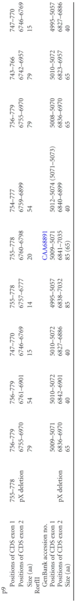
p9 Positions of CDS exon 1 755–778 756–779 756–779 747–770 755–778 755–778 754–777 756–779 743–766 747–770 Positions of CDS exon 2 pX deletion 6755–6970 6761–6901 6746–6769 6757–6777 6760–6798 6759–6899 6755–6970 6742–6957 6746–6769 Size (aa) 79 54 15 14 20 54 79 79 15 RorfII GenBank accession no. CAA68891 Positions of CDS exon 1 5009–5071 5010–5072 5010–5072 4995–5057 5009–5071 5012–5074 (5071–5073) 5008–5070 5010–5072 4995–5057 Positions of CDS exon 2 pX deletion 6836–6970 6842–6901 6827–6886 6838–7032 6841–7035 6840–6899 6836–6970 6823–6957 6827–6886 Size (aa) 65 40 40 85 85 (65) 40 65 65 40 aNewly identified auxiliary ORFs in PPA-F3 and PH969 sequences were searched from 10 full-length sequencees of PTLV-3 genomes available in GenBank, as described in Materials and Methods. The nucleotide location of each coding sequence (CDS) is indicated for the different PTLV-3 strains. The size of the corresponding predicted protein and its GenBank accession numbe r, if available in the databases, are also indicated. When needed, minor adjustments were made to the online coding sequences and amino acids sizes, but original numbers are indicated in parentheses.
FIG 3p5 is a cytoplasmic and nuclear protein. (A) p5 amino acid sequence (PPA-F3 strain). (B) HeLa cells transfected with 400 ng of an HA-p5-encoding vector. Cells were observed as described in Materials and Methods. Bar, 10m.
New PTLV Auxiliary Proteins
on May 14, 2018 by B-ON FCCN TRIAL
http://jvi.asm.org/
GE Healthcare). Membranes were then developed by using the ECL Plus kit (GE Healthcare).
Luciferase assays.A total of 3⫻105HeLa cells were transfected (Ef-fectene; Qiagen) with the STLV-3 LTR-Luc (50 ng) reporter plasmid (38) together with the STLV-3 molecular clone (50 ng) or STLV-3 Tax-3 (50 ng) plasmid (66) and increasing amounts (100 to 500 ng) of STLV-3 auxiliary protein-encoding plasmids. When required, cells were trans-fected with the luciferase–RxRE-3 (25 ng) reporter plasmid together with Rex-3–His- and p8-His-encoding plasmids. Transfections were carried out in the presence of the phRG-TK vector (10 ng) in order to normalize the results for transfection efficiency. Reporter activities were assayed at 40 h posttransfection by using the dual-luciferase reporter assay system (Promega). Luminescence measurements were assessed on a Glomax mi-croplate luminometer (Promega).
Transformation of Rat-1 fibroblasts in soft agar.HA-p5-, p8-His-, p9-His-, RorfII-His-, Tax-3–His-, and APH-3–His (75)-encoding se-quences were inserted into the pCSEF-IRES-bsd lentiviral vector, kindly provided by M. Fujii (76). Lentiviral vectors were transfected into 2⫻106 293T cells together with pCAG-HIVgp and pCMV-VSV-G-RSV-Rev to produce viral particles. The production of lentiviral vectors, stable trans-duction of Rat-1 cells, and transformation assays were previously de-scribed (77).
Nucleotide sequence accession numbers.The new STLV-3 mRNA coding sequences characterized in the present study have been deposited in GenBank with the following accession numbers: KP187845 (p5PPA-F3), KP187846 (p8PPA-F3), KP187847 (p9PPA-F3), KP187848 RorfIIPPA-F3, KP187849 (p5PH969), and KP187850 (p8PH969).
RESULTS
Presence of four auxiliary mRNAs in STLV-3-infected or
-trans-fected cells.
Van Brussel et al. previously demonstrated the
pres-ence of an auxiliary mRNA, named
RorfII
, in cells chronically
infected with STLV-3
PH969.
RorfII
encodes an 85-amino-acid-long
protein (
78
). Given the importance of HTLV-1 auxiliary proteins
in the viral cycle and in viral persistence
in vivo
, we sought to
search for and characterize other STLV-3 auxiliary transcripts.
First, RNA was extracted from both PH969-infected cells (
22
)
and 293T cells transfected with an STLV-3
PPA-F3molecular clone
(
24
,
66
). Next, a series of RT-PCR experiments was performed by
using forward primers located in the 5
=
LTR of STLV-3 or before
the splice donor site of the
tax/rex
mRNA and reverse primers
before the splice acceptor site of the
tax/rex
mRNA (
Fig. 1A
). This
strategy was designed to avoid amplification of
tax/rex
mRNA,
which is likely to be more abundant than other pX transcripts
(
79–81
). This allowed us to amplify several doubly or singly
spliced STLV-3 mRNA species. All bands visible on the agarose
gels were cut and sequenced. Sequences corresponding to ORFs of
⬍
40 amino acids were excluded from subsequent analyses. Three
ORFs corresponding to putative open reading frames (
Fig. 1B
to
D
) were discovered, in addition to the previously described ORF
RorfII. These ORFs encode putative proteins of 5 kDa, 8 kDa, and
9 kDa and were therefore named p5, p8, and p9, respectively (
Fig.
1B
and
E
). Sequencing of these new mRNA species allowed
in silico
prediction of the locations of the putative splice donor and
accep-tor sites (
Fig. 1F
). mRNAs encoding p8 were also amplified from
PH969 RNA samples (data not shown).
A second series of RT-PCR experiments was then performed
by using splice-specific primers (
Fig. 1G
). Consistent with our
initial results,
p5
,
p8
, and
p9
mRNAs were present in 293T cells
transfected with the STLV-3 molecular clone (
Fig. 1H
). As
con-trols,
RorfII
mRNA (
78
) as well as
tax/rex
mRNA were also
ampli-fied. mRNA encoding the short version of Rex (tRex-3), which
was previously described in HTLV-2- and HTLV-1-infected cells,
was also amplified (
82
,
83
). As an internal control,
gapdh
was also
amplified by RT-PCR.
p5
and
p8
are expressed
in vivo
in infected nonhuman
pri-mates.
We next sought to confirm these results by using
ex vivo
samples. PBMCs were obtained from a single
P. hamadryas papio
baboon naturally infected with STLV-3 (
24
) and from a
nonin-fected baboon used as a negative control (
Fig. 2A
). RNA was
ex-tracted, and RT-PCR was performed as described above. These
experiments allowed us to demonstrate the presence of
p8
and
p5
transcripts (
Fig. 2B
), while
RorfII
- and
p9
-specific signals were not
detected (data not shown). As controls,
tax/rex-3
and
tRex-3
mRNAs were also amplified (
Fig. 2B
). Amplification of
gapdh
transcripts by RT-PCR demonstrated that both samples contained
amplifiable mRNA (
Fig. 2B
, bottom right).
p9
and
RorfII
are reexpressed after
in vitro
culture of
pri-mary PBMCs.
We then hypothesized that
p9
and
RorfII
might be
expressed following viral reactivation, as previously reported for
HTLV-1 and -2 transcripts (
83
,
84
). To test this hypothesis,
PBMCs obtained from the same animals were cultured in the
pres-ence of IL-2 and PHA for 24 to 72 h before RT-PCR was
per-formed. Indeed,
p9
and
RorfII
transcripts were detected after 72 h
of
ex vivo
culture (
Fig. 2C
), and their presence was confirmed by
sequencing (data not shown).
Altogether, these experiments demonstrated that the STLV-3
pX region encodes transcripts that are expressed
in vitro
and
in
vivo
.
In order to determine whether STLV-3 p5, p8, p9, and RorfII
may be homologues of the HTLV-1 p12, p13, or p30 protein, their
cDNAs were cloned into expression vectors and transfected into
HeLa cells. In addition, 10 full-length PTLV-3 sequences
depos-ited in GenBank, corresponding to 4 HTLV-3 and 6 STLV-3
iso-lates of the A, B, and D subtypes, were analyzed (
Tables 1
and
2
).
This allowed us to perform amino acid sequence comparisons. Of
note, the HTLV-3 Pyl43 provirus contains a deletion in its pX
region that ablates most putative ORFs in this domain (
20
).
p5 sequence analysis and intracellular localization.
As shown
in
Fig. 1E
,
p
5 arises from a doubly spliced mRNA. The protein,
en-coded by ORF V using the Tax AUG initiation codon, is 52 aa in
length. Its sequence does not resemble those of other HTLV-1
auxil-iary proteins (
Fig. 3A
). Amino acid sequence comparisons revealed
that the p5 sequence is present in HTLV-3 and STLV-3 strains,
inde-pendently of the viral subtype, with conservation ranging from 75 to
94% identity (
Table 1
). The HA-p5 (
Fig. 3B
) construct was then
transfected into human (HeLa) or simian (Cos) cells. p5 localized
diffusely in the nucleus and cytoplasm in both cell types.
Colocaliza-tion between p5 and a specific organelle was not observed (data not
shown). As a control, HA-p5 protein expression was determined by
Western blot analysis (see
Fig. 7A
and data not shown).
p9 induces a loss of reticulum labeling, similarly to HTLV-1
p12.
p9 arises from a singly spliced mRNA from ORF II (
Fig. 1E
),
as is the case for RorfII. This peculiar splicing of p9, with the first
exon close to the 5
=
-LTR 3
=
end, is reminiscent of bovine leukemia
virus (BLV) G4 (
85
). p9 is a 79-amino-acid-long protein that is
translated after initiation at the Gag AUG start codon. The p9
protein contains the first 8 amino acids of the Gag precursor, a
putative nuclear export signal (NES), 2 putative classic SH3
li-gand-binding sites (PXXP motif), and a 44-amino-acid-long
main that is shared with RorfII and contains 2 leucine-rich
do-mains (
Fig. 4A
). This observation is reminiscent of the HTLV-1
Turpin et al.
940 jvi.asm.org Journal of Virology January 2015 Volume 89 Number 2
on May 14, 2018 by B-ON FCCN TRIAL
http://jvi.asm.org/
FIG 4The STLV-3 p9 protein induces a loss of reticulum labeling similarly to the HTLV-1 p12 protein. (A) Amino acid sequence of p9 (PPA-F3 strain). The dashed box represents the predicted nuclear export signal (LxxxLxxLxL), and boxes show leucine-rich domains. Predicted SH3 ligand-binding sites (PXXP) are shown in boldface type and are underlined with a dashed line. (B and C) HeLa (B) and Cos (C) cells were transfected with 400 ng of HA-p9-, p9-His-, and/or p12-HA-encoding vectors. Thirty-six hours later, cells were observed as described in Materials and Methods. White arrows indicate p9- or p12-expressing cells and the corresponding loss of the calreticulin signal. Yellow arrows indicate cells expressing low levels of p12-HA or HA-p9 and the corresponding calreticulin signal. (B and C, top and middle) Images of relevant optical slices of a z-stack acquisition of the width of the studied cells. To emphasize the loss of the calreticulin signal, maximum-intensity z-projections were realized on all signals. (Bottom) Quantitative p9-His and p12-HA colocalization analysis done by using the JACoP tool (ImageJ software) to measure Pearson’s correlation coefficient. Bar, 10m. (D) Western blot analysis of p12-HA and HA-p9 expression. HeLa cells were transfected with 1.5g of HA-tagged-protein-expressing plasmid or the pSG5M empty vector. Cell lysates (50g) were subjected to electrophoresis and probed with anti-HA or anti-beta-actin antibodies.
on May 14, 2018 by B-ON FCCN TRIAL
http://jvi.asm.org/
p12 protein, which contains four SH3 domains and two putative
leucine zipper-like motifs. Analysis of GenBank sequences showed
that ORF II, which encodes p9, is present in different subtype B
strains, but the corresponding ORF displays a premature stop
codon in subtype A and D sequences (
Table 1
). Colocalization
experiments were then performed with human (HeLa) and simian
(Cos) cells transfected with p9 expression plasmids. When
ex-pressed at low levels, HA-p9 had no impact on calreticulin (ER
marker), while high HA-p9 expression levels led to decreased
cal-reticulin staining on the entire volume of the cells, as
demon-strated by the z-projection of HA and calreticulin signals (
Fig. 4B
and
C
, top, white arrows [high p9 expression] and yellow arrows
[low p9 expression], and right [z-projections]), similarly to
HTLV-1 p12 expression (
Fig. 4B
and
C
, middle, white arrows
[high p12 expression] and yellow arrows [low p12 expression]).
As a control, a histidine-tagged p9 construct was transfected.
His-p9 demonstrated a similar phenotype (data not shown). We
also coexpressed STLV-3 p9 and HTLV-1 p12 and quantitatively
assessed whether they colocalized or not. Image analysis
demon-strated colocalization of both proteins in the cytoplasm of
trans-fected cells (Pearson’s coefficient [
r
]
⫽
0.891 and 0.896 for HeLa
and Cos cells, respectively) (
Fig. 4B
and
C
, bottom). Thus, p9, like
p12, is located in the endoplasmic reticulum. As a control for
protein expression, p12 and p9 Western blot analyses were
per-formed (
Fig. 4D
). Altogether, these results suggest that although
their respective sequences show little similarity, p9 and p12 may
be functionally related.
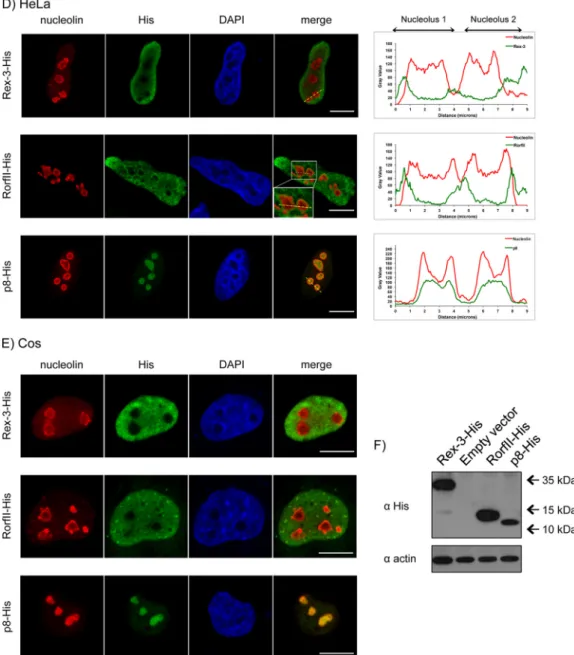
Rex-3, RorfII, and p8 share the N-terminal domain but do
not have a similar localization.
STLV-3 p8 is a 63-aa-long
pro-tein, encoded by ORF I (
Fig. 1E
), whose sequence is present in all
HTLV-3/STLV-3 subtypes. Sequence analyses revealed that p8
shares its N-terminal arginine/lysine-rich 21-aa sequence with
both Rex and RorfII (
Fig. 5A
to
C
). Based on previous HTLV-1
Rex studies, it could be envisioned that this domain contains a
putative NLS/nucleolar localization signal (NoLS) and an
RNA-binding domain (
86
,
87
). Of note, Rex also contains a putative
NES and two multimerization sequences. We performed a
side-by-side comparison of p8, Rex-3, and RorfII localizations (
Fig. 5D
and
E
). Although the 21-aa N-terminal domain is common to the
3 proteins, only p8 localizes to the nucleolus, while Rex-3 was
found in the nucleus of HeLa cells (
Fig. 5D
, right) and Cos cells
(
Fig. 5E
). In HeLa cells, the RorfII localization in the nuclei and
around the nucleoli is reminiscent of that of the HTLV-2 p10
accessory protein, which also contains the HTLV-2 Rex
N-termi-nal 21 aa (
88
). A similar pattern was observed in Cos cells,
al-though the RorfII signal was also observed in the cytoplasm. p8
protein localization was confirmed by using either histidine- or
HA-tagged constructs (data not shown). As a control, HTLV-1
Rex that was also cloned into the same expression vector
demon-strated a nucleolar localization (data not shown). Western blots
demonstrated that all proteins were expressed (
Fig. 5E
).
Alto-gether, these experiments demonstrate that although the STLV-3
Rex-3, RorfII, and p8 proteins share a domain that was reported to
be important for nucleolar import in HTLV-1, only p8 was found
in the nucleolus. Thus, we hypothesize that either a second NLS
domain is required for p8 nucleolar localization or protein folding
impairs the function of this domain in the context of the
full-length Rex-3 and RorfII proteins.
p8 contains two independent nucleolar localization signals.
HTLV-1 p30, a repressor of viral expression, also localizes within
the cell nucleolus. Given the role of p30 in the viral cycle, we
sought to determine the function of STLV-3 p8. p8 sequence
anal-ysis revealed the presence of numerous arginine residues located
between amino acids 1 and 31 (
Fig. 5C
). We obtained a series of
deletion constructs encompassing either amino acids 1 to 21
(pu-tative NLS), amino acids 22 to 63 (containing additional
argi-nines), or amino acids 31 to 63 as a control (
Fig. 6A
, left).
Immu-nofluorescence experiments were then performed after transient
transfection of HeLa cells (
Fig. 6A
, right). We observed that the
presence of either the deletion construct encompassing aa 1 to 21
or the deletion construct encompassing aa 22 to 63 allowed p8
localization in the nucleoli, while deletion of amino acids 1 to 30
strongly impaired this localization. This indicates that the
de-letion constructs encompassing aa 1 to 21 (containing five
ar-ginines) and aa 22 to 31 (containing two arar-ginines) both harbor
a NoLS. However, amino acids 22 to 31 were not sufficient for
relocalization of the proteins into the nucleoli (data not
shown), thus suggesting that this domain requires folding of
the protein for efficient import. These results suggest that p8
localization is driven by two NoLS domains present within the
regions encompassing amino acids 1 to 21 and 22 to 31.
West-ern blotting demonstrated that all proteins were expressed
(
Fig. 6B
).
To further define which arginines are required for the
localiza-tion of p8 in nucleoli, the arginine residues of either the first
pu-tative NoLS (amino acids 1 to 21) (
Fig. 6C
) or the second putative
NoLS (amino acids 22 to 63) (
Fig. 6D
) or throughout the
full-length p8 construct (
Fig. 6E
) were mutated.
Mutation of two internal arginines (R) to alanines (A) (which
are important for Rex-1 localization [
87
]) had a limited effect,
while mutation of either the first or the last arginine, in
combina-tion with the three internal ones, completely abolished p8
local-ization in the context of the construct encompassing aa 1 to 21
(
Fig. 6Ca
to
e
). We then evaluated the importance of arginine
residues present in the region spanning amino acids 22 to 30 (
Fig.
6Df
). Surprisingly, mutation of arginine to alanine at both
posi-tions had a limited impact on the construct encompassing aa 22 to
63 (
Fig. 6Df
). Thus, we combined these mutations with those of
the domain encompassing aa 1 to 21 (
Fig. 6Dg
to
j
). As described
above (
Fig. 6C
), p8 localization was driven by the presence or
absence of arginines within the domain encompassing aa 1 to 21
rather than by those within the sequence at aa 22 to 30. These
results were confirmed by using the full-length p8 protein with
arginine mutations in both domains (
Fig. 6Ek
to
n
). Consistent
with the results presented above, mutations in the sequence
spanning aa 22 to 30 had no impact, while mutation of 3 to 4
out of 5 arginines in the first 21 N-terminal amino acids
strongly impaired p8 localization. Altogether, these results
show that although the sequence spanning aa 22 to 30 has the
ability to drive localization into the nucleolus when cloned in
front of GFP, the critical NoLS sequence is present at residues 1
to 21 of the N-terminal domain of p8. As controls, expression
levels of the different p8 mutants were analyzed by Western
blotting (
Fig. 6F
to
I
).
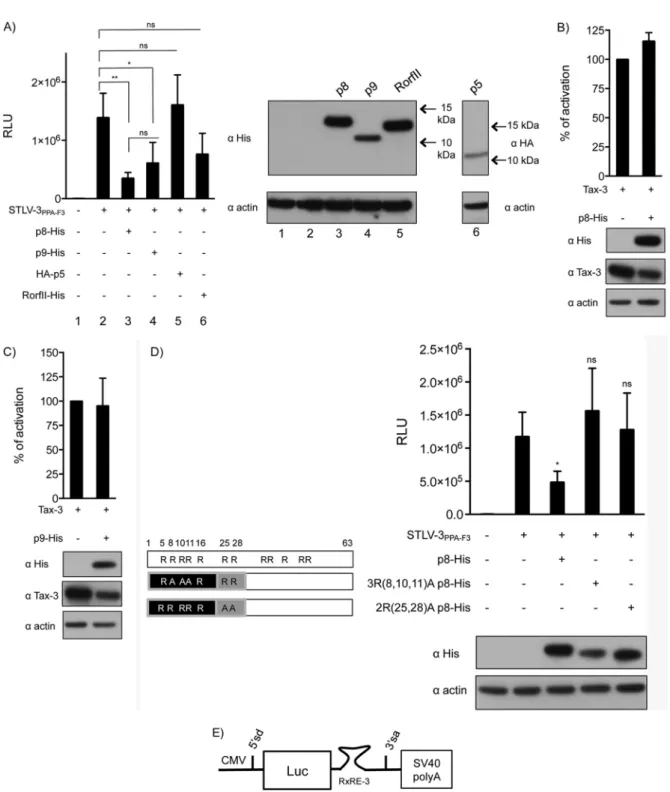
p8 and p9 repress expression from the viral LTR.
We next
wanted to determine whether STLV-3 p8, p5, or p9 could repress
viral expression similarly to HTLV-1 p30. To address this
ques-tion, we used an experimental model that was previously
devel-oped for p30 (
51
). Cells were cotransfected with the STLV-3
PPA-F3molecular clone in the presence of plasmids encoding the
dif-Turpin et al.
942 jvi.asm.org Journal of Virology January 2015 Volume 89 Number 2
on May 14, 2018 by B-ON FCCN TRIAL
http://jvi.asm.org/
FIG 5Rex, RorfII, and p8 share the same N-terminal domain but do not have a similar localization. (A) Amino acid sequence of Rex-3 (PPA-F3 strain). The internal initiation codon of tRex is indicated. Underlined sequences correspond to predicted multimerization domains (MD). (B) Amino acid sequence of RorfII (PH969 strain). (C) Amino acid sequence of p8 (PH969 strain) consisting of the first 21 amino acids of Rex-3 linked to the ORF number 1 of the pX region (X-I)-encoded sequence. (D and E) HeLa (D) and Cos (E) cells were transfected with 400 ng of a Rex-3–His-, RorfII-His, or p8-His-encoding vector, as indicated. Thirty-six hours later, cells were observed as described in Materials and Methods. Enlargement of RorfII localization is shown in the merged images in panel D. The intensity of fluorescence for each staining along the yellow line drawn on the merged images is plotted in the diagrams on the right. Bar, 10m. (F) HeLa cells were transfected with 1.5g of a Rex-3–His-, RorfII-His-, or p8-His-expressing plasmid or with the backbone vector. Cell lysates (50g) were subjected to electrophoresis and probed with antihistidine or anti-beta-actin antibodies.
on May 14, 2018 by B-ON FCCN TRIAL
http://jvi.asm.org/
ferent STLV-3 auxiliary proteins and an STLV-3
LTR-lucifer-ase reporter construct (
Fig. 7A
). Expression of either p8 or p9
led to significantly decreased luciferase activity, while p5 and
RorfII had no significant effect (
Fig. 7A
). As controls, p5, p8,
p9, and RorfII protein levels were determined (
Fig. 7A
, right).
In control experiments, p8 or p9 plasmids were cotransfected
together with Tax and the LTR reporter construct (
Fig. 7B
and
C
). As expected, the luciferase activity was not significantly
altered, thus suggesting that p8 and p9 do not prevent Tax from
recruiting the transcription machinery on the viral promoter.
Western blot analyses of p8, p9, and Tax were performed (
Fig.
7B
and
C
, bottom). Altogether, these experiments demonstrate
that at least two STLV-3 auxiliary proteins have the ability to
alter viral expression
in cellulo
.
Given that p8 was the most potent repressor of LTR activity
(
Fig. 7A
), we then sought to determine its mechanism of action.
We postulated that p8 amino-terminal arginine residues,
sim-ilarly to p30, could support its inhibitory activity. To
discrim-inate between direct and indirect (via localization) functions of
arginines, we tested two p8 constructs with mutations in either
the first or the second NoLS but which still display a nucleolar
localization. Interestingly, both p8 mutants lacked the ability
to inhibit luciferase activity (
Fig. 7D
), indicating that these
arginines are involved in the repressive function of p8
per se
,
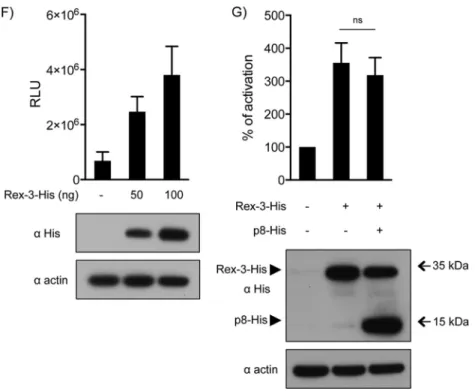
independently of their role in the control of p8 localization.
HTLV-1 Rex (Rex-1) stabilizes unspliced and singly spliced
viral mRNAs and mediates their nuclear export by binding to a
cis
-acting sequence present on the RNA called the Rex response
element (RxRE). Since STLV-3 p8 shares the first 21 aa with
Rex-3, which are critical for the ability of Rex-1 to bind the
RxRE, we tested whether p8 could compete with Rex-3 for
binding to the RxRE. As described previously (
70
), we
gener-ated a CMV–Luc–RxRE-3 reporter plasmid that contains the
luciferase coding sequence and the RxRE of STLV-3
PPA-F3be-tween a donor and an acceptor splice site (
Fig. 7E
). Since Rex
favors the export of unspliced mRNA, we hypothesized that the
overexpression of this viral protein should lead to increased
luciferase activity. This was indeed observed (
Fig. 7F
). p8
ecto-FIG 6p8 is a nucleolar protein with two independent nucleolar localization signals. (A, left) Map of the different plasmids encoding truncated p8 proteins fused to GFP. Bars represent the length of each truncated construct. (Right) p8-GFP-encoding plasmids or a control pEGFP-N1 backbone vector was transfected into HeLa cells. Thirty-six hours later, cells were observed as described in Materials and Methods. Bar, 10m. The table on the right quantitatively summarizes the intensities of the GFP signals in nucleoli (see Materials and Methods for details). (B) Western blot analysis of the p8-GFP deletion mutants. HeLa cells were transfected with 1.5g of each p8-GFP plasmid or with the pEGFP-N1 empty vector. Cell lysates (50g) were subjected to electrophoresis and probed with anti-GFP or anti-beta-actin antibodies. (C to E) Plasmids encoding p8 proteins with single or combined point mutations encompassing aa 1 to 21 (C), aa 22 to 63 or 1 to 30 (D), and aa 1 to 63 (E) were transfected into HeLa cells as described above. Localization analyses were performed as described above for panel A. (F to I) HeLa cells were transfected with 1.5g of the p8-GFP constructs or with the pEGFP-N1 backbone vector. Cell lysates (70g) were subjected to electrophoresis and probed with anti-GFP and anti-beta-actin. Western blot analysis of p8-GFP mutants with deletion constructs encompassing aa 1 to 21 (F), aa 22 to 63 (G), aa 1 to 30 (H), and aa 1 to 63 (I). The letters correspond to the plasmids used in panels C to E.Turpin et al.
944 jvi.asm.org Journal of Virology January 2015 Volume 89 Number 2
on May 14, 2018 by B-ON FCCN TRIAL
http://jvi.asm.org/
pic expression had no effect on Rex-3-mediated mRNA export,
thus suggesting that p8 does not compete with Rex for RxRE
binding (
Fig. 7G
). Western blot analyses demonstrated that all
proteins were expressed (
Fig. 7F
and
G
, bottom).
STLV-3 auxiliary proteins are unable to promote cell
prolif-eration.
Finally, we tested whether p5, p8, p9, and RorfII could
transform cells by using an assay of colony formation in soft agar
(CFSA). Based on our previous data suggesting that Tax-3 is
phe-notypically related to Tax-1, we hypothesized that Tax-3 could
transform cells
in vitro
. Tax-1 and Tax-3 were therefore used as
positive controls in these experiments. We also examined
whether APH-3 could transform cells. As expected, Tax-3
ex-pression led to a high number of colonies in soft agar, while
none of the STLV-3 auxiliary proteins (p5, p8, p9, and RorfII)
FIG 6continuedNew PTLV Auxiliary Proteins
on May 14, 2018 by B-ON FCCN TRIAL
http://jvi.asm.org/
FIG 7p8 and p9 repress expression from the viral LTR. (A) HeLa cells were transfected with an STLV-3 LTR-Luc construct together with either an empty vector or the STLV-3 molecular clone and p8-His, p9-His, RorfII-His, or HA-p5, as indicated. (Left) Results are shown as mean RLU (relative light units) with standard deviations. (Right) Levels of expression of the viral constructs were determined by Western blot analysis using anti-His or an anti-HA antibodies, as indicated. (B and C) HeLa cells were transfected with an STLV-3 LTR-Luc construct together with Tax-3 and p8-His (B) or p9-His (C), as indicated. Forty hours later, luciferase activity was measured and normalized as 100% activity for the Tax-3 condition without p8-His or p9-His. Levels of expression of the viral constructs were determined by Western blot analysis using anti-His and anti-Tax-3 antibodies. As a control, the membrane was probed with an anti-actin antibody. (D) HeLa cells were transfected with an STLV-3 LTR-Luc construct together with either an empty vector or the STLV-3 molecular clone and wild-type p8 or p8 constructs with a mutation in the first NoLS [3R(8,10,11)A p8-His, equivalent to the construct inFig. 6Ek] or in the second NoLS [2R(25,28)A p8-His, equivalent to the construct inFig. 6El]. Luciferase activity was measured at 40 h posttransfection. Data are presented as the means and standard deviations from 3 independent experiments. (E) Schematic representation of the CMV–Luc–RXRE-3 reporter plasmid. (F) HeLa cells were transfected with the CMV–Luc–RXRE-3 construct together with 50 or 100 ng of the Rex-3–His construct or the backbone plasmid. Data in panels D and F are presented as mean relative light units and standard deviations from 3 independent experiments. (G) HeLa cells were transfected with the CMV–Luc–RXRE-3 construct together with 50 ng of the Rex-3–His construct or an empty vector and 1,000 ng of p8-His. Luciferase activity was measured 40 h later and normalized. In panels D, F, and G, levels of expression of the different constructs were determined by Western blot analysis using an anti-His antibody (ⴱ,P⬍0.05;ⴱⴱ,
P⬍0.01; ns, nonsignificant [determined by attest]).
Turpin et al.
946 jvi.asm.org Journal of Virology January 2015 Volume 89 Number 2
on May 14, 2018 by B-ON FCCN TRIAL
http://jvi.asm.org/
led to a significant increase in cell transformation (
Fig. 8
).
In-terestingly, APH-3 expression led to a number of colonies that
were significantly different from the background. Thus, these
experiments demonstrated that Tax-3 and, to a lesser extent,
APH-3 have oncogenic properties but that STLV-3 auxiliary
proteins do not have a direct role in cellular transformation.
DISCUSSION
HTLV-1 and HTLV-2 pX domains contain several ORFs that
en-code auxiliary proteins that play critical roles in the viral cycle and
infectivity (
45
,
88–92
). HTLV-1
p12
(initially named
Rof
),
p13
,
and
p30
(or
Tof
) mRNAs were discovered in HTLV-1-infected cell
lines, and their expression was confirmed in cells transfected with
an HTLV-1 molecular clone or in
ex vivo
samples (
82
,
92
,
93
).
However, the expression of these proteins
in vivo
was proven only
indirectly (
94
). Furthermore, HTLV-2 pX encodes the p10, p11,
and p28 auxiliary proteins. HTLV-2 p10 was initially thought to
be an HTLV-1 p12 homologue. However, it does not have the
same localization and does not bind to the same cellular proteins
(
88
). While HTLV-2 p28 and HTLV-1 p30 could be functional
counterparts, they also display low overall amino acid sequence
conservation (
88
). Importantly, the level of expression of HTLV-1
and HTLV-2 auxiliary transcripts is low and is tightly regulated
in
vivo
and
in vitro
(
79
,
81
,
93
).
HTLV-1 strains are classified into seven subtypes (subtypes A
to G) (
10
), while HTLV-2 strains belong to three subtypes
(sub-types A to C). HTLV-1 molecular clones and most HTLV-1
sam-ples used originate from Japan, the Caribbean region, or South
America and are therefore very likely to belong to subtype A. Thus,
the presence of p13- and p30-encoding ORFs in HTLV-1 genomes
belonging to other subtypes has not yet been tested. Previous
stud-ies demonstrated that a significant number of strains from Japan
and Argentina encode a truncated p12 protein that lacks the
fourth SH3 sequence but retains the other domains (
95
).
In silico
analyses performed on a full-length subtype B provirus (EL)
showed 13% variability in its p12 sequence, with conservation of
the calcineurin-binding motif (
96
,
97
). One study also reported a
truncated p12 protein in West and Central African STLV-1 strains
(
97
). However, none of those studies tested whether the HTLV-1/
STLV-1 p12 variants have the same properties as the prototype
sequence.
Regarding HTLV-2, the search for pX transcripts was
per-formed by using a single cell line (MoT) infected with subtype A or
by using a molecular clone derived from this cell line (
88
).
In silico
analyses suggested that the ORFs encoding p10 and p11 are also
FIG 7continuedFIG 8Colony formation assay. Rat-1 cells were stably transduced with lenti-viral vectors expressing the p5, p8, p9, and RorfII proteins as well as APH-3. Cells were plated in soft agar, and colonies were counted 3 weeks later. p8, p9, and RorfII were tested by using Tax-1 as a positive control, while p5 and APH-3 were compared with Tax-3. The data are presented as mean CFU and standard errors of the means from 3 independent experiments performed in triplicate for each construct.
New PTLV Auxiliary Proteins