Vol.58, n.6: pp. 913-922, November-December 2015 http://dx.doi.org/10.1590/S1516-89132015060319
ISSN 1516-8913 Printed in Brazil
BRAZILIAN ARCHIVES OF BIOLOGY AND TECHNOLOGY
A N I N T E R N A T I O N A L J O U R N A L
Molecular Cloning and Sequencing of Alkalophilic
Cellulosimicrobium cellulans
CKMX1 Xylanase Gene
Isolated from Mushroom Compost and Characterization of
the Gene Product
Abhishek Walia
1*, Preeti Mehta
2, Shiwani Guleria
3, Anjali Chauhan
3and C.K. Shirkot
3 1Department of Microbiology; DAV University; Jalandhar, Punjab – India. 2DBT-IOC Centre for BioenergyResearch, Indian Oil R&D Centre, Haryana, India. 3Department of Basic Sciences (Microbiology Section); Dr. YS Parmar University of Horticulture and Forestry; Nauni, Solan – India
ABSTRACT
A xylanolytic bacterium was isolated from mushroom compost by using enrichment technique. Results from the metabolic fingerprinting, whole-cell fatty acids methyl ester analysis and 16S rDNA sequencing suggested the bacterium to be Cellulosimicrobium cellulans CKMX1. Due to the xylanolytic activity of this bacterium, isolation and characterization of the xylanase gene were attempted. A distinct fragment of about 1671 bp was successfully amplified using PCR and cloned into Escherichia coliDH5α. A BLAST search confirmed that the DNA sequence
from the amplified fragment was endo-1, 4-beta-xylanase, which was a member of glycoside hydrolase family 11. It showed 98% homology with Cellulosimicrobium sp. xylanase gene (Accession no. FJ859907.1) reported from the gut of Eisenia fetida in Korea. In silico physico-chemical characterization of amino acid sequence of xylanase showed an open reading frame encoding a 556 amino acid sequence with a molecular weight of 58 kDa and theoretical isolectric point (pI) of 4.46 was computed using Expasy’s ProtParam server. Secondary and homology based 3D structure of xylanase was analysed using SOPMA and Swiss-Prot software.
Key words: Cellulase free xylanase, Cellulosimicrobium cellulans, E.coli, Cloning, Gene
*Author for correspondence: [email protected]
INTRODUCTION
Xylan is the major hemicellulosic constituent of hard and soft wood, and is the next most abundant renewable polysaccharide after cellulose. This complex heteropolysaccharide consists of a main
chain of 1,4-β-D-xylose monomers and short chain
branches consisting of O-acetyl, α
-L-arabinofuranosyl and α-D-glucuronyl residues.
Xylanases and associated debranching enzymes produced by a variety of microorganisms including bacteria, yeast and filamentous fungi, bring about the hydrolysis of hemicelluloses
(Maheshwari et al. 2000). Xylanolytic enzymes are receiving increasing attention because of their potential application in pulp bleaching (Golugiri et al. 2012; Singh et al. 2013) and bioconversion of lignocelluloses into feedstocks and fuels (Kim et
al. 2000). The xylan degrading system includes
endo-1,4-xylanases (1,4-β-xylan xylanohydrolase; EC 3.2.1.8), which release long and short xylo-oligosaccharides, and other xylanases that attack only longer chains, and β-D-xylosidase (1,4-β-xylan xylohydrolase; EC 3.2.1.37), which remove
D-xylose residues from short
Recent advances in molecular biology and genetic engineering in the last two decades have opened up the areas of application of gene cloning and recombinant DNA technology for the construction of genetically modified microbial strains with selected enzyme machinery. To ensure the commercial utilization of hemicellulosic residues in the pulp and paper industries, the production of higher xylanase yields at low capital cost is required. In this respect, isolation and cloning of the xylanase gene represents an essential step in
the engineering of the most efficient
microorganisms (Hernandez et al. 2008; Deesukon
et al. 2011). Several reports describe the
production, purification, partial characterization, molecular cloning, sequencing and expression of the alkaline xylanase gene from alkalophilic
Bacillus sp. strain C-125 in non-cellulase
producing E.coli carrying a plasmid pCX311 (Liu
et al.2010; Verma and Satyanarayana, 2012; Driss
et al. 2012). Such studies are essential to produce a
more-efficient xylanase producer, which will allow improvement of paper quality (Walia et al. 2015). Furthermore, biochemical studies on
xylanase-secreting and non-secreting
microorganisms could lead to better understanding of the xylanase secretory process and the development of cloning strategies that would guarantee secretion of desired products. Various
molecular and biotechnological aspects of
xylanase producing microorganisms, such as the regulation of xylanase biosynthesis at the molecular level, and newer strategies, such as use of gene cloning, protein engineering, and site-directed mutagenesis for obtaining xylanase with novel properties, have been described in detail by
Kulkarni et al.(2003).
Thus, the aim of this work was to isolate, clone and sequencing of xylanase gene and physico-chemical characterization of deduced amino acid
sequence of xylanase gene by using Expasy’s
ProtParam server. Secondary and homology based 3D structure of xylanase was also analysed and
compared with some other commercially
important xylanases using significant
improvement in protein secondary structure prediction by consensus prediction from multiple alignments (SOPMA) and Swiss-Prot software.
MATERIAL AND METHODS
Strain isolation
Alkalophilic Cellulosimicrobium cellulans
CKMX1 was previously isolated from mushroom compost (Walia et al. 2013). Xylan degrading bacteria were isolated by the enrichment technique. The most predominant bacterial colonies capable of good growth on basal salt medium (BSM) with the following composition
(g/L): Na2HPO4, 6.0; KH2PO4, 3.0; NaCl, 0.5;
NH4Cl, 1.0, 1 M MgSO4 (2 mL) and 1 M CaCl2
(0.1 mL) were picked and purified. The bacterial culture was grown and maintained in BSM, pH 8.0, containing 0.5% xylan. The bacterial culture was maintained in 30% glycerol at -20°C.
Identification of bacteria
The bacterium was identified using colony
morphology observation, biochemical tests,
whole-cell fatty acid methyl ester analysis and 16S rDNA sequencing.
Bacterial DNA extraction
Bacterial isolate was grown in nutrient broth at 35°C overnight and the cells were harvested by centrifugation at 5,000×g for 5 min. DNA was isolated from these cells by using Real genomic DNA extraction kit (Taiwan), which was suspended in 100 µL of elution buffer and quantified on 1% agarose gel. The total genomic DNA was kept at -20°C before use (Sambrook and Russel 2001).
PCR amplification of 16S rDNA and sequence determination
Species level identification of strain was conducted by 16S rDNA sequence comparison. PCR reaction was carried out in 20 µL reaction containing ~50ng of template DNA, 20 pmoles of
each primer fC1
(5´-GCAAGTCGAGCGGACAGATGGGAGC-3´)
and reverse primer rC2
(5´-AACTCTCGTGGTGTGACG GGCGGTG-3´),
Primers for cloning of xylanase gene
PCR Primers to amplify the xylanase gene were designed based on the endo-1,4-beta-xylanase
gene sequences from different Cellulosimicrobium
sp. DNA sequences of xylanases were obtained from GenBank (www.ncbi.nlm.nih.gov) and were
aligned using ClustalW (Larkin et al. 2007) and
CLC sequence viewer (www.clcbio.com).
Conserved regions were detected.
i) Primers for cloning and sequencing of partial
xylanase gene
Following set of primers were designed on the basis of gene sequences of earlier known xylanases submitted in NCBI database:
Primer set 1:
These primers were designed using known consensus sequences of xylanase:
Forward XylF: 5’-cgtcggcttcgcgctcgaccc-3’ (21
mer)
Reverse XylR: 5’-cggtgatgcgcacgtccacgcc-3’ (22
mer)
ii) Primers for cloning and sequencing of
complete xylanase gene Primer set 1:
These primers were designed from the closest match of known partial sequence of xylanase CKMX1:
Forward XylCompF: 5’
-atgaccaggaccatctggagacgacc-3’ (26 mer)
Reverse XylCompR: 5’
-tcaggcgacctcgcaggcggcaccgtcg-3’ (28 mer)
The above primers were custom synthesized and supplied in lyophilized form by ITC Promega. Primers were regenerated by suspending in autoclaved double distilled water to make 1.0 mL of 1 pM primer before use.
Xylanase gene isolation
PCR was performed using the genomic DNA isolated from the bacterium as a template. PCR reaction were cycled 30 times as denaturation was 94ºC for 45 s, annealing was 45ºC for 1 min and extension was 72ºC for 2 min followed by final extension at 72ºC for 10 min.
Cloning and transformation
Purified PCR products (amplicons) was ligated into pGEM-T easy cloning vector as per
manufacturer’s instructions (pGMET- clone kit,
Promega) and transformed into E. coli DH5α
competent cells using the heatshock method. Successful transformants were selected using blue/white screening. Plasmids were extracted
using a plasmid extraction kit (Axygen PrepTM
Plasmid Mini prep Kit), according to
manufacturer’s instruction from the culture with
positive colony PCR results verified by restriction digest, and was sent for sequencing by the commercial sequencing facility (Xcleris lab).
Plate assay for screening recombinants
The recombinants containing desired PCR product inserts were screened for the expression of xylanase activity by the Congo-red assay described by Teather and Wood (1982). The
recombinant E. coli clones containing the
recombinants were overlaid with 0.5% oat-spelt xylan, dissolved in solid LB and then incubated at 37ºC overnight. Then, the plates were stained with 1% Congo-red for 30 min and destained with 1 M NaCl. Positive clones were identified by a zone of clearance around xylanase-expressing clones truncated.
Sequencing and phylogenetic analysis of xylanase gene of C. cellulans CKMX1
The sequence was aligned with corresponding sequences of 16S rRNA from the database using
BLAST from the website
http://www.ncbi.nlm.nih.gov/blast (Altschul et
al.1997). Multiple alignments were generated by
the MULTALIN program from the website: http://prodes.toulouse.inra.fr/multialin/multialin.ht ml (Corpet 1998). The phylogenetic trees were constructed after truncating the sequences to the length of shortest sequence in a given alignment data by neighbor-joining algorithm using PHYLIP
Package (Felsenstein et al. 1993). The stability
among clades of a phylogenetic tree was assessed by taking 1000 replicates of the data set and was
analyzed using the programs SEQBOOT,
DNADIST, NEIGHBOR and CONSENSE of the PHYLIP package. Tree was viewed with the help
of TreeView from the website
http://taxonomy.zoology.gla.ac.uk/rod/treeview.ht ml (Page 1996).
Physico-chemical characterization of amino acid sequence of xylanase
Amino acid sequence of xylanase gene of C.
cellulans CKMX1 was deduced by using xylanase gene sequence of strain CKMX1 and translates into protein sequence by expasy protein software. Theoretical isoelectric point (pI), molecular
residues, instability index (Guruprasad et al.
1990), aliphatic index (Ikai 1980) were computed
using the Expasy’s ProtParam server (Gasteiger et
al. 2005) (http://us.expasy.org/tools/
protparam.html). Amino acid composition of the protein sequences could reveal their nature; hence,
amino acidcomposition was also computed.
Secondary structure prediction
SOPMA was employed for calculating the secondary structural features of the selected target protein sequences considered for this study (Geourjon and Deleage 1995).
Homology-based structural model of xylanase Homology-based structural model of xylanase
from C. cellulans CKMX1 was built using
SWISS-MODEL server
(http://swissmodel.expasy.org/). Exo-beta-1,
4-glucanase from Cellulomonas fimi (PDB ID:
3CUG, 69.16% sequence identity) was used as template to build the structural model of xylanase. The model was visualised using PyMOL (Delano 2002) The PyMOL Molecular Graphics System (2002) found online (http://pymol.org)].
RESULTS
Isolation and identification
Xylanolytic bacteria was previously isolated from the enriched culture on basal salt media (BSM) containing xylan as the sole carbon source and
identified as C. cellulans CKMX1 according to
morphological, biochemical, whole-cell fatty acid methyl ester analysis and 16S rDNA sequence (Walia et al. 2013; 2014).
Phylogenetic analysis of isolate CKMX1 according to 16S rDNA sequence
Universal primers were used successfully to amplify 16S rDNA from bacterial isolate CKMX1, yielding an amplicon of the expected size, i.e., ~1136 bp. The sequence of 16S rDNA from CKMX1 was then analyzed using BLASTn analysis (http://www.ncbi.nlm.nih.gov/blast) and
was found to have 97% homology with several C.
cellulans strains reported earlier. The 16S rDNA sequence of CKMX1 was also compared with the corresponding sequences of eight different
Cellulosimicrobium sp. reported earlier. Sequence
analysis revealed that CKMX1 belonged to C.
cellulans strain CKMX1 as it showed maximum
homology (97%) with C. cellulans strain AMP-11
(Accession no. HM104377).
To trace out the evolutionary patterns of the test isolate and to determine the relationship with other selected sequences at NCBI, a phylogenetic tree was also constructed using the neighbour-joining (J) method of mathematical averages among 16S rDNA sequence of CKMX1 and the corresponding
sequence of eight different Cellulosimicrobium sp.
strain CKMX1 was united with quite high statistical support by the bootstrap estimates for 1,000 replications. The resulting phylogenetic tree
also verified CKMX1 as C. cellulans as it
clustered closely with C. cellulans with high
(80%) boot strap value. The 16S rDNA sequence of the strain has been deposited in the GenBank database under accession number JN135476.
Detection and isolation of xylanase gene from genomic DNA of C. cellulans CKMX1 using specific primer based PCR method
Xylanase gene specific primers were designed for the isolation of partial xylanase i.e., XylF and XylR, as mentioned in the materials and methods section, were used for the detection and
amplification of partial xylanase gene from C.
cellulans CKMX1. PCR reaction resulted in the amplification of ~564 bp fragment as expected for partial xylanase gene. The amplified fragment was then excised from the gel and was used for cloning into pGEM-T easy vector.
DNA sequencing and cloning of PCR product into pGEM-T easy vector system
The purified PCR product was ligated to pGEM-t easy vector and finally transformed to competent
E. coli (DH5α) cells. Positive clone containing
insert was identified using colony PCR. Plasmid was then isolated from the selected positive colony and was sequenced. Sequence of 564 bp corresponding to partial xylanase gene of CKMX1 was obtained. The partial xylanase gene sequence of the strain was deposited in the GenBank database under accession number HF546135.
Amplificantion and sequencing of complete xylanase gene from C. cellulans CKMX1
After obtaining the partial sequence of xylanase
gene from C. cellulans CKMX1, primers were
product of ~1671 bp was obtained in PCR amplification using designed primers (Fig. 1).
Figure 1 - Agarose gel electrophoresis showing the isolation of DNA and amplification of complete xylanase gene from C. cellulans
CKMX1. Lane M: 100 bp Marker, Lane 1: Showing ~1671 bp amplification.
PCR product was then cloned to pGEM-T easy
vector system and transformed to competent E.
coli (DH5α) cells. Clones containing insert was
then identified using colony PCR. Plasmid was then isolated from selected positive colony and
was sequenced. Sequence of 1671 bp
corresponding to complete xylanase gene of
CKMX1 was obtained.
Sequence analysis of complete xylanase gene of
C. cellulans CKMX1
Sequence of complete xylanase gene of C.
cellulans CKMX1 was analysed with
corresponding sequences of nineteen different
sequences of xylanase gene from
Cellulosimicrobium sp. reported earlier. Sequence
analysis revealed that xylanase gene of C.
cellulans CKMX1 showed maximum homology
(98%) with xylanase gene of Cellulosimicrobium
sp. (Accession no. FJ859907.1) reported from the
gut of Eisenia fetida in Korea, followed by 75.6%
homology with Streptomyces rameus strain L2001
(Accession no. KC011007).
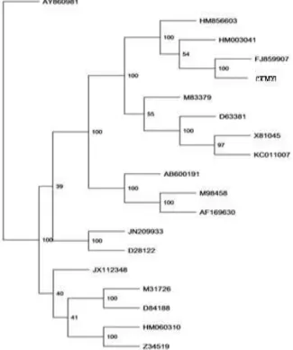
C. cellulans CKMX1 was united with quite high statistical support by the bootstrap method and value inferred higher than 40% are only presented in Figure 2. Phylogenetic tree based on complete xylanase sequences (Fig. 2) validated that the
xylanase gene of C. cellulans CKMX1 clustered
closely with xylanase gene of other reported
isolates of Cellulosimicrobium sp. with high boot
strap value (100%). The complete xylanase gene sequence of the strain has been deposited in the GenBank database under accession number HG425182. Multiple sequence alignment of xylanase gene of strain CKMX1 with closely related strain HY-13 was done by ClustalW alignment software. Nucleotide sequence of CKMX1 was then used to deduce the amino acid sequence of the xylanase protein using expasy protein translation tool (http://us.expasy.org/ tools.html). Amino acid sequence corresponding to xylanase protein from CKMX1 was also aligned with the corresponding amino acid sequence from
C. Cellulans strain HY-13 reported from the gut of
E. fetida in Korea.
Figure 2 - Neighbour-joining phylogenetic tree based on complete xylanase gene sequence data of C. cellulans CKMX1 and related strains. Isolate used in the study is in boldface.
Physico-chemical characterization of amino acid sequence of xylanase
Theoretical isoelectric point (pI), molecular weight, total number of positive and negative residues, instability index, and aliphatic index were computed using the Expasy’s ProtParam server (http://us.expasy.org/tools/protparam.html).
Amino acid composition of the protein sequences could reveal their nature; hence, amino acid composition was also computed (Table 1).
Maximum percentage composition of alanine was 15.1%, followed by threonine 11.2%, glycine 10.4%, valine 7.9% and so on. The amino acid composition of xylanase polypeptide showed that it contained 63 negatively charged residues (Asp+Glu) and 37 positively charged residues
(Arg+Lys). The xylanases from different
microorganisms showed significant similarity
when theoretical protein parameters were
compared (Table 2). The pI values of all protein sequences were in the range of 4.32-9.57, indicating that all considered xylanase sequences
were acidic, except C. flavigena XIB (9.57),
C. flavigena DSM 20109 (8.94), Cellvibrio gilvus
ATCC 13127 (8.70) and Bacillus subtilis (9.18),
Pedobacter saltans DSM 12145 (9.16). The
xylanase of C. cellulans CKMX1 had the highest
pI value of 4.66, which showed the xylanase sequence was acidic.
Table 1 - Deduced amino acid composition of xylanase of C. cellulans CKMX1.
Amino acid No. Percentage composition (%)
Ala (A) 84 15.1
Arg (R) 28 5.0
Asn (N) 12 2.2
Asp (D) 42 7.6
Cys (C) 5 0.9
Gln (Q) 23 4.1
Glu (E) 21 3.8
Gly (G) 58 10.4
His (H) 10 1.8
Ile (I) 18 3.2
Leu (L) 34 6.1
Lys (K) 9 1.6
Met (M) 4 0.7
Phe (F) 20 3.6
Pro (P) 24 4.3
Ser (S) 29 5.2
Thr (T) 62 11.2
Trp (W) 15 2.7
Tyr (Y) 14 2.5
Val (V) 44 7.9
Pyl (O) 0 0.0
Sec (U) 0 0.0
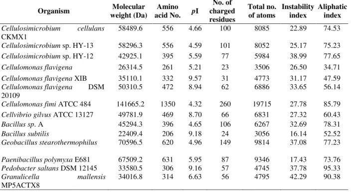
Table 2 - Theoretical protein parameters of few xylanases calculated using bioinformatics tools.
Organism Molecular
weight (Da)
Amino acid No. pI
No. of charged residues
Total no. of atoms
Instability index
Aliphatic index
Cellulosimicrobium cellulans
CKMX1
58489.6 556 4.66 100 8085 22.89 74.53
Cellulosimicrobium sp. HY-13 58296.3 556 4.59 101 8052 25.17 75.23
Cellulosimicrobium sp. HY-12 42925.1 395 5.59 77 5984 38.99 77.65
Cellulomonas flavigena 26314.5 261 5.21 23 3506 26.50 34.71
Cellulomonas flavigena XIB 35110.1 332 9.57 31 4773 31.17 47.59
Cellulomonas flavigena DSM 20109
50310.5 472 8.94 62 6886 33.65 56.14
Cellulomonas fimi ATCC 484 141665.2 1350 4.32 260 19715 27.78 85.79
Cellvibrio gilvus ATCC 13127 49781.9 469 8.70 66 6831 27.32 60.43
Bacillus sp. A 45294.3 396 4.65 106 6267 32.69 78.31
Bacillus subtilis 22409.4 206 9.18 24 3056 16.14 52.52
Geobacillus stearothermophilus 70596.5 620 4.96 149 9814 37.08 77.23
Paenibacillus polymyxa E681 67509.2 631 5.95 87 9346 17.43 73.76
Pedobacter saltans DSM 12145 33580.5 306 9.16 57 4745 37.78 95.33
Granulicella mallensis
MP5ACTX8
34016.8 314 6.63 56 4795 42.29 90.38
Instability index showed that all the considered sequences were classified as stable with values
ranging from 16.14 to 38.99, except Granulicella
mallensis MP5ACTX8 (42.29). The xylanase
sequence of C. cellulans CKMX1 showed
temperature range, including the xylanase
sequence of C. cellulans CKMX1, which showed
74.53 aliphatic index.
Secondary structure prediction
Significant improvement in protein secondary structure prediction by consensus prediction from
multiple alignments (SOPMA) was employed for
calculating the secondary structural features of the selected target protein sequences considered for this study. The secondary structure indicates whether a given amino acid lies in a helix, strand or coil. Secondary structure features as predicted
using SOPMA are represented in Table 3. The
results revealed that random coils dominated among secondary structure elements, followed by
alpha helix, extended strand and beta turn in C.
cellulans CKMX1.
Table 3 - Calculated secondary structure elements of C. cellulans CKMX1.
Secondary structure C. cellulans CKMX1 (%)
Alpha helix (Hh) 27.70
310 helix (Gg) 0.00
Pi helix (Ii) 0.00
Beta bridge (Bb) 0.00
Extended strand (Ee) 19.24
Beta turn (Tt) 7.19
Bend region (Ss) 0.00
Random coil (Cc) 45.86
Ambigous states 0.00
Other states 0.00
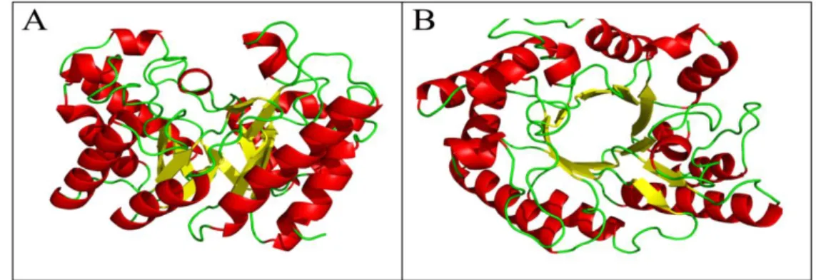
Homology based 3D structure prediction
The molecular model of the endo-1,4-β xylanase is
described as consisting of an alpha helix and several beta pleated sheets with a compact structure and is shown in Figure 3.
Figure 3 - Homology based 3D structure of xylanase proteins by using Swiss-Prot model A) Side view B) Top view.
DISCUSSION
Several recombinant xylanases from bacteria, yeast and fungi have been cloned and expressed in
E. coli by construction of genomic libraries, followed by expression cloning. In this study, a
highly xylanolytic C.cellulans CKMX1 was
isolated from mushroom compost. Cellulomonas
sp. is known to produce and secrete a variety of hydrolytic enzymes, and thus can utilize various complex carbohydrates in their natural habitats such as soil. Xylanases are produced in various amounts by bacteria and filamentous fungi to degrade xylan as a source of energy and have potential application in a wide range of industrial
processes (Kulkarni et al. 2003; Keshwani and
Cheng 2009). Xylanases encoding genes from
Cellulomonas species have been cloned and
sequenced (Bhalero et al. 1990). Multiple
xylanases may be produced by the organisms to enhance the utilization of xylan (Wong et al. 1988).
In the present study, C. cellulans CKMX1 had
unique ability to produce cellulase-free xylanase
(Walia et al. 2012; Walia et al. 2013 and 2014).
The results also showed the xylan hydrolysis by
endoxylanases (1,4-β-D-xylan xylanohydrolase
E.C.3.2.1.8). Therefore, C. cellulans CKMX1
isolated from mushroom compost was selected for further identification of xylanse encoding gene responsible for xylanase production. The PCR
results obtained showed that the C. cellulans
gene (Fig. 1). The nucleotide sequence of 1671 bp amplicon shared 98% homology to xylanase gene of Cellulosimicrobium sp. HY-13
endo-beta-1,4-xylanase (xylK-1) gene, (Accession no.
FJ859907), thus confirming the isolation of
xylanase gene from C. cellulans CKMX1 (Fig. 1).
A positive clone, indicating that the clone contained an insert of 1671 bp in size with a 58% G+C content, in conformity with the high G+C levels was found in nucleotide sequences from
other Cellulosimicrobium sp., e.g., endo-beta-1,
4-xylanase gene from Cellulosimicrobium sp. HY-13
(Kim et al. 2009). The conserved domain search of
the xylanases gene across the Cellulosimicrobium
sp. and other bacterial species indicates the significance of enzyme in xylan hydrolysis. The mature protein consisted of amino acids 556 with calculated molecular weight of 58 KDa. To-date, many xylanases genes have been cloned from
different microorganisms, including Actiomadura
sp. S14 (Thayat et al.2011), Paenibacillus sp.
12-11 (Zhao et al.2011) and Streptomyces sp. S27 (Li
et al. 2009). However, this study was to clone a
xylanase gene from C. cellulans CKMX1 and a
very little information was available regarding
cellulase-free xylanases from C. cellulans
CKMX1. Further, the work regarding
hyperxylanolytic production could be done in vivo.
Amino acid composition determines the
fundamental properties of the enzyme (Arora et al.
2009). The amino acid composition of xylanase sequences is represented in Table 1. The pI values of all protein sequences were in the range of 4.32-9.57, indicating that all considered xylanase
sequences were acidic except C. flavigena XIB
(9.57), C. flavigena DSM 20109 (8.94), Cellvibrio
gilvus ATCC 13127 (8.70), B. subtilis (9.18) and
Pedobacter saltans DSM 12145 (9.16). The
xylanase enzyme of C. cellulans CKMX1 had the
pI value of 4.66 which showed that the xylanase
sequence was acidic (Arora et al. 2009). The
calculated isoelectric point (pI) will be useful because at pI, solubility is least and mobility in an electro focusing system is zero. The instability index, which gives clue about the stability of a
protein in vitro, can be calculated using Expasy’s
ProtParam server (Geourjon and Deleage 1995). All the considered sequences were classified as stable with values ranging from 16.14 to 38.99,
except Granulicella mallensis MP5ACTX8
(42.29). The xylanase sequence of C. cellulans
CKMX1 (22.89) indicated a stable protein (Table 2).
The aliphatic index (AI), which is defined as the relative volume of a protein occupied by aliphatic side chains, is regarded as a positive factor for the increase of thermal stability of globular proteins. It can be calculated by using equation, i.e., Aliphatic index= X(Ala) + a*X(Val) + b*X(Leu)) + b*X(Ile) and Expasy’s ProtParam server
(Gasteiger et al. 2005). Aliphatic index for the
xylanase sequences ranged from 34.71-95.33 (Table 2). The very high aliphatic index of all xylanase sequences indicated that these xylanases could be stable for a wide temperature range,
including the xylanase sequence of C. cellulans
CKMX1, which showed 74.53 aliphatic index
(Arora et al. 2009). The secondary structure
indicates whether a given amino acid lies in a helix, strand or coil. Secondary structure features as predicted using SOPMA are represented in
Table 3. The results revealed that random coils
dominated among the secondary structure
elements, followed by alpha helix, extended strand and beta turn. The molecular model of the
endo-1,4- β xylanase is described as consisting of an
alpha helix and several beta pleated sheets with a compact structure and is shown in Figure 3. This is the first report describing cellulase-free
xylanase by C. cellulans CKMX1 isolated from
mushroom compost. Coupled with the small compact structure of the enzyme allowed it to degrade the xylan without damaging the cellulose fibres in the cellulose-hemicellulose matrix. This enzyme was also used in pulp and paper industry and showed positive results.
CONCLUSION
An endo-1,4-beta-xylanase gene from
actinomycetes was isolated and characterized. The
bacterium was identified as C. cellulans CKMX1
based on the morphological, biochemical
characterisation, whole cell fatty acid methyl ester analysis and 16S rDNA sequence. The xylanase gene of this bacterium, however, showed higher
similarity to Cellulosimicrobium sp. deposited in
the NCBI database.
ACKNOWLEDGMENTS
REFERENCES
Altschul SF, Thomas LM, Alejandro AS, Jinghui Z, Zheng Z, Webb M, et al. Gapped BLAST and PSIBLAST: a new generation of protein database search programs. Nucleic Acids Res. 1997; 25: 3389-3402.
Arora N, Banerjee AK, Mutyala S, Murty US. Comparative characterization of commercially important xylanase enzymes. Bioinformation. 2009; 3(10): 446-453.
Bhalerao J, Patki AH, Bhave M, Khurana I, Deobagkar DN. Molecular cloning and expression of a xylanase gene from Cellulomonas sp. into Escherichia coli. Appl Microbiol Biotechnol. 1990; 34: 71-76.
Corpet F. Multiple sequence alignment with hierarchial clustering. Nucleic Acids Res. 1988; 16: 10881-10890.
Deesukon W, Nishimurab Y, Haradab N, Sakamotob T, Wasana S. Purification, characterization and gene cloning of two forms of a thermostable endo-xylanase from Streptomyces sp. SWU10. Proc Biochem. 2011; 46: 2255-2262.
DeLano WL. "The PyMOL Molecular Graphics System." DeLano Scientific LLC, San Carlos, CA, USA. http://www.pymol.org. 2002.
Driss D, Bhiri F, Ghorbel R, Chaabouni SE. Cloning and constitutive expression of His-tagged xylanase GH 11 from Penicillium occitanis Pol6 in Pichia pastoris X33: purification and characterization.
Protein Express Purif. 2012; 83: 8-14.
Felsenstein J. PHYLIP (Phylogeny Inference Package) version 3.5c. Department of Genetics, University of Washington Seattle. 1993.
Gasteiger E, Hoogland C, Gattiker A, Duvaud S, Wilkins MR, Appel RD, et al. Protein identification and analysis tools on the ExPASy server. In: Walker JM. The proteomics protocols handbook. Human Press; 2005. pp. 571-607.
Geourjon C, Deleage G. SOPMA: significant improvements in protein secondary structure prediction by consensus prediction from multiple alignments. Comput Appl Biosci. 1995; 11(6): 681-684.
Goluguri BR, Thulluri C, Cherupally M, Nidadavolu N, Achuthananda D, Mangamuri LN, et al. Potential of thermo and alkali stable xylanases from Thielaviopsis basicola (MTCC-1467) in biobleaching of wood Kraft pulp. Appl Biochem Biotechnol. 2012; 167: 2369-2380.
Gomez LD, Steele-King CG, McQueen-Mason SJ. Sustainable liquid biofuels from biomass: the writing's on the walls. The New Phytologist. 2008; 178(3): 473-485.
Guruprasad K, Reddy BV, Pandit MW. Correlation between stability of a protein and its dipeptide composition: a novel approach for predicting in vivo stability of a protein from its primary sequence.
Protein Eng. 1990; 4(2): 155-161.
Hernández A, López JC, Arenas M, Santamaría R, Díaz M, Fernández-Abalos J M, et al. Xylan-binding xylanase Xyl30 from Streptomyces avermitilis: cloning, characterization, and overproduction in solid-state fermentation. Intern Microbiol. 2008; 11: 133-141.
Ikai AJ. Thermostability and aliphatic index of globular proteins. J Biochem. 1980; 88(6): 1895-1898.
Jeong KJ, Lee PC, Park IY, Kim MS, Kim SC. Molecular cloning and characterization of an endoxylanase gene of Bacillus sp. in Escherichia coli. Enzyme Microb Technol. 1998; 22: 599-605.
Keshwani DR, Cheng JJ. Switchgrass for bioethanol and other value-added applications: a review.
Bioresour Technol. 2009; 100: 1515-1523.
Kim DY, Han MK, Park DS, Lee JS, Oh HW, Shin DH, et al. Novel GH10 xylanase, with a fibronectin type 3 domain, from Cellulosimicrobium sp. strain HY-13, a bacterium in the gut of Eisenia fetida. Appl Environ Microbiol. 2009; 75(22): 7275-7279.
Kim JH, Kim SC, Nam SW. Constitutive overexpression of the endoxylanase gene in Bacillus subtilis. J Microbiol Biotechnol.2000; 10: 551-553. Kulkarni N, Shendye A, Rao M. Molecular and
biotechnological aspects of xylanases. FEMS Microbiol Rev. 2003; 23: 411-456.
Larkin MA, Blackshields G, Brown NP, Chenna R, McGettigan PA, McWilliam H, et al. ClustalW and ClustalX version 2. Bioinformatics. 2007; 23: 2947-2948.
Li N, Shi P, Yang P, Wang Y, Luo H, Bai Y, et al. Cloning, expression, and characterization of a new
Streptomyces sp. S27 xylanase for which xylobiose is the main hydrolysis product. Appl Biochem Biotechnol. 2009; 159: 521-531.
Liu Wanli, Shi Pengjun, Chen Qiang, Yang Peilong, Wang Guozeng, Wang Yaru, et al. Gene cloning, overexpression, and characterization of a xylanase from Penicillium sp. CGMCC 1669. Appl Biochem Biotechnol. 2010: 162: 1-12.
Maheshwari R, Bharadwaj G, Bhat MK. Thermophilic fungi: their physiology and enzymes. Microbiol Mol Biol Rev. 2000; 64: 461-488.
Page RDM. TREEVIEW: An application to display phylogenetic trees on personal computers. Comput Appl Biosci.1996; 12: 357-358.
Saha BC. Hemicellulose bioconversion. J Indust Microbiol Biotechnol. 2003; 30: 279-291.
Singh V, Pandey VC, Agrawal S. Potential of Laceyella sacchari strain B42 crude xylanase in biobleaching of kraft pulp. Afr J Biotechnol. 2013; 12(6): 570-579. Teather RM, Wood PJ. Use of Congo
red-polysaccharide interactions in enumeration and characterization of cellulolytic bacteria from the bovine rumen. Appl Environ Microbiol. 1982; 43: 777-780.
Thayat S, Peechapack S, Kenji M, Fusako K, Kosum C. Cloning of a thermostable xylanase from
Actinomadura sp. S14 and its expression in
Escherichia coli and Pichia pastoris. J Biosci Bioeng.
2011; 111: 528-536.
Verma D, Satyanarayana T. Molecular approaches for ameliorating microbial xylanases. Bioresource Technol. 2012; 17: 360-367.
Walia A, Mehta P, Chauhan A, Shirkot CK. Production of alkalophilic xylanases by Paenibacillus polymyxa
CKWX1 isolated from decomposing wood. Proc Natl Acad Sci India Sect B Biol Sci. 2012; 83(2): 215-223. Walia A, Mehta P, Chauhan A, Shirkot CK.
Optimization of cellulase-free xylanase production by alkalophilic Cellulosimicrobium sp. CKMX1 in solid-state fermentation of apple pomace using central composite design and response surface methodology.
Ann Microbiol. 2013; 63: 187-198.
Walia A, Mehta P, Chauhan A, Kulshrestha S, Shirkot CK. Purification and characterization of cellulase-free low molecular weight endo β-1, 4 xylanase from an alkalophilic Cellulosimicrobium cellulans CKMX1 isolated from mushroom compost. World J Microbiol Biotechnol. 2014; 30: 2597-2608.
Walia A, Mehta P, Guleria S, Shirkot CK. Improvement for enhanced xylanase production by
Cellulosimicrobium cellulans CKMX1 using Central Composite Design of Response Surface Methodology. 3Biotech. 2015; DOI: 10.1007/s13205-015-0309-2.
Walia A, Mehta P, Guleria S, Shirkot CK. Modification in the properties of paper by using cellulase-free xylanase produced from alkalophilic
Cellulosimicrobium cellulans CKMX1 in biobleaching of wheat straw pulp. Can J Microbiol. 2015; DOI: 10.1139/cjm-2015-0178.
Wong KKY, Tan LUL, Saddler JN. Multiplicity of β -1,4 xylanase in microorganisms: functions and applications. Microbiol Rev. 1988; 52: 305-317. Zhao Y, Meng K, Luo H, Yang P, Shi P, Huang H, et
al. Cloning, expression, and characterization of a new xylanase from alkalophilic Paenibacillus sp. 12-11. J Microbiol Biotechnol. 2011; 21: 861-868.