J of Evolution of Med and Dent Sci/ eISSN- 2278-4802, pISSN- 2278-4748/ Vol. 4/ Issue 30/ Apr 13, 2015 Page 5093
ELECTROCARDIOGRAPHIC AND ECHOCARDIOGRAPHIC CHANGES IN
CHRONIC OBSTRUCTIVE PULMONARY DISEASE (COPD) OF DIFFERENT
GRADES OF SEVERITY
Suma K. R1, Srinath S2, Praveen3
HOW TO CITE THIS ARTICLE:
Suma K. R, Srinath S, Praveen. Electrocardiographic and Echocardiographic Changes in Chronic Obstructive Pulmonary Disease (COPD) of Different Grades of Severity. Journal of Evolution of Medical and Dental Sciences 2015; Vol. 4, Issue 30, April 13; Page: 5093-5101, DOI: 10.14260/jemds/2015/744
ABSTRACT: BACKGROUND: Chronic obstructive pulmonary disease (COPD) is a major cause of chronic morbidity and mortality throughout the world. Pulmonary hypertension is the major cardiovascular complication of COPD; this is associated with right ventricular dysfunction and corpulmonale which has a poor prognosis. The detection of right ventricular (RV) hypertrophy in electrocardiography (ECG) has a high specificity but very low sensitivity. 2-D echocardiography can be used to assess right ventricular dimensions and the presence of pulmonary artery hypertension in patients with COPD. AIMS: This study was undertaken to observe the electrocardiographic and echocardiographic changes in COPD patients with different grades of severity as assessed clinically and through spirometry and correlate the findings with duration and severity of the disease. METHODS: 50 patients with COPD were randomly selected over a period of 2 years. All cases were studied clinically, and underwent chest X-ray, electrocardiography, echocardiography, and spirometry. The severity of the disease was graded according to BTS (British thoracic society)
guidelines. Statistical analysis of correlation was done with Pearson s test and Chi square test, and statistical significance was taken a p < 0.05. RESULTS: Mean age was 59.9+/- 10.4 years, with male preponderance. Mean duration of disease was 5.71 years, with mean exposure to smoking of 23.2 +/- 3.6 pack years. ECG findings that showed significant correlation with severity of disease were p pulmonale, right axis deviation, right bundle branch block, right ventricular hypertrophy and poor R wave progression. Only right axis deviation increased significantly with duration of disease. Among echocardiographic findings, R.A (right atrial)/R.V dilatation, R.V. failure, pulmonary hypertension and corpulmonale also showed significant correlation with severity of disease. With respect to duration of disease, R.V. dilatation, pulmonary hypertension and corpulmonale showed significant increased occurrence with increasing duration of disease .Diagnosis of corpulmonale clinically was 36%, by ECG was 44% and by echocardiography was 54%. CONCLUSION: COPD is more common in males in 5th to
7th decade of life, with a smoking history of more than 20 pack years. Most patients have moderate to
severe disease at presentation. The occurrence of ECG and echocardiographic findings increase as severity and duration of disease increases, and echocardiography detects more number of patients with R. V. dysfunction than ECG or clinical methods.
KEYWORDS: COPD, ECG, echocardiography, corpulmonale.
INTRODUCTION: Chronic obstructive pulmonary disease is a major cause of chronic morbidity and mortality throughout the world. It is the 4th leading cause of death and will be the 5th leading cause of
DALYs (Disability adjusted life years) lost world wide in 2020, accounting for enormous socio economic burden.1 COPD is characterized by slowly progressive airflow obstruction resulting in
J of Evolution of Med and Dent Sci/ eISSN- 2278-4802, pISSN- 2278-4748/ Vol. 4/ Issue 30/ Apr 13, 2015 Page 5094 complication.2 The global initiative for COPD (GOLD) defines COPD as a disease state characterized by
airflow limitation that is not fully reversible. It is usually progressive and associated with an abnormal inflammatory response of lungs to noxious particles or gases.1 Pulmonary hypertension is
the major cardiovascular complication of COPD, and occurs in those who have marked airflow limitation (FEV1 {forced expiratory volume in 1st second} <25% predicted) and also have chronic
hypoxemia. This is associated with right ventricular dysfunction and corpulmonale which has a poor prognosis.1,3 Right ventricular dysfunction is common in patients with COPD, and occurs in up to 50%
of the patients with moderate to severe COPD, and portends a higher mortality rate. Its recognition and treatment may lead to prolonged survival and improved quality of life.4
The detection of RV hypertrophy in ECG has a high specificity but very low sensitivity, a normal ECG does not rule out the presence of pulmonary hypertension in COPD5. The ECG
abnormalities are usually less pronounced in COPD than other forms of pulmonary hypertension because of the relatively modest degree of pulmonary hypertension and effects of hyperinflation.6
2-D echocardiography can be used to assess right ventricular dimensions and wall thickening and right ventricular volume overload in patients with COPD and also the presence of pulmonary artery hypertension. 2D echocardiography may be technically difficult in patients with COPD because of increase in retrosternal air which transmits sound waves poorly, but an adequate exam can be obtained in 80% of the patients.4
This study was undertaken to observe the electrocardiographic and echocardiographic changes in COPD patients with different grades of severity as assessed clinically and through spirometry.
METHODOLOGY: 50 patients with COPD, both male and female, who were admitted in our institution, were randomly selected over a period of 2 years, between January 2011 to December 2013.Patients with other pulmonary pathologies like bronchial asthma, tuberculosis, pneumo-coniosis, restrictive lung diseases, and cardiac disease of congenital, rheumatic, ischemic and hypertensive heart diseases were excluded.
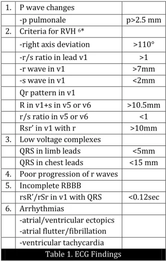
All cases were studied clinically, and underwent chest X-ray, electrocardiography, echocardiography, and spirometry. Routine investigations like complete blood count, renal function test, sputum for acid fast bacilli (AFB), and gram stain were also done. The severity of the disease was graded according to British thoracic society (BTS) guidelines.7 The findings of ECG (Table 1) and
echocardiography (table 2) were recorded. A 12 lead ECG was taken in all patients under study and following points were noted,8,9 All patients were subjected to echocardiographic examination in 2D
and M mode, to note the presence of pulmonary hypertension, RV hypertrophy, RV dilatation and RV failure.7,10
J of Evolution of Med and Dent Sci/ eISSN- 2278-4802, pISSN- 2278-4748/ Vol. 4/ Issue 30/ Apr 13, 2015 Page 5095 60% of predicted. 60% of the patients had severe airflow obstruction at the time of presentation, 36% had moderate disease and only 4% had mild disease. The mean duration of tobacco use was 23.2 pack years, ranging from 5 to 45 pack years. Majority of the patients had a tobacco exposure history of 20 to 29 pack years (32%), pts less than 10 pack years history was only 2%. 22% had 10 -19 pack years, 18% had 30-39 pack years, and 10% had >40 pack years of exposure. Majority of the patients with severe disease (70% i.e. 21 out of 30 pts) had history of greater than 20 pack years of tobacco exposure .Among patients with 10-19 pack years, 8% had moderate and 14%had severe disease.16% of the patients with moderate disease had > 20 pack years of exposure.
Only 2% had <10 pack years of exposure and had moderately severe disease. All patients in this study had history of breathlessness at presentation (100%), 96% of patients had cough with sputum at presentation. 38% of the patients presented with edema.24% had fever, and 8% had reduced urine output. The most common sign at presentation was tachypnea in 70%, followed by epigastric pulsations in 58%. 36% of the patients had evidence of congestive cardiac failure like, raised JVP (jugular venous pulse), edema and hepatomegaly. 32% of patients had loud p2, suggestive of pulmonary hypertention, 30% had parasternal heave, the clinical evidence of right ventricular hypertrophy, and 26% had cyanosis, clubbing or both which is evidence of hypoxic state, and 2% had CO2 narcosis.
In chest X-ray, 80% of the patients had features of emphysema. 64% had increased bronchovascular markings suggestive of chronic bronchitis. X –ray evidence of pulmonary hypertension, i.e., prominent pulmonary conus or right descending pulmonary artery >16 mm was present in 30% of patients. Cardiomegaly was seen in 20%.
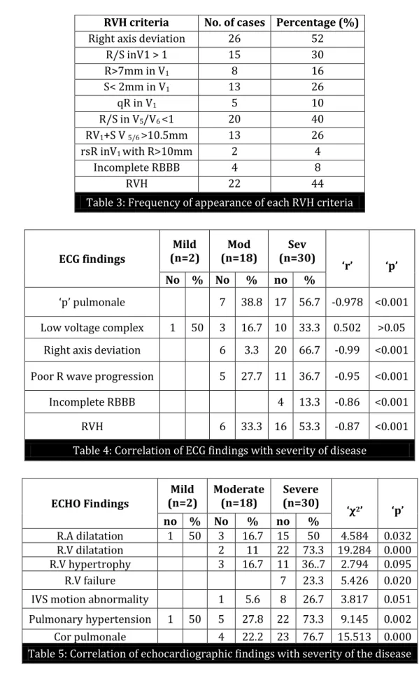
Analysis of ECG findings showed that 44% of the patients had ECG evidence of right ventricular hypertrophy (RVH) in the study. The most common RVH criteria in these patients was right axis deviation, present in 100% of the patients, followed by R/S in V 5/6 < 1 in 90%.48% 0f the
patients in this study had P pulmonale. Low voltage complexes and poor progression of R wave, which are characteristic ECG changes in emphysema were found in 28% and 32% of the patients respectively.1 patient had complete RBBB(right bundle branch block), and 1 had multiple atrial ectopics. (Table 3). Correlation of ECG findings with severity of the disease (Table 4) showed that the
findings of p pulmonale, RAD (Right axis deviation), poor R wave progression, incomplete RBBB and RVH correlate significantly with severity of the disease. (p<0.05).
Correlation of ECG findings with duration of symptoms showed that the most common ECG finding in patients with 1- years duration of symptoms was p pulmonale % . % of the patients in 1-5 years duration group had ECG evidence of RVH.50 % of the patients in 6-10 year group had ECG evidence of RVH.80% of the patients in >10 year duration group had ECG evidence of RVH. 10% of the patients in 6-10 years group had incomplete RBBB, and 20% in >10 year group had incomplete RBBB. Only right axis deviation had significant correlation with duration of the disease (p<0.05).
J of Evolution of Med and Dent Sci/ eISSN- 2278-4802, pISSN- 2278-4748/ Vol. 4/ Issue 30/ Apr 13, 2015 Page 5096 in the 1-5 year duration group had evidence of corpulmonale. 71.4% of the patients in 6- 10 years group had evidence of corpulmonale and in patients with > 10 years of symptoms 100% of them had corpulmonale. The frequency of occurrence of echocardiographic features of pulmonary hypertension in 1-5, 6-10 and > 10 years groups were 45.2%, 71.4% and 90% respectively. Features of RV failure was present in 9.7%, 21.4% and 20% in 1-5, 6-10 and >10 years groups respectively.
The echocardiographic findings of RV hypertrophy and corpulmonale correlated significantly with the duration of disease (p<0.05).Correlation of echocardiographic findings with severity of the disease (Table 5) showed that the echocardiographic signs of R.A dilatation, R.V. dilatation, R.V failure, pulmonary hypertension and corpulmonale correlated significantly with the severity of the disease (p< 0.05).
DISCUSSION: In this study, males form 84% of the patients, comparable to other studies11,12, the
higher incidence is attributable to smoking. All the females had history of exposure to smoke while cooking with dried cow dung or wood. As with previous studies 2, 11, 13, 14, the maximum number of
patients were in the age group of 50-70 years, with a mean duration of symptoms of 5.71 years, and 60% of them were having severe disease(FEV1<40%) with majority of them having a mean of 23.2
pack years of tobacco exposure.
Dyspnea and cough with expectoration was the commonest presenting symptom (50%) and most of them had signs of hyperinflation with reduced breath sounds. Signs of RVH and pulmonary hypertension were found in 30% and 32% respectively as in previous studies.11,13
Among ECG findings, 44% of the patients in this study had RVH, which varies widely in different studies depending on the criteria used, and number of patients with corpulmonale of varying etiologies.13,15,16 Among the different RVH criteria, right axis deviation, R/S in V5/V6 <1, R/S in
V1>1 were the commonest ECG changes, which according to different studies were important criteria or RVH.48% of the patients had p- pulmonale which according to some can be taken as indirect evidence of RVH.8 On correlating the ECG findings with severity of the disease, it was found that,
incidence of all ECG findings increased as the severity of the disease increased .Statistical correlation was found with p pulmonale, right axis deviation, incomplete RBBB, and RVH which was significant.(i.e. p<0.05).
Among the ECG findings of emphysema, low voltage complexes and poor progression of r wave, increased in incidence with increasing severity, but not statistically significant. Other studies correlating ECG findings with severity of the disease have also made similar observations. V.K Singh
et al also found increasing incidence of p pulmonale, R/S in V5/V6 <1, QRS axis >90° RV6<5mm, with
increasing severity of the disease. M.K Tandon also found increasing incidence of p pulmonale, right axis deviation of QRS, and dominant S in V5/6 with increasing severity as defined by FEV1/FVC.17,18 On
correlating the ECG findings with duration of symptoms, p pulmonale, right axis deviation, and RVH and incomplete RBBB, increased with the duration of the disease, statistical significance was found only with right axis deviation.(p<0.05).11,13
In the analysis of ECHO findings, our study showed 54% of the patients had echocardiographic evidence of corpulmonale, comprising of R.V. dilatation, R. V. hypertrophy, R. A. dilatation, or evidence of R. V. failure, or inter ventricular septum motion abnormality .Similar incidences were found in some previous studies 19, 20. On correlating the echocardiographic findings
J of Evolution of Med and Dent Sci/ eISSN- 2278-4802, pISSN- 2278-4748/ Vol. 4/ Issue 30/ Apr 13, 2015 Page 5097 All the findings had statistically significant correlation with severity, except R.V. hypertrophy, and inter ventricular wall motion abnormality, probably because of relatively lesser number of patients in moderate group, and variation in measurement of R.V wall thickness due to the presence of trabaculae, and difficulty in differentiating it from surrounding structures. Studies by Higham et al showed pulmonary hypertension in 43% in moderate and 68% in severe group, and a study by N.K. Guptha, Rithesh Kumar Agarwal showed 54.5% in moderate, 60% in severe and 100% in very severe groups, as compared to 28% and 73% in our study, the higher occurrence in severe group is probably due to higher percentage of our patients were in severe group.2,21 On correlating echocardiographic
findings with duration of disease, statistically significant correlation was found R.V dilatation, pulmonary hypertension and corpulmonale.
In this study, a diagnosis of corpulmonale could be made in 36% of patients by clinical methods, 44% by electrocardiographic methods and 54% by echocardiographic methods. This shows that echocardiography can detect more number of patients with corpulmonale in COPD and is similar to previous studies,19,22,23 This is because, clinical signs of R.V dysfunction are difficult to detect in
COPD due to lung hyperinflation and posterior rotation of heart. ECG criteria for detecting R.V hypertrophy have a reasonably high specificity but relatively low sensitivity. Echocardiography also has its difficulties in COPD patients due to over inflation of lungs which reduces the probability of getting reliable measurements.20 But most studies report that adequate examination can be obtained
in more than 70% of the patients. Many studies have proved that echocardiography is more sensitive than electrocardiography in detecting R.V dysfunction in COPD.23,24 Therefore early and periodic
echocardiography in COPD patients can help in detection of pulmonary hypertension and cor pulmonale in early stages thereby, ensuring adequate treatment thus reducing morbidity in COPD.
REFERENCES:
1. Global Initiative for Chronic Obstructive Lung Disease – Global Strategy for Diagnosis, Management, and Prevention of Chronic Obstructive Pulmonary disease.
http.//www.goldcopd.com (accessed. 09-08-2004).
2. M. A. Higham, D. Dawson, J. Joshi, P. Nihoyanno Paulos, N W Morell. Utility of echocardio-graphy in assessment of pulmonary hypertension secondary to COPD. Eur. Respir. J. 2001; 17: 350-355. 3. John R. Reilly Jr., Edwin K Silverman, Steven D. Shapiro. Chronic Obstructive Pulmonary
disease. Chapter . (arrison s Principles of )nternal Medicine th Edition.
Dennis L. Kasper, Anthony S. Fauci, Dan L. Longo, E Braunwald, Stephen L. Hauser, J. Larry Jameson. McGraw Hill, (USA) 2005: 1547-1554.
4. James R Klinger, Nicholas S Hill. Right ventricular dysfunction in chronic obstructive pulmonary disease, Evaluation and management. Chest 1991; 99: 715-23.
5. Emmanuel Weitzenblum. Chronic corpulmonale. Heart 2003; 89: 225-230.
6. Vallerie V. McLaughlin, Stuart Rich. Cor-pulmonale. Chapter 54. Heart Disease – A text book of Cardiovascular Medicine. Eugene Braunwald, 6th edition, W. B. Saunders Company,
Philadelphia. 2001: 1936-1954.
7. William MacNee. Chronic bronchitis and emphysema. Chapter . Crofton and Douglas Respiratory Diseases. Anthony Seaton, Douglas Seaton, Gordon Leitch, 5th edition. Black well
J of Evolution of Med and Dent Sci/ eISSN- 2278-4802, pISSN- 2278-4748/ Vol. 4/ Issue 30/ Apr 13, 2015 Page 5098 8. S. Padmavathi and Veena Raizada. Electrocardiogram in chronic corpulmonale. British Heart
Journal 1972; 34: 658-667.
9. John H. Phillips, Goerge E. Burch, Problems in the diagnosis of corpulmonale. Am. Heart J. 1963; 6(6): 818-832.
10. Salcedo. The right atrium and The Right ventricle. Chapter 14. Atlas of Echocardiography. Salcedo. 2nd edition, W. B. Saunders Company. Philadelphia. 1985: 281-292.
11. J. C. Banergae. Natural history and sympotomatology of chronic cor pulmonale. Indian Journal of Chest Disease, 1965; VIII: 174-181.
12. G. Chappel. The electrocardiogram in chronic bronchitis and emphysema. Bri. Heart. J. 1966; 28: 517-522.
13. Gupta S, Khastgir T, Gupta MP, Sethi KK, Manoharan S. Clinical, Haemodynamic and Echocardiographic study in chronic corpulmonale. JAPI 1989; 37(6): 373-376.
14. Putnik M, Povazan D, Vindisjesic M. Electrocardiography and echocardiography in the diagnosis of chronic corpulmonale (Article in Serbo Croatian (Roman)). Med Pregl, 1998; 51 (11): 528-31.
15. Marvin L. Murphy, Fred Hutcheson. The electrocardiographic diagnosis of right ventricular hypertrophy in chronic obstructive pulmonary disease. Chest. 1974; 65: 622-627.
16. Noble O. Fowler, Clarke Daniels, Ralph Scott, Benigno SF, Mosche Gueron. The electrocar diogram in corpulmonale with and without emphysema. The American Journal of Cardiology, 1965; 16 : 500-505
17. V. K. Singh and S. K. Jain. Effects of Airflow limitation on the electrocardiogram in COPD. Indian Journal of chest diseases and Allied sciences, 1989; 31(1): 1-8.
18. Tandon MK. Correlation of electrocardiographic features with airway obstruction in chronic bronchitis. Chest 63 (2) : 146-148
19. Himelmann RB, Struve SN, Brown JK, Namnum P, Schiller NB. Improved recognition of cor pulmonale in patients with severe chronic obstructive pulmonary disease. Am J. Med. 1988; 84: 891-898.
20. N. Danchin, A Cornette, A Henriquez. J. P. Godenir, G. Ethevenot, J. M. Polu, P. Sadant. Two dimensional echocardiographic assessment of the right ventricle in patients with chronic obstructive lung disease. Chest 1987; 92(2): 229-233.
21. N. K. Gupta, Ritesh kumar Agrawal, A. V. Srivastav and M. L. Ved Echocardiographic evaluation of heart in chronic obstructive pulmonary disease patient and its co-relation with the severity of disease. Lung India. 2011 Apr-Jun; 28(2): 105–109.
22. Oswald Mammosser M, Oswald T, Nyankiye E, Dickele McCrange D, Weitzenblum E. Non- invasive diagnosis of pulmonary hypertension in chronic obstructive pulmonary disease Comparison of ECG, radiological measurements, echocardiography and myocardial scintigraphy. Eur. J. Respir Dis. 1987; 71(5): 419-29.
23. Putnik M, Povazan D, Vindisjesic M. Electrocardiography and echocardiography in the diagnosis of chronic cor pulmonale (Article in Serbo Croatian (Roman)). Med Pregl, 1998; 51(11): 528-31.
J of Evolution of Med and Dent Sci/ eISSN- 2278-4802, pISSN- 2278-4748/ Vol. 4/ Issue 30/ Apr 13, 2015 Page 5099 1. P wave changes
-p pulmonale p>2.5 mm 2. Criteria for RVH 6*
-right axis deviation >110°
-r/s ratio in lead v1 >1
-r wave in v1 >7mm
-s wave in v1 <2mm
Qr pattern in v1
R in v1+s in v5 or v6 >10.5mm r/s ratio in v5 or v6 <1
Rsr in v with r >10mm 3. Low voltage complexes
QRS in limb leads <5mm QRS in chest leads <15 mm 4. Poor progression of r waves
5. Incomplete RBBB
rsR /rSr in v with QRS <0.12sec 6. Arrhythmias
-atrial/ventricular ectopics -atrial flutter/fibrillation -ventricular tachycardia
Table 1. ECG Findings
*presence of any of the above criteria is suggestive, but presence of 2 or more criteria is diagnostic.
1. Pulmonary artery diameter
2. Evidence of pulmonary hypertension on M mode examination of pulmonary valve
-a wave -EF slope
-Midsystolic notch, flutter
3. RV hypertrophy -Thickness of anterior wall & septum>6mm
4. RV dilatation -Diastolic dimention>25mm
5. RA dilatation
6. RV failure
-Tricuspid regurgitation -RV wall motion abnormalities -Dilatation of IVC and hepatic vein
7. Corpulmonale
-RV dilatation -RVH
-RV failure
J of Evolution of Med and Dent Sci/ eISSN- 2278-4802, pISSN- 2278-4748/ Vol. 4/ Issue 30/ Apr 13, 2015 Page 5100 RVH criteria No. of cases Percentage (%)
Right axis deviation 26 52
R/S inV1 > 1 15 30
R>7mm in V1 8 16
S< 2mm in V1 13 26
qR in V1 5 10
R/S in V5/V6 <1 20 40
RV1+S V 5/6 >10.5mm 13 26
rsR inV1 with R>10mm 2 4
Incomplete RBBB 4 8
RVH 22 44
Table 3: Frequency of appearance of each RVH criteria
ECG findings
Mild (n=2)
Mod (n=18)
Sev (n=30)
r p
No % No % no %
p pulmonale 7 38.8 17 56.7 -0.978 <0.001
Low voltage complex 1 50 3 16.7 10 33.3 0.502 >0.05
Right axis deviation 6 3.3 20 66.7 -0.99 <0.001
Poor R wave progression 5 27.7 11 36.7 -0.95 <0.001
Incomplete RBBB 4 13.3 -0.86 <0.001
RVH 6 33.3 16 53.3 -0.87 <0.001
Table 4: Correlation of ECG findings with severity of disease
ECHO Findings
Mild (n=2)
Moderate (n=18)
Severe (n=30)
χ2 p
no % No % no %
R.A dilatation 1 50 3 16.7 15 50 4.584 0.032 R.V dilatation 2 11 22 73.3 19.284 0.000 R.V hypertrophy 3 16.7 11 36..7 2.794 0.095
R.V failure 7 23.3 5.426 0.020
IVS motion abnormality 1 5.6 8 26.7 3.817 0.051
Pulmonary hypertension 1 50 5 27.8 22 73.3 9.145 0.002
Cor pulmonale 4 22.2 23 76.7 15.513 0.000
J of Evolution of Med and Dent Sci/ eISSN- 2278-4802, pISSN- 2278-4748/ Vol. 4/ Issue 30/ Apr 13, 2015 Page 5101
AUTHORS:
1. Suma K. R. 2. Srinath S. 3. Praveen
PARTICULARS OF CONTRIBUTORS:
1. Associate Professor, Department of General Medicine, Sri Siddhartha Medical College, Tumkur.
2. Professor, Department of General Surgery, Sri Siddhartha Medical College, Tumkur.
3. Assistant Professor, Department of General Medicine, Sri Siddhartha Medical College, Tumkur.
FINANCIAL OR OTHER
COMPETING INTERESTS: None
NAME ADDRESS EMAIL ID OF THE CORRESPONDING AUTHOR:
Dr. Suma K. R, Associate Professor,
Department of General Medicine, Sri Siddhartha Medical College, Agalakote, Tumkur, Karnataka, India. E-mail: [email protected]