CHANGES IN CORTICAL RELATIVE POWER IN
PATIENTS SUBMITTED TO A TENDON TRANSFER
A pre and post surgery study
Julio Guilherme Silva
1,2, Irocy Guedes Knackfuss
3, Cláudio Elidio Portella
1,
Sergio Machado
1,2, Bruna Velasques
1,2, Victor Hugo do Vale Bastos
1,2,
Ricardo de Andrade Queiroz
2, Marco Antonio Orsini Neves
4, Mariana Pacheco
2,
Camila Ferreira Vorkapic
1, Mauricio Cagy
1,2, Roberto Piedade
1,2, Pedro Ribeiro
1,2ABSTRACT - The aim of this study is analyze possible modifications in the cerebral cortex, through quanti-tative electroencephalography (qEEG) in patients submitted to a tendon transfer procedure (posterior tib-ialis) by the Srinivasan’s technique. Four subjects (2 men and 2 women), 49.25 age average (SD±21.4) were studied. All subjects have been through surgical procedure due to leprosy and had, at least, two years of drop foot condition. The qEEG measured the electrocortical activity (relative power) between 8 and 25 Hz frequencies pre and post surgery. A paired t test analyzed all data (p≤0,05). The results show significant al-terations in the alpha relative power, electrodes F7 (p=0.01) and F8 (p=0.021). Altogether, based on find-ings of the current literature, we can conclude that thetendon transfer procedure suggests electrocortical alterations sensitive to specific qEEG bands.
KEY WORDS: qEEG, tendon transfer, relative power, drop foot, leprosy.
Alterações na potência relativa cortical em pacientes submetidos a transferência de tendão: es-tudo pré e pós-cirurgico
RESUMO - O objetivo deste estudo é analisar possíveis modificações no córtex cerebral, através da electren-cefalografia quantitativa (EEGq), em pacientes submetidos a um procedimento de transferência de ten-dão (tibial posterior) pela técnica de Srinivasan. Quatro sujeitos (2 homens e 2 mulheres), com média de idade de 49,25 anos (±21,4 DP) foram estudados. Todos os sujeitos realizaram o procedimento cirúrgico devido a hanseníase e tinham, pelo menos, dois anos de pé caído. O EEGq mediu a atividade electrocorti-cal (potencia relativa)entre freqüências de 8 e 25 Hz, no pré e pós-operatório. Um teste t pareado anali-sou todos os dados (p≤0,05). Os resultados mostram alterações significativas na potência relativa em alfa, nos elétrodos F7 (p=0,01) e F8 (p=0,021). Baseados em recentes achados na literatura, podemos concluir que o procedimento de transferência de tendão sugere alterações eletrocorticais sensíveis às freqüências específicas do EEGq.
PALAVRAS-CHAVE: EEGq, transferência tendinosa, potencia relativa, pé caído, hanseníase.
1
Laboratory of Brain Mapping and Sensory-Motor Integration, Institute of Psychiatry, Rio de Janeiro RJ, Brazil (IPUB / UFRJ); 2
IBBN - Instituto Brasileiro de Biociências Neurais; 3Department of Orthopedics, Medicine School (FM-UFRJ); 4Department of Neurology
Universidade Federal Fluminense, Niteroi RJ, Brazil.
Received 21 September 2006, received in final form 17 January 2007. Accepted 21 March 2007.
Dr. Julio Guilherme Silva - Rua Filgueiras Lima 68 / 104 - 20950-050 Rio de Janeiro RJ - Brasil. E-mail: [email protected]
The peripheral neuropathy is one of the most im-portant consequences of leprosy, because of the my-elinization and demymy-elinization reactions that occur
in the infection process by the Mycobacterium
lep-rae1
. Such nervous lesion promotes deficits in the mo-tor and sensory systems, especially in hands and feet2.
The deformities caused by the muscular unbalance in these corporal segments become a point of study for health professionals working with leprosy. In the
orthopedics field, one of the procedures for treat-ing this type of muscular impairment is the tendon transfer3
. This surgical procedure objects the rees-tablishment of the segment functionality undergo-ing paralysis. In the thirties, Ober4
most frequently utilized is the one that uses the tib-ialis posterior muscle. It is also often indicated in the cases of peroneal nervous lesions, which causes the drop foot. Srinivasan5
has developed the most uti-lized posterior tibialis tendon transfer technique. The aim of this procedure is to restore active ankle dorsi-flexion6
. Few investigations observed the re-learning of the movements after transfer procedures, espe-cially through quantitative electroencephalography (qEEG) analysis of the possible cortical alterations be-tween the pre and post operative times7,8.
Some experiments have tried to elucidate the dy-namics of cortical activity when individuals incorpo-rate motor procedures9,10
. However, such investiga-tions aiming at explaining the neuroplasticity mech-anisms during the processes of functional recovery and motor re-learning of the dorsiflexion are still scarce. Particularly in patients submitted to tendon
transfer for drop foot, as function of leprosy8
. Os-man et al.11 analyzed the cortical lateralization
para-digm during imagination of feet movements. During the task, subjects had to imagine the hands and feet moving randomly. The results showed that the task of imagining movements was not related to the lat-eralization paradigm, although, paradoxically, while imagining feet movements, they observed an activity increase in somatossensory areas. This suggests that imagining and actually elaborating movements pro-duces muscular activity11. In EEG studies, alpha and
beta ranges are related to activities in the somatos-sensory cortex and novelty learning process. Some in-vestigations have demonstrated the correlation be-tween the alpha band (8-13 Hz) and cognitive aspects such as attention while performing or re-learning a motor task12,13
. The beta range (13-30 Hz) has a di-rect relationship with motor and somesthesia pro-cesses14
. For this reason, the aim of this study was to analyze, through qEEG, possible modifications in the electrocortical activity, specifically in the alpha and beta bands/ranges, in patients submitted to the tib-ialis posterior tendon transfer.
METHOD
Sample– Four subjects were selected (2 men and 2 women). They were patients from the service of Orthope-dics and Traumatology of the Hospital Universitário Clem-entino Fraga Filho / Universidade Federal do Rio de Janei-ro (HUCFF/UFRJ) with dJanei-rop foot due to lepJanei-rosy. All subjects had a lesioned in the right foot and lacked both dorsiflex-ion and eversdorsiflex-ion movements. The age of the patients varied from 20 to 71 years (average 49.75 / SD±21,4). A question-naire was given in order to identify and exclude subjetcs. An anamnesis identified biological determinants that could alter qEEG, such as: fatigue, medication, hours of sleep,
body temperature, and blood pressure. Subjects were in-structed not to smoke, drink coffee and caffeine or xan-thine-containing soft drinks, not to ingest alcohol at least for 10 hours before the exam and have at least 8 hours of sleep. Subjects who had suffered previous physiotherapeu-tic and orthopedical treatment for correction of the drop foot were excluded from the study. Other exclusion crite-ria were: plantar ulcers and rigid deformities of the foot, motor deficitsor use of psychotropic or psychoactive drugs. The entire experimentalprotocol was approved by the Re-search Ethic Committee of HUCFF / UFRJ under the number 228/04 with CIC 193/04.
Experimental protocol – Electroencephalographic anal-ysis observed the possible modifications in the electrical ac-tivity between the pre and post surgery times. Two signal acquisition phases were considered for the development of this experimental design. The pre operative phase con-sisted of a qEEG of five blocks and ten trials, during which, each subject tried to accomplish a dorsiflexion movement in the paralyzed foot. After the surgical intervention, all subjects were immobilized for 6 weeks. At withdrawal of imobilization the subjects were submitted in the same day, a second qEEG was performed while subjects attempted to make the same movement. At the pre and post surgery phases, the electroencephalographic analysis had a total duration of 20 minutes, 10 minutes with eyes open and 10 minutes with closed eyes.The task (dorsiflexion) was done in 5 blocks of 10 movements for each condition (open eyes/ closed eyes). Pre surgery phase scalp signals were captured when the investigator requested the subjects to perform the task and post surgery was synchronized by a goniom-eter which marks the qEEG data.Two-minutes of interval were given between blocks to avoid muscular fatigue. To mark the beginning of the dorsiflexion movement, it was stipulated that the task would begin after the examiner touches the lateral part of the subject’s left forearm. All subjects accomplished the dorsiflexion movement at the maximum of the limit range.
Thigh and leg areas were supported in their posterior phases so that the foot was in suspension and the knee ex-tended, placing the foot in a neutral position. The goniom-eter was fixed to the ankle, respecting all anatomical points for its placement, as follows: the superior arm of the goni-ometer placed in the border of the fibula, and the inferior one put at the external border of the foot, aligned the di-aphysis of the 5th metatarsus. In the central axis of the
goni-ometer, located laterally on the ankle, a potentiometer was coupled to the EEG device, enabling the observation of the movement onset. In the experiment, the goniometer simply registers the dorsiflexion movement and does not consid-er the degree precision of the articular amplitude. For the quantification of functional analysis, we adapted the eval-uation model of tendon transfers proposed by Yeap15. The
questionnaire was composed of 7 categories with differ-ent values which are: pain, need of orthesis, normal shoes, functional outcomes, muscle power, degree of active dor-siflexion and foot posture.
During the acquisition of the signs, lights were reduced to minimize visual artifacts. Subjects sat down comfortably in a chair with arm supports, reducing the action of the mus-cular devices in the reception of the EEG sign. A Braintech 3000 (EMSA - Medical Instruments, Brazil) device was used to acquire all electroencephalographic data.This system uses an analogical-digital converter plate (A/D) of 32 chan-nels, 12 bit resolution, put in a slote ISA of a Pentium III processor of 750 Hz. For the electrodes, a cap whose place-ment obeys the international10-20 system was used, includ-ing the electrodes of both auricular references16. The size
of the cap used accorded to each subject’s cranial perim-eter (caps of varied sizes). The electrophysiological signs were filtered between 0,01 low) and 100 Hz (pass-high) having a sample tax of 200 Hz. The software for ac-quisition is called EEG-reception (Emsa-DELPHI 5.0), with a filter Notch of 60 Hz, and yet filters with cut of 0.3 Hz (pass-high) and 25 Hz (pass-low). The sign acquired in a specific electrode will be the result of the difference between the electrical potential of the scalp and the pre-established ref-erence. All electrode impedances were kept below 5 kΩ. The signal was amplified with a gain of 22.000, analogical-ly filtered between 0.01 Hz (high-pass) and 100 Hz (low-pass), and sampled at 240 Hz using a Braintech-3000TM
(EM-SA-Medical Instruments, Brazil) EEG acquisition system. The EEG was recorded by means of the software ERP Acquisi-tion (Delphi 5.0TM, USA), developed at the Brain Mapping
and Motor Sensory Integration Lab - Universidade Federal do Rio de Janeiro, employing the following digital filters:
notch (60 Hz), high-pass of 0.3 Hz and low-pass of 30 Hz. The signals acquired (peak to peak) were smaller than 100 µV (total amplitude). The parameters of qEEG were ex-tracted corresponding to the dorsiflexion movement phas-es before and after the tendon transfer surgical procedure. Muscular artifacts were extracted as followed: (1) visual in-spection to guarantee a specific selection of the valid pas-sages, and (2) an automatic algorithm device rejected all values beyond 100 µV. Subsequently, EEG signals were pro-cessed by the Neurometrics Program (NxLink, Ltd., USA), which calculated relative power measures from alpha and beta bands.
Spatial location of electrodes and frequency bands - In this study frontal and parietal electrodes were selected. The frontal area was detached due to the relation of these with the motivation processes, task organization and atten-tion at execuatten-tion of the voluntary movement17,18. The
inclu-sion of parietal electrodes justifies by sensorial control and space mechanisms that occur in such area19. Based on these
evidences, for this experiment the pairs of electrodes F3-FZ, F7-FZ, F8-FZ, F4-FZ, P3-PZ, P4-PZ at relative power of the al-pha and beta bands (8-30 Hz) were selected.
Statistical analysis - Since electrodes occupy a space po-sition differentiated in the scalp, we opted for an indepen-dent statistical analysis. EEG data were measured in two different times: before and after the surgery. All data were previously log transformed (log 10) and statistically analyzed by a paired t-test.
RESULTS
The results obtained were divided into 2 catego-ries: behavioral and electrophysiological.
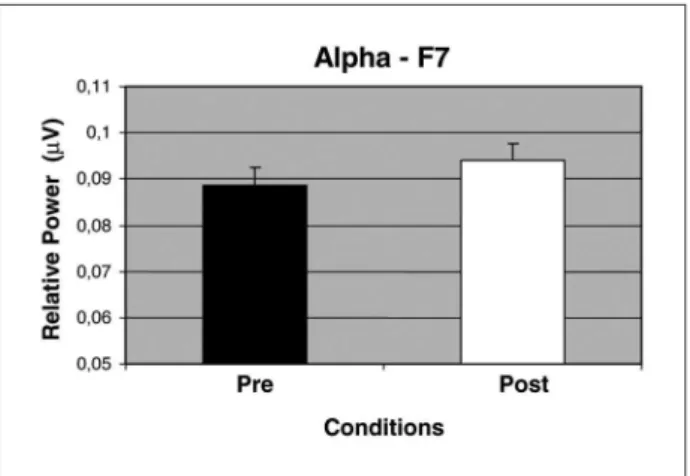
In the present experiment, statistical analysis dem-onstrated a significant difference between the two experimental times (pre and post surgery) in the al-pha band, in electrodes F7 (p=0.01) and F8 (p=0.021). The Figures 1 and 2 show the electrophysiological outcomes for the alpha frequency band. No signifi-cant results were found at beta band.
DISCUSSION
The present study aimed at observing electrocor-tical changes after the tendon transfer surgery. Es-sentially, behavioral and cortical modifications were expressed in terms of Yeap3 and alpha relative power,
respectively. The behavioral variable included motor performance, while electrophysiological variables, represented by the qEEG, examined electrocortical alterations.
Fig 1. Alpha relative power variation before and after the sur-gery, in the F7 electrode.
Behavioral results (dorsiflexor function) - Several studies nowadays have shown that the tibialis poste-rior tendon transfer produces significantly functional results in the treatment of drop foot3,8,15
. In the pres-ent study, subjects were analyzed by the functional evaluation Yeap scale for tendinous transfers, before and after the surgery. Half of the subjects were clas-sified as “good” and the other half as “poor”, for functions of daily activities before the surgery. Af-ter the surgery, even with edema and hypotrophy of the posterior muscular group of the leg and the transferred muscle, subjects considered the surgery’s initial result as satisfactory, especially concerning the capacity to make the dorsiflexion movement again and foot positioning. Therefore, according to the Yeap scale, two subjects developed from “good” to “excellent” even with some small functionary march-ing deficits and two subjects developed from “poor” to “good”.
Usually after the surgery, subjects tend to lose some degree of muscular power. Due to the lesion characteristics, the mechanisms of re-establishment of the dorsiflexion function express the motor re-learning process. This consolidation needs proprio-ceptive information which is integrated to other internal sensorial inputs. These inputs are obtained from previous experiences like specific task
memo-ries and formation of new motor patterns18. In the
tibialis posterior tendon transfer, the dorsiflexion is present, even in small movement degrees. It is at an angle great enough to promote pace function im-provement. Some studies have demonstrated that the ankle can be considered a functional unit when it comes to walking, with a dorsiflexion mean of
ap-proximately 6 to 10 degree3. Although the surgery
presents good results regarding the foot dorsiflex-ion functdorsiflex-ion, it might act better when a concomi-tant post surgery physiotherapeutic program is ini-tiated. Through specific exercises to gain range of motion and muscular power, the gait can also have benefits.
Electrophysiological results (qEEG and motor re-learning) – According to our results, an increase in alpha relative power in the electrodes F7 and F8 (Figs 1 and 2) was observed. The alpha band is an important qEEG variable, representing general de-mands of effort when performing a task with some level of activation, attention and cognition20.
Spe-cifically, there is an inversely proportional relation-ship between alpha power increase and activation level of the involved area13
. The results indicate that the tendon transfer procedure and the return of the
dorsiflexion function promote electrocortical altera-tions. The alpha power increase in F7 electrode might represent a neuronal specialization that occurred after the tendon transfer procedure; suggesting a greater mental effort and attention in an attempt to
perform the new movement20
. In addition, it might
be considered a pre learning stage21. Our outcomes
are in agreement with neuroplasticity theories, re-garding the motor restoration mechanisms of foot movement after the surgery. Experimental models have proved the consolidation of retaining specific information arriving from motor tasks by synapitic modifications22. A few investigators affirm that the
dorsiflexion movement itself activates several motor areas representing specific muscular groups involved with gait23,24
. Recent fMRI demonstrated that at the moment of dorsiflexion movement, the contralateral areas of the primary motor cortex, supplementary motor area, ipsilateral thalamus, basal ganglia, pre-frontal cortex and ipsilateral cerebellum are all active in healthy individuals23
.
The alpha relative power increase in the F8 elec-trode, situated in the contralateral motor cortex of the active member during the task to the responsible for the task, seems to represent an intrinsic process of cortical activity modulation. Therefore, we have concluded that the results indicate an integration processes between the planning areas of the right (F8) and left (F7) hemisphere. It is possible that such effects result of an alteration in the activation state, due to neuronal deactivation of the motor area
re-garding the hemispheres25. Consequently, the alpha
relative power increase in the F8 electrode suggests a reduced neuronal recruitment processing the activity in such hemisphere. Neurons of the right hemisphere might be in state of readiness, possibly due to cogni-tive and sensory processes, and neurons of the left hemisphere could be involved in the planning task18.
Hence making the cognitive and sensory aspects in-fluence the oscillation of alpha power in the motor areas. Experimental models attribute alterations in the somesthesic inputs, which occur during the task to proprioceptive impulses. The somatossensory pa-rameters related to the foot are essential for the mo-tor planning improvement. A few experiments have demonstrated that such proprioceptive mechanisms are specific of these somatossensory cortical areas, specifically, hands and feet25-27
. However, the real meaning of cortical activity changes is still far from being elucidated28.
due to a higher attention level for the accomplish-ment of the dorsiflexion moveaccomplish-ment; indicating that cognitive aspects can influence modulations of such
a specific movement in the postoperative phase29.
Even with neuroimage progress, just a few experi-mental models have been formulated to discuss the real alterations of the sensory-motor areas immedi-ately after the surgery, focusing, instead, on specific rehabilitation therapies24,30. Such a model might fill
out the emptiness in the literature concerning the functional recovery and the neuroplastic processes.
Based on the present findings, it is not unreason-able to conclude that the tendon transfer procedure itself promotes electrocortical alterations, although future studies on distinct neural plasticity investiga-tions before and after such surgical procedure, are still needed to replicate these outcomes, specifically during functional recovery and motor re-learning programs on patients suffering from droop foot due to leprosy.
REFERENCES
1. Freedman VH, Weinsten DE, Kaplan G. How Mycobacterium leprae
in-fects peripheral nerves. Lepr Rev 1999;70:136-139.
2. Van Brakel WH, Anderson AM, Wörpel FC et al. A scale to assess ac-tivities of daily living in persons affected by leprosy. Lepr Rev 1999;70: 314-323.
3. Yeap JS, Singh R, Brich D. A method for evaluating the results of ten-don transfers for foot drop. Clin Orthop Relat Res 2001;383:208-213. 4. Ober FR. Tendon transplantation in lower extremity. N Eng J Med 1933;
209:52-59.
5. Srinivasan H, Mukherjee SM, Subramaniam RA. Two-tailed transfer of tibialis posterior for correction of drop-foot in leprosy. J Bone Joint Sur-gery 1968;50:623-628.
6. Vertullo CJ, Nunley JA. Acquired flatfoot deformity following poste-rior tibialis tendon transfer for peroneal nerve injury: a case report. J Bone Joint Surgery 2002;84:1214-1217.
7. Mckay DR, Ridding MC, Thompson PD, Miles TS. Induction of persis-tent changes in the organization of the human motor cortex. Exp Brain Research 2002;143:342-349.
8. Silva JG, Knackfuss IG, Portella CE, et al. EEG spectral coherence at pa-tients submitted to tendon transfer surgery: study pre- and post-sur-gery. Arq Neuropsiquiatr 2006;644:73-77.
9. Andres FG, Mima T, Schulman AE, Dichgans J, Hallett M, Gerloff C. Functional coupling of human cortical sensorimotor areas during bi-manual skill acquisition. Brain 1999;122:855-870.
10. Ungerleider LG, Doyon J, Karni A. Imaging brain plasticity during mo-tor skill learning. Neurobiol Learn Mem 2002;78:553-564.
11. Osman A, Muller KM, Syre P, Russ B. Paradoxal lateralization of brain potentials during imagined foot movement. Brain Res Cogn Brain Res 2005;24:727-731.
12. Smith ME, McEvoy LK, Gevins A. Neurophysiological indices of strate-gy development and skill acquisition. Cogn Brain Res 1999;7:389-404. 13. Neuper C, Grabner RC, Fink A, Nuebauer AC. Long-term stability and
consistency of EEG event-related (de-)synchronization across different cognitive task. Clin Neurophysiol 2005;116:1681-1694.
14. Alegre M, Labarga A, Gurtubay IG, Iriarte J, Malanda A, Artieda J. Beta electroencephalograph changes during passive movements: sensory af-ferences contribute to beta event-related desynchronization in humans. Neurosci Lett 2002;331:29-32.
15. Yeap JS, Brich R, Singh D. Long-term results of tibialis posterior trans-fer for drop-foot. Int Orthop 2001;25:114-118.
16. Jasper H. The ten-twenty electrode system of the international federa-tion. EEG Clin Neurophysiol 1958;10:371-375.
17. Babiloni C, Miniussi C, Babiloni F, et al. Sub-second ‘‘temporal atten-tion’’ modulates alpha rhythms: a high-resolution EEG study. Cogn Brain Res 2004;19:259-268.
18. Klimesch W, Doppelmayr M, Russegger H, Pachinger T, Schwaiger J. Induced alpha band power changes in the human EEG and attention. Neurosci Lett 1998;244:73-76.
19. Smyrnis N, Thelerits C, Evodkimidis I, Muri RM, Karandreas N. Sin-gle-pulse transcranial magnetic stimulation of parietal and prefrontal areas in memory delay arm poiting task. J Neurophysiol 2003;89:3344-3350.
20. Neuper C, Pfurtscheller G. Event-related dynamics of cortical rhythms frequency-specifity features an functional correlates. Int J Psychophysiol 2001;43:41-58.
21. Bastos VH, Machado D, Cunha M et al. Medidas eletroencefalográfica durante a aprendizagem de tarefa motora sob efeito do bromazepam. Arq Neuropsiquiatr 2005;63:443-451.
22. Luft AR, Buitrago MM, Ringer T, Dichgans J, Schulz JB. Motor skill learning depends on protein synthesis in motor cortex after training. J Neurosci 2004;24:6515-6520.
23. Sahyoun C, Floyer-Lea A, Johansen-Berg H, Matthews P. Towards an understanding of gait control: brain activation during the anticipation, preparation and execution of foot movements. Neuroimage 2004;21: 568-575.
24. Dobkin BH, Firestine A, West M, Saremi K, Woods R. Ankle dorsiflex-ion as an fMRI paradigm to assay motor control for walking during re-habilitation. Neuroimage 2004;23:370-381.
25. Pfurtscheller G, Lopes da Silva F. Event-related EEG/MEG synchro-nization and desynchrosynchro-nization: basic principles. Clin Neurophysiol 1999;110:1842-1857.
26. Cassim F, Szurhaj W, Sediri H, et al. Brief and sustained movements: differences in event-related (de)synchronization (ERD / ERS) patterns. Clin Neurophysiol 2000;111:2032-2039.
27. Pfurtscheller G, Aranibar A. Event-related cortical desynchronization detected by power measurement of scalp EEG. Electroencephalogr Clin Neurophysiol 1997;42:817-826.
28. Alegre M, Gurtubay IG, Labarga A, et al. Alpha and beta oscillatory ac-tivity during a sequence of two movements. Clin Neurophysiol 2004; 115:124-130.
29. Serrien DJ, Pogosyan AH, Brown P. Cortico-cortical coupling patterns during dual task performance. Exp Brain Res 2004;157:79-84. 30. Ward N, Brown N, Thompson A, Frackowiak R. Neural correlates of