ACOUSTIC NEUROMA
VESTIBULAR SCHWANNOMA
Surgical results on 240 patients operated
on dorsal decubitus position
Arquimedes Cavalcante Cardoso
1, Yvens B. Fernandes
2,
Ricardo Ramina
2, Guilherme Borges
3ABSTRACT - Objective: To evaluate the result of the surgical treatment of vestibular schwannoma (VS) op-erated in dorsal decubitus (mastoid position). Method: 240 patients with a VS underwent a retrosigmoid craniotomy for tumor resection in dorsal decubitus (mastoid position). The function of 7th and 8th cranial
nerves was monitored during surgery and the opened internal auditory canal (IAC) was reconstructed us-ing a vascularized dura flap, muscle and fibrin glue. Results: Complete tumor removal was achieved in 99% of the cases, with a mortality of 1.6%. The facial nerve function was preserved in 85% of cases and hearing in 40% of the patients (with preoperative hearing) with tumors of up 1.5 cm in diameter. The in-cidence of cerebrospinal fluid leak was 5.8% and meningitis 2.9%. Venous air embolism was registered in 3% of cases; it was not associated to mortality. Conclusion: Surgical removal of VS in dorsal position has several advantages; the morbidity and mortality are very low.
KEY WORDS: acoustic neuroma, vestibular schwannoma, mastoid position, venous air embolism.
Neurinoma do acústico (schwannoma do vestibular): resultados do tratamento cirúrgico de 240 pacientes operados na posição de decúbito dorsal
RESUMO - Objetivo: Avaliar o resultado do tratamento cirúrgico de pacientes portadores de schwannoma do vestibular (SV) operados em decúbito dorsal (posição de mastóide). Método: 240 pacientes foram sub-metidos a craniotomia retrosigmóide na posição de mastóide. A função do VII e VIII nervos cranianos foi monitorizada durante a cirurgia e a reconstrução da abertura do conduto auditivo interno foi realizada com retalho vascularizado de dura-mater, músculo e cola de fibrina. Resultados: A exérese foi completa em 99% dos casos, com mortalidade de 1,6%. Houve preservação da função do nervo facial em 85% dos casos e da audição em 40% dos pacientes com audição prévia e tumores menores de 1,5 cm. A incidência de fístula liquórica foi 5,8% e meningite 2,9%. Embolia gasosa foi registrada em 3% dos casos, não asso-ciada à mortalidade. Conclusão: O tratamento cirúrgico dos SV utilizando-se a posição de mastóide tem várias vantagens, com baixa morbidade e mortalidade.
PALAVRAS-CHAVE: neurinoma do acústico, schwannoma vestibular, posição de mastóide, embolia gasosa.
Departamento de Neurologia, Faculdade de Ciências Médicas, Universidade Estadual de Campinas, Campinas SP, Brasil (UNICAMP):
1Doutorando em Ciências Médicas; 2Professor Doutor; 3Livre-Docente Professor Associado. Supported by CNPq Grant # 302189/2004-1.
Received 28 November 2006, received in final form 26 February 2007. Accepted 18 April 2007.
Dr. Arquimedes Cavalcante Cardoso - Avenida Rio Poti 2061 / 504 - 64049-410 Teresina PI - Brasil. E-mail: [email protected]
The acoustic neuroma originates from the schwann cell, in the peripheral portion of superior and infe-rior vestibular nerves and also from cochlear nerve. These tumors are usually referred to as acoustic neu-roma, but the preferred term to be used is vestib-ular schwannoma (VS), once they are composed of Schwann cells and more frequently are originated in the vestibular portion of the 8th
cranial nerve1 . Pa-tients diagnosed with VS often present unilateral
hearing loss, tinnitus and dizziness2. The VS occurs in an incidence of about 1:100000 inhabitants per year. In most recent statistics an increase of such incidence has been reported due to frequent use of more sensi-tive magnetic resonance (MR) techniques, diagnosing very small tumors3
606 Arq Neuropsiquiatr 2007;65(3-A)
the diagnosis of VS is still delayed in a great num-ber of cases, and the tumors present a considerable size when a correct diagnosis is performed4,5. Three main surgical approaches: retrosigmoid-transmeatal (suboccipital), translabyrinthic and the middle cra-nial fossa or subtemporal have been used. Each one has both advantages and disadvantages, and excel-lent results have been reported with all these ap-proaches5-7.
Traditionally the suboccipital approach has been performed with the patient in a semi-sitting position. The sitting position for neurosurgical procedures has been a controversial subject among neurosurgeons and anesthesiologists. This position permits drainage of blood and cerebrospinal fluid (CSF) allowing a bet-ter visualization and dissection of the cranial nerves, easier hemostasia and reduces venous bleeding. The main disadvantages of this position are the increased risk of venous air embolism and hemodynamic insta-bility, especially in elderly patients8-12.
The aim of this study was to evaluate the results of surgical treatment of VS in patients operated on through the retrosigmoid-transmeatal approach, in the dorsal decubitus position (mastoid position).
METHOD
In this study 240 consecutive patients with VS operat-ed on at the Hospital das Clinicas - UNICAMP, SP and at the Instituto de Neurologia de Curitiba, PR, between January 1990 and December 2005, were retrospectively evaluated. This study was approved by the Research Ethics Committee of both institutions.
In this study they were included 240 patients (238 with tumors sporadic, unilateral and 2 neurofibromatosis type 2 patients, operated of just one on the sides) operated on dorsal decubitus position (mastoid position) through the retrosigmoid-transmeatal approach (Fig 1). The age of the patients ranged between 16 and 81 years. No difference was observed regarding the gender or the side of the af-fected ear.
Tumor size was classified according to the Hannover grading system13 (Fig 2), in which tumor extension classes
were described as follows: T1, purely intrameatal; T2, intra-extrameatal; T3a, filling the cerebellopontine cistern; T3b, reaching the brainstem; T4a, compressing the brainstem; and T4b, severely dislocating the brainstem and compress-ing the fourth ventricle.
The size of the lesions is demonstrated in Table 1. Audiometric evaluation with discrimination comput-erized tomography (CT) and MR were performed in all cases before surgery. Intraoperative monitoring of facial nerve and cochlear nerve function was carried out by the group of neurophysiology. The facial nerve was monitored through electrodes put into the facial muscles (orbicular-is occulli and or(orbicular-is) that were stimulated mechanically or electrically during dissection of the tumor. The cochlear nerve was monitored by recording evoked potentials of the brainstem5.
Besides routine neuroanaesthesia monitorization pa-rameters an ultrasonic precordial Doppler sensor was used to detect venous air embolism and a right atrial catheter was placed to remove air bubbles if necessary.
Surgical technique – Retrosigmoid craniotomy with a diameter of about 5 cm was employed in all cases. The du-ra mater is opened padu-rallel to the sigmoid and tdu-ransversal sinuses. CSF is drained from the cerebellomedullary cistern to expose the cerebellopontine angle with minimal cere-bellar retraction. The dura mater covering the internal au-ditory canal (IAC) is cut forming a dura flap attached over the jugular foramen region. This vascularized dura flap is used to cover the cranial nerves and reconstruct the IAC at the end of surgery. Microsurgical tumor excision with use of microsurgical instruments, preserving the arachnoid plane to preserve the cranial nerves, brain stem and vessels was the surgical technique used. Bipolar coagulation is careful utilized to avoid cranial nerves injury by heating. After to-tal resection of the tumor and careful hemostasia, the IAC is covered by the dura flap and a piece of muscle and fibrin glue is placed over it to avoid CSF leak. The dura is sutured with running stitches and covered with surgicel and fibrin glue. The bone flap is replaced, the subcutaneous tissue and the skin are closed with separated sutures.
Patients were evaluated regarding clinical and surgi-cal complications: related to positioning, detection of
ve-Fig 1. Dorsal decubitus (mastoid) position.
Table 1. Size of the tumor, according to the Hannover classi-fication.
Size N %
T1 12 5.00
T2 41 17.08
T3 a 21 8.75
T3 b 24 10.00
T4 a 63 26.25
T4 b 79 32.92
nous air embolism and its effects, presence of CSF leak and meningitis and preservation of the cranial nerve’s function. The function of the facial nerve was evaluated through the scale of House and Brackmann14 and the auditory
func-tion through the audiometric classificafunc-tion of Hannover4.
All patients had a postoperative MR on average 4 months after surgery.
RESULTS
Most patients presented tumors larger than 3.0cm in (166 cases). The function of the facial nerve could be preserved in 85% of patients with preoperative fa-cial function, regardless the size of the lesion (Grades 1 to 3 House and Brackmann)14
. In patients with tu-mors smaller than 3.0 cm in diameter preservation of the facial nerve function was possible in 100% of the cases. Hearing preservation was possible in 8 (40 %) of the 20 patients with preoperative hearing with tu-mors of up to 1.5 cm in diameter.
Four patients died in these series (1.6 %). Three patients presented large tumors, 5.0 cm in diameter (T4b). Mortality was caused by a hematoma in the tumor bed in two cases and thrombosis of the bas-ilar artery in the other two. Causes of basbas-ilar artery thrombosis were damage to the vertebral artery dur-ing dissection at C1-C2 region in one of the patients and inadequate positioning with exaggerated head rotation in the other patient.
Cerebrospinal fluid leak occurred in 14 patients (5.8%). Seven (2.9%) developed meningitis, treated with antibiotics. Compressive dressing and lumbar
drainage was used to treat the CSF fistula. Surgical closure of CSF leak was necessary in 5 patients.
Postoperative control MR was carried out in all cases, the facial nerve function was evaluated and when the cochlear nerve could be preserved the hearing function was tested. Total removal of the lesion was obtained in 238 cases (99 %). In two cases of cystic tumors the control MR revealed recurrence of the lesion.
In this series 36 patients had postoperative le-sion of the facial nerve. In two of them suture of the stumps of the facial nerve was accomplished in the same surgical act and the remaining of the patients was submitted to a facial-hypoglossal nerve anasto-mosis. 16 patients with anatomical lesion were op-erated among 7-10 days after the surgery and in 18 patients with preservation of the facial nerve, how-ever with facial paralysis, the reconstruction was ac-complished 1 year after the surgery. A functional im-provement (grade 3 or 4) was postoperatively ob-served in all these patients.
The incidence of venous air embolism detected through Doppler was 3%, however, it was not asso-ciated with morbidity or mortality in this series. Ex-cluding the patient with vertebral artery lesion, no further complications were related to the position-ing of the patient.
DISCUSSION
The centers with extensive experience in the
sur-Fig 2. MRI scans showing a T4a VS13:
608 Arq Neuropsiquiatr 2007;65(3-A)
gery of VS generally opt for four categories of surgi-cal access: (1) suboccipital access, (2) translabyrinthic access, (3) middle cranial fossa access and (4) eclectic access. Excellent results have been published by the use of these four approaches15-21
.
In our series the retrosigmoid-transmeatal access was used in all patients. According to our experience this approach allows an adequate exposure to VS of all sizes, is quickly performed, permits preservation of hearing, easy identification of the facial nerve in-side the IAC, adequate controlling of vascular and nervous structures of the posterior cranial fossa and brainstem and primary closure of dura mater22
.
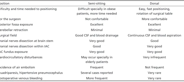
The positions used for resection of the VS through the retrosigmoid-transmeatal access are: semi-sitting, ventral and park bench. The position that we use in this study is dorsal with the patient’s head turned in order to expose the posterior cranial fossa and the mastoid. The advantages of this position are related to the easy of positioning, to avoid hemodynamic instability, the comfort of the surgeon and the sig-nificant reduction of the risk of venous air embolism. These advantages are present principally regarding the semi-sitting position, despite there being several proponents of this last position16,22,23.
The sitting position has been controversial since it was first introduced by De Martel in Paris, in 1913, to operate a brain tumor surgery with local anesthesia. Due to hydrostatic gravity effect, an accumulation of blood in the extremities as high as 1500 mL occurs producing systemic decrease in blood pressure8. This position makes the surgical field clear of blood and
CSF. The most feared complication of this position is venous air embolism. Air embolism may occur in any operation with an open vein and a gradient of venous pressure between the surgical site and the heart8-12. The incidence in the horizontal position is, however, lower.
Series comparing the prognosis of patients oper-ated on in sitting and horizontal position, conclude that despite the venous air embolism being an im-portant factor in surgery of posterior cranial fossa, the current methods of monitoring allow an early detection and prompt therapy of venous air embo-lism12,23. Nevertheless, we have to consider that not all departments have such modern methods of moni-toring. The incidence of complications of right atrial catheter may be high and the volume of air removed through it can be minimal9-12,23
.
There are other potential complications associ-ated with the position of the patient during the VS surgery. Orthopedic, peripheral nerve and cutaneous lesions, have been described in all surgical procedures and may be avoided with careful positioning of the patient23. Other reasons to avoid the sitting position are the risk of quadriparesis, increased risk of hyper-tensive pneumocephalus, post-operative bleeding, tongue and larynx edema8,9,23
. The advantages and disadvantages8-12,14,22,23 of both positions are showed on Table 2.
The main goals of the surgery include complete excision of the lesion in one stage, without mortality and preservation of the neurological function, espe-cially of the 7th nerve and hearing.
Table 2. Semi-sitting X dorsal position, advantages and disadvantages.
Position Semi-sitting Dorsal
Dificulty and time needed to positioning Difficult specially in obese patients, more time needed
Easy, fast positioning, rotation of surgical table For the surgeon Not confortable More confortable
Posterior fossa exposure Excellent Excellent
Cerebellar retraction Minimal Minimal
Surgical field Good CSF and blood drainage Continuous CSF and blood aspiration Cranial nerves dissection at brain stem Very good Good
Cranial nerves dissection within IAC Good Very good
IAC fundus exposure Very good Very good
Cardiocircullatory disturbances May occur specially in elderly patients
Very infrequent
Incidence of air embolism Frequent Not frequent Quadriparesis, hipertensive pneumocephalus Several cases reported Very rare Postoperative venous bleeding More frequent Very rare
In this series the tumor was larger than 3.0 cm in most patients (166 cases). The facial nerve function could be preserved (Grades 1 to 3 House and Brack-mann)14
in 85% of patients with preoperative facial function, regardless of the size of the lesion. In pa-tients with tumors smaller than 3.0 cm in diameter, the preservation of the facial nerve occurred in 100% of the cases. These data are comparable with results published by very experienced authors15-19,24.
Early diagnosis of small and intracanicular VS has increased due improvement of radiological di-agnostic methods. Often these patients present use-ful hearing of the side affected by the tumor mak-ing hearmak-ing preservation an objective of treatment. There are also some reports of hearing preservation in large tumors4,17-19,25,26
.
Our strategy is to try to preserve the cochlear nerve and hearing in every case with preoperative cochlear nerve function. Even the hearing classified as non useful is still many times superior to that one obtained with cochlear implants and it is far superior to that obtained with brain stem implants. It is also possible that preservation of cochlear nerve will in the future, with new technology, allow patients to recover hearing5,22
.
CSF leak and its association with the risk of menin-gitis remain as important issues in the surgical treat-ment of VS. An incidence between 0 and 30%, with an approximate rate of 12% is reported27. With the use of fibrin glue and reconstruction of the IAC with dura mater flap and muscle filling, we were able to reduce the incidence of CSF fistula in comparison with previous report27.
In the last 20 years, radiosurgery and stereotatic radiosurgery have been used to treat VS as a less in-vasive alternative to the surgical treatment. Some au-thors regard it as the first option to tumors smaller than 3 cm2,28 . No tumor can be cured with this meth-od and the objective of treatment is to obtain “con-trol of tumor growth”. In our opinion, radiotherapy/ radiosurgery should be only exceptionally indicated in elderly patients or patients without clinical condition for radical removal and cure of this benign tumor.
REFERENCES
1. Roswell E, Dilys P. Summary: vestibular schwannomas (acoustic neu-roma). Consensus Development Conference. Conference Proceedings. Neurosurgery 1992;30:962-964.
2. Pollock BE, Driscoll CLW, Foote RL, et al. Patient outcomes after ves-tibular schwannoma management: a prospective comparison of micro-surgical resection and stereotactic radiosurgery. Neurosurgery 2006;1: 77-85.
3. Haines SJ, Levine SC. Intracanalicular acoustic neuroma:early surgery for preservation of hearing. J Neurosurg 1993;79:515-520.
4. Matthies C, Samii M. Vestibular schwannomas and auditory function: options in large T3 and T4 tumors? Neurochirurgie 2002;48:461-470. 5. Ramina R, Fernandes YB, Borges G, et al. Neurinoma do acústico
(schwannoma do vestibular). Tópicos em Neurocirurgia. Rio de Janei-ro: Revinter, 2001:59-64.
6. Ojemann RG. Management of acoustic neuromas (vestibular schwanno-mas). Clin Neurosurg 1993;40:498-535.
7. Koerbel A, Gharabaghi A, Safavi-Abbasi S, Tatagiba M, Sammi M. Evo-lution of vestibular schwannoma surgery: the long journey to current success. Neurosurg Focus 2005;18:24.
8. Albin MS, Babinski M, Maroon JC, Jannetta PJ. Anesthetic management of posterior fossa surgery in the sitting position. Acta Anaesth Scand 1976;20:117-128.
9. Standefer M, Bay JW, Trusso R. The sitting position in neurosurgery: a retrospective analysis of 488 cases. Neurosurgery 1984;14:649-658. 10. Matjasko J, Petrozza P, Cohen M, Steinberg P. Anesthesia and surgery
in the seated position: analysis of 554 cases. Neurosurgery 1985;17: 695-702.
11. Young ML, Smith DS, Murtagh F, Vasquez A, Levitt J. Comparison of surgical and anesthetic complications in neurosurgical patients expe-riencing venous air embolism in the sitting position. Neurosurgery 1986;18:157-161.
12. Duke DA, Lynch JJ, Harner SG, Faust RJ, Ebersold MJ. Venous air em-bolism in sitting and supine patients undergoing vestibular schwanno-ma resection. Neurosurgery 1998;42:1282-1287.
13. Matthies C, Samii, M. Management of 1000 vestibular schwanno-mas (acoustic neuroschwanno-mas): clinical presentation. Neurosurgery 1997;40: 1-10.
14. House JW, Brackmann DE. Facial nerve grading system. Otolaryngol Head Neck Surg 1985;93:146-147.
15. Siqueira JM, Souza OG. Preservation of the facial nerve in large acoustic neuromas using the combined translabyrinthine-subocciptal approach. Arq Bras Neurocirurg 1992;11:77-80.
16. Samii M, Matthies C. Management of 1000 vestibular schwannomas (acoustic neuroma): surgical management and results with an empha-sis on complications and how to avoid them. Neurosurgery 1997;40: 11-23.
17. Bento RF, Miniti A, Bogar P. Experiência em 115 casos de cirurgia para exérese de neurinoma do acústico. Rev Bras Otorrinolaringol 1995; 61:204-217.
18. Ebersold MJ, Harner SG, Beatty CW, Harper Jr CM, Quast LM. Current results of the retrosigmoid approach to acoustic neurinoma. J Neuro-surg 1992;76:901-909.
19. Ojemann RG. Retrosigmoid approach to acoustic neuroma (vestibular schwannoma). Neurosurgery 2001;48:553-558.
20. Day JD, Chen DA, Arriaga M. Translabyrinthine approach for acoustic neuroma. Neurosurgery 2004;54:391-396.
21. Sanna M, Russo A, Falcioni M, Taibah A, Agarwal, M. Enlarged trans-labyrinthine approach for the management of large and giant acoustic neuromas: a report of 175 consecutive cases. Ann Otol Rhinol Laryn-gol 2004;113:319-328.
22. Ramina R, Maniglia JJ, Meneses MS, et al. Acoustic neurinomas diag-nosis and treatment. Arq Neuropsiquiatr 1997;55:393-402.
23. Black S, Ockert DB, Oliver Jr WC, Cucchiara RF. Outcome following posterior fossa craniectomy in patients in the sitting or horizontal po-sitions. Anesthesiology 1988;69:49-56.
24. Wiegand DA, Fickel V. Acoustic neuroma: the patient´s perspective: subjective assessment of symptoms, diagnosis, therapy and outcome in 541 patients. Laryngoscope 1989;99:179-187.
25. Gardner G, Robertson JH. Hearing preservation in unilateral acoustic neuroma surgery. Ann Otol Rhinol Laryngol 1988;97:55-66.
26. Silveira RL, Gusmão SNS, Ferraz FAP, Cabral G Filho, Tazinaffo U. Per-spectivas atuais sobre a preservação da audição na cirurgia do neuri-noma do acústico. J Bras Neurocirurg 1996;7:5-18.
27. Brennan JW, Rowed DW, Nedzelski JM, Chen JM. Cerebrospinal fluid leak after acoustic neuroma surgery: influence of tumor size and sur-gical approach on incidence and response to treatment. J Neurosurg 2001;94:217-233.