Arq Bras Cardiol 2003; 81: 359-62.
Cavalcanti et al Morphometric and topographic study of coronary ostia
3 5 9
Universidade Federal de Pernambuco, Recife
Mailing address: Jennecy Sales Cavalcanti - Rua Félix de Brito Melo, 912/501 Cep 51020-260 - Recife, PE, Brazil - E-mail:
English version by Stela Maris C. e Gandour
Arq Bras Cardiol, volume 81 (nº 4), 359-62, 2003
Jennecy Sales Cavalcanti, Natália Corrêa Vieira de Melo, Renata Simões de Vasconcelos
Recife, PE - Brazil
Morphometric and Topographic Study of Coronary Ostia
Original Article
The great importance of coronary catheterization for diagnostic and therapeutic purposes has currently motiva-ted several studies on the anatomic position of coronary ostia. In most investigations, 2 coronary ostia, one right and one left, were observed located in the 2 aortic sinuses (sinuses of Valsalva) closer to the pulmonary trunk 1-6.
Adequate irrigation of the heart depends fundamental-ly on the good morphological conditions of the right and left coronary arteries. These arteries have the peculiarity of being the only ones filled during the diastolic phase of car-diac rhythm. For this to happen properly, some conditions, such as anatomic integrity of the aortic valve, absence of valvular malformations and anatomic malformations of the coronary arteries that result in a reduction in blood flow to the myocardium, are required 7-15.
Other factors may be involved in the possible reduc-tion in coronary blood flow, such as variareduc-tions in the posi-tion of the coronary ostia in relaposi-tion to the aortic leaflets and the diameter of the ostia 16-19.
Considering the hypotheses formulated by these au-thors that changes in coronary flow may be due to changes in diameter, position, and anatomic relations of the coronary ostia, this study aimed at investigating these factors, em-phasizing their possible implications in functional order.
Methods
Fifty-one hearts with their great vessels attached and fixed in 10% formalin were used in this study. This autopsy material originated from white and nonwhite adults of both sexes, whose cause of death was not heart disease, from the Department of Anatomy of the Center of Biological Scien-ces of the Federal University of Pernambuco (UFPE).
The hearts were dissected, the pericardium involving the root of the aorta was removed, and the origin of the right and left coronary arteries was isolated. Then, the as-cending aorta was transversally sectioned approximately 1 cm above the commissures of the aortic leaflets. Next, the aorta was longitudinally opened at the level of the posterior aortic sinus (noncoronary sinus) to enable the visualization and analysis of the right and left aortic leaflets and their res-pective coronary ostia. In addition, the coronary arteries were sectioned at the level of their origins in the aortic wall (juxtamural portion of the coronary arteries).
Objective - To investigate the morphometric and to-pographic aspects of coronary ostia, correlating them with the aortic leaflets.
Methods - Fifty-one hearts with the great vessels atta-ched were analyzed in this study. The ascending aorta was transversally sectioned 1 cm above the commissures of the aortic leaflets. The right and left coronary ostia were analy-zed, as were the distances from these ostia to the bottom of the aortic sinuses and to the commissures of the aortic leaflets.
Results - The left coronary ostium was located below the intercommissural line in 42% of cases, above that line in 40% of cases, and at the level of that line in 18% of cases. The mean distance from the left coronary ostium to the bottom of the corresponding sinus was 12.6±2.61 mm. The right coronary ostium was located below the intercommissural line in 60% of cases, above that line in 28% of cases, and at the level of that line in 12% of cases. The mean distance from the right coronary ostium to the bottom of the correspon-ding aortic sinus was 13.2±2.64 mm. The mean diameters of the left and right coronary ostia were 4.75±0.93 mm and 3.46±0.94 mm, respectively. The mean diameters of the jux-tamural portion of the left and right coronary arteries were 3.75±0.79 mm and 2.9±0.73 mm, respectively. In one case, both ostia were located in the left coronary sinus.
Conclusion - The left coronary ostium may be located either above or below the intercommissural line. The right coronary ostium is predominantly located below the inter-commissural line. The coronary ostia have reduced diame-ters as compared with the juxtamural diamediame-ters of their respective coronary arteries.
3 6 0
Cavalcanti et al
Morphometric and topographic study of coronary ostia
Arq Bras Cardiol 2003; 81: 359-62.
The coronary ostia in relation to the right and left aortic leaflets were identified and, with the aid of a millimeter ruler and a pachymeter, the following measurements were taken: the diameters of the coronary ostia, the diameters of the juxtamural portion of the coronary arteries, and the distance from the coronary ostia to the bottom of the aortic sinus and to the commissures of the aortic leaflets. To measure these parameters, one extremity of the pachymeter was placed in the median region of the ostium and the other extremity was displaced to the bottom of the aortic sinus, and then, to the commissure to the left of the ostium and to the right of the same ostium. The values found in each measurement were listed in tables and statistically analyzed.
The relation of the coronary ostia with the intercom-missural line was also observed, and the hearts were grou-ped according to the following parameters: ostium above the intercommissural line, below the intercommissural line, and at the level of the intercommissural line.
Results
All hearts studied had trileaflet aortic valves. In 50 of the 51 hearts studied, the coronary arteries originated from the respective aortic sinuses; in one of the hearts, however, both arteries originated from the left aortic sinus. Data obtained from the latter heart were not included in the calcu-lation of the means of the results in this study. No isolated nourishing artery of the right ventricular infundibulum (conus artery) was observed.
The relation between the location of the left and right coronary ostia and the intercommissural line in each heart is shown in table I. Figure 1 depicts the coronary ostia located below and at the level of the intercommissural line.
The results of the measurements of the distances from the left and right coronary ostia to the commissures located to the right and left of the referred ostia are shown in table II. Considering this same table, on average, the left coronary ostium was located in the central region of the correspon-ding aortic sinus, while the right coronary ostium was dis-placed to the right in the respective aortic sinus.
The mean distance from the left coronary ostium to the bottom of the corresponding aortic sinus was 12.6±2.61 mm. The mean distance from the right coronary ostium to the bottom of the respective aortic sinus was 13.2±2.64 mm.
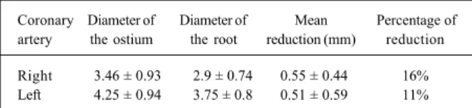
The results of the measurements of the diameters of the coronary ostia, as well as the diameters of the juxtamural portion of the corresponding coronary arteries, are shown in table III. A mean reduction of 0.51 mm (11%) was observed in the diameter of the left coronary ostium and of the left juxta-mural portion. A mean reduction of 0.55 mm (16%) was ob-served in the diameter of the right coronary ostium and of the right juxtamural portion.
In 22% of the sample (11 hearts), the diameter of the right coronary ostium was greater than that of the left one. In 76% of the cases (38 hearts), the diameter of the left coronary ostium was greater than that of the right one, and only in 2% of the sample (1 heart), the 2 coronary ostia had equal diameters.
In 8 hearts (16%), the diameter of the juxtamural por-tion of the right coronary artery was greater than that of the left coronary artery. In 41 cases (82%), the diameter of the juxtamural portion of the left coronary artery was greater than that of the right coronary artery. In only 1 heart (2%) were the diameters equal.
Discussion
Our study, which comprised a series of 51 hearts, confirmed that the 2 coronary ostia are related to the right
Table I - Location of the coronary ostia in relation to the intercom-missural line in each heart
Coronary ostium Frequency %
Right and left below 16 32%
Right and left above 9 18%
Right above, left below 3 6%
Right below, left above 8 16%
Right at the line, left below 2 4%
Right below, left at the line 6 12%
Right at the line, left above 3 6%
Right above, left at the line 2 4%
Right and left at the line 1 2%
Fig.1 - Fhotograph of the aortic valve showing the location of the coronary ostia in regard the intercommissural line. A: right and left ostia below the intercommissural line; B: right and left ostia at the level of the intercommissural line.
Arq Bras Cardiol 2003; 81: 359-62.
Cavalcanti et al Morphometric and topographic study of coronary ostia
3 6 1
and left leaflets of the aortic valve 1-6. In our case series, only
1 heart did not fit into the above cited pattern, having the 2 ostia at the level of the left aortic sinus. This variation was also found in 1 of the 100 hearts studied by Jatene et al 16.
Previous studies emphasize the limitations in the measurements of cardiac valves during autopsy, because a certain degree of retraction and stiffness of the structures analyzed always exists, due to the rigor mortis and the fixa-tion process 20. However, Tei et al 21 compared 3 groups of
materials and observed similar values in hearts not fixed in formalin, hearts fixed in formalin, and hearts assessed on echocardiograms. Similar results have also been reported by Scholz et al 22 and Maron et al 23 when studying cardiac
mass, valvular circumferences, and ventricular thickness. Of the 100 ostia analyzed in the present study, 51% we-re located below the intercommissural line, 34% above it, and 15% at its level, ie, 25 of the 50 hearts analyzed had 1 or both ostia above the intercommissural line. These results are similar to those of Vlodaver et al 6 who reported that 22 of
50 hearts studied had 1 or both ostia above the intercom-missural line. On the other hand, our results do not agree with those obtained by Turner and Navaratnam, 1 who
found 16% of the ostia above the intercommissural line, and with the results obtained by Muriago et al, 18 who
found only 13% of the ostia above that line.
In regard to the left coronary ostium, our results differ from those by Muriago et al, 18 who reported a left coronary
ostium below the intercommissural line in 60% of their case series, above that line in 22%, and at the level of the inter-commissural line in 9%. In regard to the right coronary os-tium, these same authors reported that the right coronary ostium was below the intercommissural line in 78% of their case series, above that line in 13%, and at the level of that line in 9%. Our findings also differ from those reported by these authors regarding the location of both ostia in each heart. According to their study, both ostia were below the intercommissural line in 57% of cases, and both ostia were above that line in only 9% of cases.
Turner and Navaratnam 1 reported that from the
func-tional point of view it would be advantageous that both co-ronary ostia were above the intercommissural line, which, according to the authors, would make the coronary blood flow easier during ventricular systole and would also pre-vent the obstruction of the ostia when the aortic valve was opened. We do not agree with these authors, because it is well known in cardiac physiology that blood flows to the coronary arteries mainly during the diastolic phase of the cardiac cycle.
The studies by Muriago et al 18 indicated that the
coro-nary ostia are not located in the center of each aortic sinus, but, on the contrary, they are displaced to the commissure located to the right of the ostia. Their results are similar to ours in regard to the location of the right coronary ostium; however, it is worth emphasizing that, in our material, the left coronary ostium was located almost in the central region of the corresponding aortic sinus, which is in accordance with the studies reported by Turner and Navaratnam1 and
McAlpine 24. Our results showed that, on average, the right
coronary ostia are located in the right side of the correspon-ding aortic sinus, in contrast with the fincorrespon-dings of Turner and Navaratnam, 1 who reported that both coronary ostia are
lo-cated in the central region of their respective aortic sinuses. McAlpine 24 proposed that the location of the right
co-ronary ostium in the right half of the corresponding aortic sinus is appropriate, because the right coronary artery is destined to pass around the tricuspid valve, therefore ha-ving a more direct course than if it originated in the left or medial portion of the sinus. According to that author, dis-placement of the left coronary ostium to the right would be more appropriate because the anterior and left parts of the heart are supplied by the left coronary artery. Still regarding this same point, Muriago et al 18 added that the more central
location of the left coronary ostium is justified, because, after its origin, the left coronary artery heads to the space between the pulmonary trunk and the left auricle, branching right after that.
No previous relevant studies on the diameters of the co-ronary ostia and the juxtamural portions of the coco-ronary ar-teries have been found nor have records of the correlation between these 2 measurements. We observed that, in 76% of our sample, the diameter of the left coronary ostium was grea-ter than that of the right coronary ostium, and, in 82% of ca-ses, the diameter of the juxtamural portion of the left coronary artery was greater than that of the right coronary artery.
We believe that these data are important for clinical and surgical practice, because it is well known that changes in coronary flow may be caused by modifications in the diame-ter, position, and anatomic relations of the coronary ostia.
In conclusion, we observed that, after studying the pa-rameters described, the left coronary ostium is more medial-ly located in regard to the intercommissural distance than the right coronary ostium, which is displaced to the right side. In regard to the intercommissural line, most coronary ostia were located below the intercommissural line or level Table III - Diameters of the coronary ostia and the roots of the
corresponding coronary arteries (mm)
Coronary Diameter of Diameter of Mean Percentage of artery the ostium the root reduction (mm) reduction
Right 3.46 ± 0.93 2.9 ± 0.74 0.55 ± 0.44 16% Left 4.25 ± 0.94 3.75 ± 0.8 0.51 ± 0.59 11%
Table II - Mean values of the distances between the coronary ostia and the commissures of the aortic leaflets (mm)
Coronary Comissure Mean distance Mean point of the
ostium intercommissural
distance
Left Commissure to the left 11.5 ± 3.64 11.3 Commissure to the right 11.2 ± 2.56
3 6 2
Cavalcanti et al
Morphometric and topographic study of coronary ostia
Arq Bras Cardiol 2003; 81: 359-62.
with it. The diameters of the left coronary ostium and of the left juxtamural portion of the coronary artery were greater than those of the right coronary ostium and of the right
jux-tamural portion of the coronary artery. The diameters of the coronary ostia are smaller than those of the respective juxta-mural portions of the coronary arteries.
1. Turner K, Navaratnam V. The positions of the coronary arterial ostia. Clin Anat 1996; 9: 376-80.
2. Anderson RH, Becker AE. An Integrated Text and Colour Atlas. London: Churchill Livingstone 1980.
3. Navaratnam V. Design of heart valves: a review. Clin Anat 1993; 6: 327-32. 4. Reid K. The anatomy of the sinus of Valsalva. Thorax 1970; 25: 79-85. 5. Roberts JP. Arteries, veins and lymphatic vessels of the heart. In A.A. Luisada
(ed.) Development and Structure of the Cardiovascular System Chapter 6. New York: McGraw Hill 1961.
6. Vlodaver Z, Neufield HN, Edwards JE. Coronary Arterial Variations in the Nor-mal Heart and in Congenital Heart Disease. New York: Academic Press 1975. 7. Choo SJ, Mcrae G, Olomon JP, et al. Aortic root geometry: pattern of differences
between leaflets and sinuses of Valsalva. J Heart Valve Dis 1999; 8: 407-15. 8. Yacoub MH, Kilner PJ, Birks EJ, et al. The aortic outflow and root: a tale of
dyna-mism and crosstalk. Ann Thorac Surg 1999; 68(suppl): S37-43.
9. Arai AE, Epstein FH, Bove KE, et al. Visualization of aortic valve leaflets using black blood MRI. J Magn Reson Imaging 1999; 10: 771-7.
10. Heusch A, Quagebeur J, Paulus A, et al. Anomalous origin of all coronary arteries from the pulmonary trunk. Cardiology 1997; 88: 603-8.
11. Lerer PK, Edwards WD. Coronary arterial anatomy in bicuspid aortic valve: ne-cropsy study of 100 hearts. Br Heart J 1981; 45: 142-7.
12. Hutchins GM, Nazararian IH, Bulkley BH. Association of the left dominant co-ronary arterial system with congenital bicuspid aortic valve. Am J Cardiol 1978; 42: 57-9.
13. Johnson AD, Detwiler JH, Charles BH. Left coronaey artery anatomy in patients with bicuspid aortic valves. Br Heart J 1978; 40: 489-93.
14. Gonzales-Angulo A, Reyes HA, Wallace AS. Anomalies of the origin of the
co-References
ronary arteries (special reference to single coronary artery). Angiology 1966; 17: 96-103.
15. Schlesinger MJ, Zoll PM, Wessler S. The conus artery: a third coronary artery. Am Heart J 1949; 38: 823-36.
16. Jatene MB, Monteiro R, Guimarães MH, et al. Avaliação da valva aórtica. Estudo anatômico em 100 corações humanos normais. Arq Bras Cardiol 1999; 73: 75-86. 17. Leguerrier A, Calmat A, Honnart F, et al. Anatomic variation of the aortic
corona-ry openings. Bull Assoc Anat (Nancy) 1976; 60: 721-31.
18. Muriago M, Sheppard MN, Ho SY, et al. Location of the coronary arterial orifices in the normal heart. Clin Anat 1997; 10: 297-302.
19. Brewgr RJ, Deck JD, Capati B, et al. The dynamic aortic root. Cardiovasc Surg 1976; 72: 413-17.
20. Curt HJV, Sanches PCR, Carvalhal SS. Rigor Mortis Cardíaco. Arq Bras Cardiol 1985; 45: 439-46.
21. Tei C, Pilgrim JP, Shah PM, Ormiston JA, Wong M. The tricuspid valve annulus study of size and mottion in normal patients with tricuspid regurgitation. Circu-lation 1982: 66: 665-71.
22. Scholz DG, Kitzman DW, Hagen PT, Ilstrup DM, Edwards WD. Age-related changes in normal hearts during the first 10 decades of life. Part I: a quantitative anatomic study of 200 specimens from subjects from birth to 19 years old. Mayo Clin Proc 1988; 63: 126-36.